2-Pyrazoline-5-One Derivative Suppresses Cell Proliferation and Induces Apoptosis With Upregulation of Bax in Breast Cancer Cells.
The most common cancer in women worldwide is still breast cancer. Among its subtypes, luminal A and triple-negative breast cancers present significant therapeutic challenges due to intrinsic drug resistance and absence of effective targeted treatments. The pharmacological potential of heterocyclic compounds has garnered more attention in recent years, and structures like pyrazolines, hydrazones, and triazoles are important in the development of new drugs. Compounds incorporating these scaffolds have been shown in numerous studies to have strong anticancer action. In this regard, pyrazoline derivatives have become a viable field of study for focused treatment approaches. This study focused on evaluating the cytotoxic potential of the 2-pyrazoline-5-one derivative compound against luminal-A (MCF-7) and triple-negative (MDA-MB-231) breast cancer cell lines. The CCK-8 test was used to assess the impact on cell viability. Additionally, Annexin V/PI staining was used to identify the apoptotic effects, and fluorescence microscopy was used for analysis. The wound healing experiment was used to measure the capacity of cells to migrate, and migration process was monitored following scratch formation. The colony formation assay was used to assess the potential for cell proliferation, and clonal growth capacities were compared. Bax protein levels in compound-treated cancer cells were quantified by ELISA to investigate the apoptotic mechanism. IC50 values in MCF-7 and MDA-MB-231 cell lines were used to evaluate the cytotoxic effects of the 2-pyrazoline-5-one derivative. The compound exhibited strong cytotoxic activity and reduced cell viability in both cell lines in a concentration-dependent manner, with IC 50 values of 13 µM in MCF-7 cells and 16 µM in MDA-MB-231 cell. Furthermore, the compound effectively induced apoptosis and significantly suppressed colony formation and cell migration in both cell lines. Additionally, it showed a concentration-dependent rise in Bax protein levels, suggesting a pro-apoptotic action. These results confirm that the 2-pyrazoline-5-one derivative has strong antiproliferative and pro-apoptotic activity through Bax activation, providing a possible pathway for the creation of targeted treatments for breast cancer.
Introduction
Globally, women are more frequently diagnosed with breast cancer. It remains a major public health concern despite tremendous advancements in detection and treatment. Numerous risk factors, such as feminine sex, old age, genetic changes, and familial susceptibility, are linked to its development [1]. Because of the variability of its molecular makeup, breast cancer is divided into many subtypes. These include triple‐negative, HER2‐positive, luminal A, and luminal B, all of which have been thoroughly investigated for their unique traits. Because the luminal A and B subtypes include progesterone and estrogen receptors (ER/PR), they can be treated with hormone‐based medications. Triple‐negative breast cancer (TNBC), on the other hand, is linked to a more aggressive course of the disease because it lacks these receptors [2]. Despite the fact that luminal A breast cancer is typically associated with a favorable clinical result, there are still substantial treatment hurdles, especially because of acquired drug resistance. Similarly, the lack of effective targeted hormonal treatment options continues to make treating triple‐negative breast cancer difficult [3]. As a result, there is an urgent need to create new treatment approaches that can successfully target the triple‐negative and luminal A subtypes of breast cancer.
One of the most successful approaches in modern cancer research is the identification of substances that can cause cancer cell lines to undergo apoptosis and slow the growth of tumors. As a result, the creation of new chemotherapeutic drugs has been a major focus of current medicinal chemistry research. These agents aim to achieve enhanced efficacy and reduced toxicity compared to currently available treatments. In this context, heterocyclic compounds have gained considerable attention in drug development studies owing to their diverse pharmacological activities. In particular, compounds containing rings such as pyrimidine, triazole, hydrazide–hydrazone, oxadiazole, tetrazole, thiadiazole, and pyrazoline are among the most widely used scaffolds for novel drug discovery and the design of biologically active molecules. One of these heterocyclic rings, pyrazoline has attracted considerable attention and is widely employed in the synthesis of pharmaceutical active compounds. A wide range of pharmacological activity have been described for the 2‐pyrazoline ring, which has been well studied in the literature. These include anticancer, antimicrobial, monoamine oxidase inhibitory, antidepressant, neuroprotective, anticonvulsant, analgesic, anti‐inflammatory, antioxidant, antihypertensive, antihistaminic, and antiviral effects [4,5,6,7]. Pyrazolines have attracted considerable attention due to their potential in preventing various types of cancer, including brain, bone, oral, oesophageal, gastric, hepatic, biliary, pancreatic, cervical, breast, prostate, lung, and colorectal cancers. Certain pyrazoline derivatives have also been identified as effective chemopreventive agents [6,8,9,10]. Similarly, compounds containing triazole and hydrazone functional groups have also been widely reported in the literature to possess significant anticancer potential [11,12].
One of the main ways that anticancer drugs work is by inducing apoptosis. In particular, regulating the survival and response to treatment of cancer cells depends on the mitochondrial (intrinsic) apoptotic pathway. The release of apoptotic agents from mitochondria into the cytoplasm is tightly regulated by members of the Bcl‐2 family of proteins, which are crucial regulators of the integrity of the mitochondrial outer membrane. Dysregulation of this pathway, which is often brought on by alterations in the expression or function of Bcl‐2 family proteins, promotes tumor growth and treatment resistance. The pro‐apoptotic protein BAX is one of these regulators that plays a crucial role in mitochondria‐mediated apoptosis. When BAX is activated, it causes mitochondrial outer membrane permeabilization (MOMP), which releases apoptogenic substances like cytochrome c, SMAC, and OMI. This causes caspase activation and cell death. However, anti‐apoptotic BCL‐2 family members either functionally suppress BAX or keep it in an inactive state in many cancer cells. Reduced sensitivity to chemotherapeutic drugs and resistance to apoptosis have been extensively linked to the overexpression of these anti‐apoptotic proteins. Because of this, direct BAX activation to encourage apoptosis has been suggested as a different therapeutic approach for the treatment of cancer [13].
As a result, direct pharmacological activation of BAX has become a viable substitute method for overcoming cancer cells' resistance to apoptosis. In this regard, it has been demonstrated that BTSA1, a small‐molecule BAX activator that has been pharmacologically optimized, binds specifically to the N‐terminal activation site of BAX, causes conformational changes, and encourages BAX‐dependent apoptosis. This substance has been demonstrated to display anticancer action in solid tumor cell lines such A549, HCT‐116, and PC‐3, diminish cell viability in a dose‐dependent way, and activate caspase‐3/7 in AML (human acute myeloid leukemia) and HeLa cells. Moreover, its novel analogue BTSA1.2 has demonstrated high cytotoxicity particularly in leukemia and lymphoma cells, showing a strong correlation with BAX expression levels [14,15,16,17].
The many pharmacological effects of pyrazoline‐5‐one derivatives, especially their anticancer potential, have garnered significant interest in medicinal chemistry. In this context, the compound 3‐Methyl‐4‐[(2,4‐dihydro‐4‐phenyl‐1,2,4‐triazole‐3‐thione‐5‐yl)phenylhydrazono]−2‐pyrazoline‐5‐one used in this study represents a structurally distinct and multifunctional heterocyclic scaffold. It integrates 2‐pyrazoline‐5‐one, 1,2,4‐triazole‐3‐thione, and hydrazone functional groups within a single molecular framework [4]. The coexistence of these pharmacophores is particularly noteworthy, as both hydrazone and triazole derivatives have been extensively reported to exhibit significant anticancer activities, thereby supporting the pharmacological relevance of this compound. This supports the pharmacological relevance of this compound [8,11,12,18].
Notably, this compound investigated in the present study has the same arylhydrazono‐2‐pyrazoline‐5‐one structural motif as BTSA1, which suggests that it may have BAX‐activating properties. This structural characteristic implies that the chemical may have antiproliferative and pro‐apoptotic effects on breast cancer cells, given the critical role that BAX‐mediated apoptosis plays in the killing of cancer cells.
Hence, we examined the effects of the 2‐pyrazoline‐5‐one derivative on key hallmarks of cancer, including cell proliferation, apoptosis induction, colony‐forming ability, and cell migration, using two breast cancer cell lines with distinct biological characteristics: the hormone receptor positive (Luminal‐A), less invasive MCF‐7 cell line and aggressive, metastatic MDA‐MB‐231 triple‐negative breast cancer (TNBC) cell line.
Materials and Methods
Synthesis of 3‐Methyl‐4‐[(2,4‐dihydro‐4‐phenyl‐1,2,4‐triazole‐3‐thione‐5‐yl)phenylhydrazono]−2‐pyrazoline‐5‐one and Cellular Application
The 2‐pyrazoline‐5‐one derivative was synthesized, purified, and structurally characterized in this study using the procedure outlined by Küçükgüzel ŞG et al. (2000). After then, 30 mM stock solution was made by dissolving it in 100% dimethyl sulfoxide (DMSO). For cell treatment, intermediate working solutions were prepared from the stock solution using DMEM medium. MCF‐7 and MDA‐MB‐231 breast cancer cells were treated with the compound at 10, 25, and 50 μM. Following a 24 h treatment period, the cells were collected and ready for further examination.
Cell Culture
Dulbecco's Modified Eagle Medium (DMEM) supplemented with 10% fetal bovine serum (FBS) and 1% penicillin‐streptomycin was used to sustain MCF‐7 and MDA‐MB‐231 cells. The culture media was changed three times a week while the cells were cultivated at 37°C in a humidified environment with 5% CO2.
Cell Viability and Cytotoxicity
The 2‐pyrazoline‐5‐one derivative's effects on cell survival and cytotoxicity were assessed in vitro using the CCK‐8 colorimetric test. After seeding 5 × 10³cells per well into 96‐well plates, the cells were left to adhere for a full day. Cells were then exposed to escalating doses of the compound (10, 25, and 50 μM) for a further 24 h. Untreated cells and cells treated with 0.1% DMSO made up the control groups. After the treatment period, 100 μL of new medium containing 10 μL of CCK‐8 reagent (Cell Counting Kit‐8, BMU106‐EN, Abbkine) was added to the culture medium. A microplate reader was used to measure absorbance at 450 nm following a 4 h incubation period at 37°C. Every experiment was run in triplicate (n= 3). Cell viability did not differ statistically significantly between the control groups with or without 0.1% DMSO. GraphPad Prism version 6.01 was used to calculate the half‐maximal inhibitory concentration (IC50) values.
Annexin‐V/Propidium Iodide Staining for Apoptosis Detection
MCF‐7 and MDA‐MB‐231 cells were seeded at a density of 4 × 10²cells per well into 24‐well plates with sterile round coverslips for apoptosis measurement. The cells were then incubated for 24 h at 37°C in a humidified environment with 5% CO2. The 2‐pyrazoline‐5‐one derivative was then applied to the cells for 24 h at the specified doses. After treatment, cells were stained with 10 μL FITC‐Annexin V and 2 μL PI in the dark for 30 min after being rinsed with 250 μL of 1× Annexin binding solution (FITC Annexin V Apoptosis Detection Kit, ABPBİO A026). Coverslips were mounted onto microscope slides and then apoptotic and necrotic cells were visualised under a fluorescence microscope.
Colony Formation Test
Six‐well plates were seeded with MCF‐7 (1000 cells/well) and MDA‐MB‐231 (500 cells/well) cells. The next day, the plates were treated with the 2‐pyrazoline‐5‐one derivative at concentrations between 0 and 50 µM for 24 h. Following treatment, MCF‐7 cells were incubated for 2 weeks and MDA‐MB‐231 cells for 3 weeks at 37°C in a humidified environment with 5% CO2. The medium was changed every 2 days. Following the incubation time, cells were fixed with 4% paraformaldehyde for 20 min, rinsed with PBS, then stained for 30 min at room temperature with 0.5% crystal violet. ImageJ software was used to examine the quantity and area of colonies with at least 50 cells.
Cell Migration for Wound Healing (Scratch) Assay
The migratory capacity of cells was assessed using an in vitro wound healing test. In 6‐well plates, MCF‐7 and MDA‐MB‐231 cells were planted and cultivated until they achieved 80%–90% confluence. A 20 µL pipette tip was used to make a linear scratch in each well, and detached cells were gently washed with 1X PBS. A light microscope was used to take the first pictures at 0 h. To minimize cell proliferation, control and treated wells received medium containing 1% FBS. Wound areas were reimaged with a microscope following a 24 h incubation period with the 2‐pyrazoline‐5‐one derivative. ImageJ was used to measure the scratch areas at 0 and 24 h, and the wound width was calculated.
Bax Protein Analysis
The total protein of MCF‐7 and MDA‐MB‐231 cells was measured after they were exposed to 0–50 μM of the 2‐pyrazoline‐5‐one derivative for a whole day. Bax levels were determined using a Human Bax ELISA Kit (A.B.T, ABT1315Hu). Standards were prepared by serial dilution from a 50 ng/mL stock. Microplate wells were filled with 100 µL of standards, blanks, and samples, and they were incubated for 90 min at 37°C. Following washing, 100 µL of biotinylated detection antibody was added and allowed to incubate for 60 min. Next, 100 µL of HRP conjugate was added and allowed to incubate for 30 min. After cleaning the wells and adding 100 µL of substrate solution, the reaction was halted with 50 µL of stop solution after 15 min in the dark. After measuring absorbance at 450 nm, Bax protein levels were computed using the standard curve and normalized to total protein.
Statistical Analysis
Statistical analyses were performed using GraphPad Prism version 6.01 (GraphPad Software, CA, USA). The Student's t‐test was used to evaluate the data. The results are displayed as mean ± SD. *p< 0.05, **p< 0.01, ***p< 0.001, and ****p< 0.0001 were the thresholds for statistical significance.
Results
Chemistry
The 2‐pyrazoline‐5‐one derivative, namely 3‐methyl‐4‐[(2,4‐dihydro‐4‐phenyl‐1,2,4‐triazol‐3‐thion‐5‐yl) phenylhydrazono]−2‐pyrazoline‐5‐one (Figure1), was synthesized using the procedures previously described by Küçükgüzel and colleagues [4]. Its purity was verified by thin‐layer chromatography (TLC) and microanalysis (CHNS), and its structure was characterised using FT‐IR,1H NMR,13C NMR, and EI‐mass spectroscopy.
![3‐Methyl‐4‐[(2,4‐dihydro‐4‐phenyl‐1,2,4‐triazole‐3‐thione‐5‐yl)phenylhydrazono]−2‐pyrazoline‐5‐one.](https://pub-4f2af807d45642319f8e7f07a4c88770.r2.dev/PMC13288017/JBT-40-e70975-g003.jpg)
3‐Methyl‐4‐[(2,4‐dihydro‐4‐phenyl‐1,2,4‐triazole‐3‐thione‐5‐yl)phenylhydrazono]−2‐pyrazoline‐5‐one.
Cytotoxic and Antiproliferative Effects of 2‐Pyrazoline‐5‐One Derivative
The cytotoxic effect of the 2‐pyrazoline‐5‐one derivative was evaluated in MCF‐7 and MDA‐MB‐231 cells after a 24 h treatment period at various concentrations (0–50 µM), and cell survival was determined through the CCK‐8 method. The compound markedly reduced cell viability in different cancer cell lines in a concentration‐dependent way (Figure2). In MCF‐7 cells, optical density readings gradually decreased with increasing concentrations, with statistically significant reductions particularly at 30 and 50 µM. MDA‐MB‐231 cells showed a similar concentration‐dependent drop in cell proliferation. Based on these analyses, the IC50(50% inhibitory concentration) values of the compound were calculated as 13 µM in MCF‐7 cells and 16 µM in MDA‐MB‐231 cells.
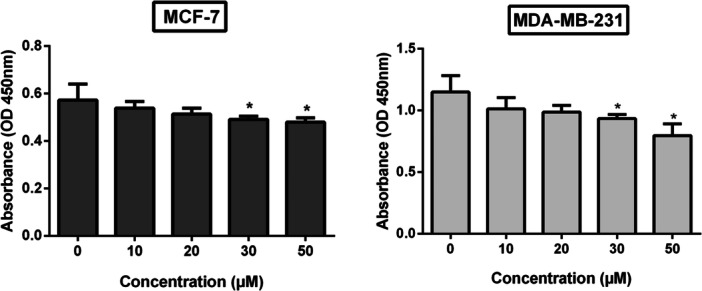
*The cell viability of both MCF‐7 and MDA‐MB‐231 cells was assessed by a CCK‐8 assay after 24 h with increasing compound concentrations. Data from three separate trials (n= 3) are shown as mean ± SD, and statistical significance in comparison to the control is highlighted (p< 0.05).
Light microscopy and the Trypan Blue exclusion experiment in breast cancer cells confirmed the compound's anti‐proliferative action (Figure3). The cytotoxicity of the compound was evaluated in MCF‐7 and MDA‐MB‐231 cells through morphological assessment and viability analysis Figure3A,B).
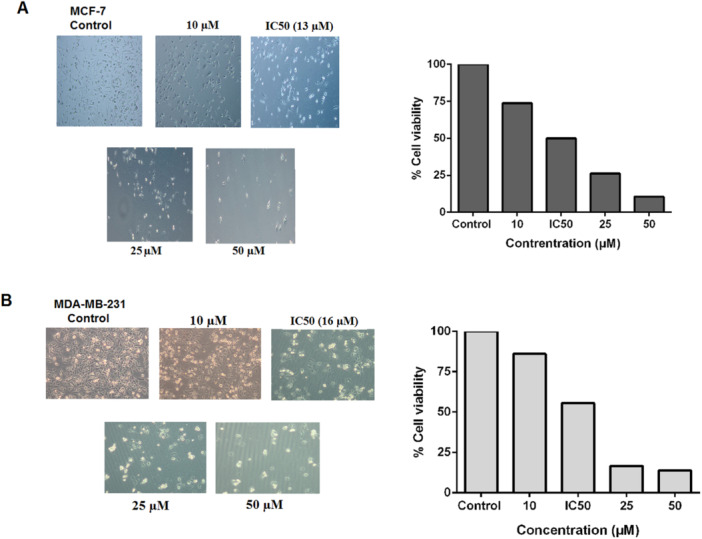
Trypan blue exclusion assay findings and typical light microscopy pictures demonstrate a dose‐dependent decrease in the viability in MCF‐7 (A) and MDA‐MB‐231 (B) cells after 24 h of treatment with the compound (10–50 µM and IC50).
In MCF‐7 cells (Figure3A), compound treatment induced dose‐dependent morphological alterations, including decreased cell density, cell rounding, detachment, and increased cellular debris compared with control cells. Cell viability decreased to 50% at the IC50concentration. Similarly, MDA‐MB‐231 cells (Figure3B) exhibited pronounced dose‐dependent cytotoxic effects, characterized by cell shrinkage, rounding, detachment, and reduced cell numbers relative to controls. Cell viability decreased to 55% as a result of the IC50concentration.
Overall, these findings indicate that the compound exerts a significant, dose‐dependent cytotoxic effect in each of the cell lines.
Apoptotic Effect of 2‐Pyrazoline‐5‐One Derivative in Breast Cancer Cells
Using the Annexin V/PI double‐staining approach, the apoptotic effects of the 2‐pyrazoline‐5‐one derivative were assessed in MCF‐7 and MDA‐MB‐231 cells. The two cell lines in the control groups showed very little Annexin V and PI positivity, suggesting that the great majority of cells were still viable. On the other hand, groups exposed to the compound at the IC50concentration showed a significant raised the number of Annexin V‐positive cells (Figure4).
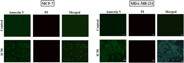
The presentation of fluorescence microscopy images of MCF‐7 and MDA‐MB‐231 breast cancer cells in control and IC50‐treated groups after they were incubated with the 2‐pyrazoline‐5‐one derivative for 24 h and then stained with Annexin V/PI (n= 3).
In MCF‐7 cells, IC50treatment caused a pronounced rise, particularly in early apoptotic cells, which are characterized by an Annexin V‐positive and PI‐negative population. Merged images clearly demonstrated an enhanced density of apoptotic cells. Likewise, in MDA‐MB‐231 cells, a significant increase in Annexin V positivity was detected following IC50treatment, and merged images showed significantly more apoptotic cells than the control group. These findings indicate that the 2‐pyrazoline‐5‐one derivative effectively induces apoptotic process in two different cancer cells and exerts its antiproliferative effect primarily by activating programmed cell death.
Inhibitory Effect of 2‐Pyrazoline‐5‐One Derivative on Colony Formation
The colony formation assay demonstrated that treatment with the 2‐pyrazoline‐5‐one derivative significantly suppressed clonogenic survival in both breast cancer cells in a concentration‐related way (Figure5). Representative images clearly show a progressive reduction in colony number with increasing concentrations of the compound.
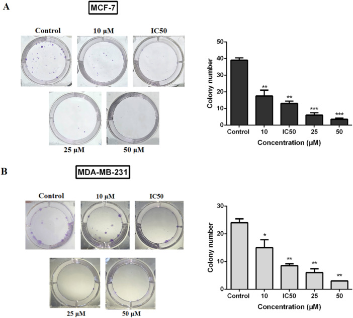
Clonogenic assay demonstrating the inhibitory effect of the 2‐pyrazoline‐5‐one derivative on long‐term survival of MCF‐7 (A) and MDA‐MB‐231 (B) cancer cells. Cells were cultured for 14–21 days (n= 2) following a 24 h treatment.p< 0.05,p< 0.01, andp< 0.001 with regard to the control.
In MCF‐7 cells, colony numbers decreased markedly starting at 10 µM and were significantly reduced at IC50and higher concentrations. At 50 µM, colony formation was nearly completely inhibited. Similarly, MDA‐MB‐231 cells displayed a notable dose‐dependent decline in colonial numbers, with substantial suppression observed at IC50and above, and minimal colony formation at 50 µM.
Overall, these findings suggest that the compound effectively inhibits a longer‐term proliferation potential and clonogenic survival of two different cancer cells, with a more pronounced suppressive effect at higher concentrations.
Cell Migration Analysis by Wound Healing (Scratch) Assay
2‐pyrazoline‐5‐one derivative significantly inhibited cell migration in both breast cancer cells. After 24 h, partial wound closure was observed in the control group of the MCF‐7 cells. However, compound treatment with IC50concentration resulted in a significant enhancement of wound width against the control group. Quantitative analysis confirmed that wound closure was significantly suppressed in IC50‐treated group (p< 0.001), indicating strong inhibition of migratory capacity (Figure6A).
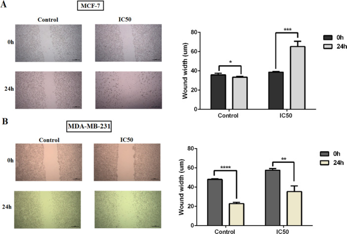
*A wound healing test was used to evaluate the impact of IC50treatment on cell migration in MCF‐7 (A) and MDA‐MB‐231 (B) cells. The related quantification of wound width (µm) and representative images for 0 and 24 h are displayed. IC50treatment significantly inhibited wound closure after 24 h (n= 3).p< 0.05,p< 0.01,*p< 0.001, andp< 0.0001.
Similarly, MDA‐MB‐231 cells displayed substantial wound closure in the control group at 24 h. However, treatment with IC50concentration significantly reduced wound closure (p< 0.01) at 24 h (Figure6B). Although MDA‐MB‐231 cells possess higher intrinsic migratory potential, the compound effectively attenuated their migration.
Effect of Treatment on Bax Protein Expression in Breast Cancer Cells
The ELISA approach was used to assess changes in Bax protein levels in the breast cancer cell lines MCF‐7 and MDA‐MB‐231. In comparison to the control group, Bax levels increased in two types of cell at all tested concentrations of the 2‐pyrazoline‐5‐one derivative (Figure7).
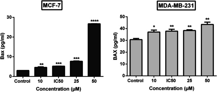
*Effect of the 2‐pyrazoline‐5‐one derivative on Bax protein levels in MCF‐7 and MDA‐MB‐231 cells after a 24 h exposure (n= 3).p< 0.05,p< 0.01,*p< 0.001, andp< 0.0001 versus to the control.
Bax expression in MCF‐7 cells increased markedly in a concentration‐dependent manner, particularly at IC50, 25 µM, and 50 µM, this increase was proved to be extremely and very highly statistically significant (p< 0.001;p< 0.0001). At the IC50concentration, Bax protein levels increased by 72% in a comparison with the control group.
In MDA‐MB‐231 cells, the increase in Bax levels was more moderate; however, it became statistically significant starting from 10 µM (p< 0.05) and showed a consistent upward trend while maintaining significance at IC50, 25 µM, and 50 µM (p< 0.01). At the IC50concentration, Bax levels increased by 23% relative to the control group, indicating a meaningful and consistent apoptotic response to the compound in MDA‐MB‐231 type cells as well.
These findings show that 2‐pyrazoline‐5‐one derivative exerts pro‐apoptotic effects in two types of cells; however, MCF‐7 cells exhibit a stronger Bax‐mediated apoptotic response than MDA‐MB‐231 cells. Overall, the outcomes suggest that the compound triggers apoptosis with upregulating BAX expression. As a result, this compound may cause cancer cells to undergo apoptosis by activating BAX. It could potentially act as a BAX activator, similar to BTSA1, in terms of its mechanism of action.
Discussion
Incidence of cancer is expected to increase significantly over the next several decades, making it the second most common cause of death globally, after cardiovascular disorders [19]. Breast cancer is the most common cancer diagnosed in women and a major contributor to cancer‐related mortality. Its molecular heterogeneity—particularly the distinction between a typical hormone receptor–positive (luminal) and triple‐negative subtypes—continues to present significant therapeutic challenges [20]. The development of novel agents capable of selectively inducing apoptosis while suppressing tumor progression remains a major focus in translational oncology.
In the present study, we demonstrate that a multifunctional 2‐pyrazoline‐5‐one derivative exerts potent anticancer activity in both luminal A (MCF‐7) and triple‐negative (MDA‐MB‐231) breast cancer cell lines. Notably, the compound displayed low micromolar IC50values (13 µM for MCF‐7 and 16 µM for MDA‐MB‐231), indicating strong cytotoxic efficacy across molecularly distinct subtypes. The slightly greater sensitivity observed in MCF‐7 cells suggests that hormone receptor–positive breast cancer cells may be more susceptible to this scaffold. This observation is consistent with previous reports describing enhanced antiproliferative activity of pyrazoline derivatives in ER‐positive models [21,22].
Importantly, the cytotoxicity observed was accompanied by classical apoptotic morphology and a marked increase in Annexin V‐positive populations, indicating that apoptosis—rather than nonspecific necrosis—constitutes the primary mechanism of cell death. Such findings are consistent with past research showing that pyrazoline‐ and hydrazone‐based heterocycles promote apoptosis [23,24]. The predominance of early apoptotic cells at IC50concentrations suggests that the compound initiates programmed cell death efficiently and at an early stage of treatment.
Beyond short‐term cytotoxicity, the compound significantly suppressed clonogenic survival in both cell lines. Since clonogenic assays reflect capacity of individual tumor cells to undergo unlimited division and contribute to recurrence, this observation is particularly relevant. The marked inhibition of colony formation at IC50and higher concentrations indicates durable antiproliferative activity and suggests that the compound may interfere with long‐term tumor sustainability. Similar sustained suppression of clonogenic potential has been reported for hybrid heterocyclic anticancer agents integrating multiple pharmacophores [25].
The therapeutic importance of these discoveries is further reinforced by the suppression of cell migration. Increased invasiveness and metastatic potential are characteristics of triple‐negative breast cancer. The observation that migration was significantly attenuated even in MDA‐MB‐231 cells underscores the compound's potential antimetastatic activity. Similar effects have been reported in previous studies showing that triazole‐ and pyrazoline‐based heterocyclic compounds can impair migration [26,27] and related malignant phenotypes in breast‐derived cancer cells.
Mechanistically, the study's most striking outcome is the significant upregulation of BAX protein levels following treatment. BAX is a central effector of mitochondrial outer membrane permeabilization, the irreversible step that commits cells to intrinsic apoptosis [28]. The observed dose‐dependent increase in Bax expression—particularly 72% elevation in MCF‐7 cells at IC50—strongly implicates activation of the mitochondrial apoptotic pathway. The comparatively attenuated increase in MDA‐MB‐231 cells (23%) may reflect the intrinsically higher apoptotic threshold and anti‐apoptotic protein dominance in TNBC cells.
The structural relevance of this finding is noteworthy. The tested compound shares the arylhydrazono‐2‐pyrazoline‐5‐one scaffold with BTSA1. BTSA1 is a direct BAX activator that has been pharmacologically optimized. It interacts with BAX's N‐terminal activation site and causes conformational activation [13]. BTSA1 and its derivatives have demonstrated pro‐apoptotic activity in hematologic and solid tumor models, typically within the low‐to‐mid micromolar range [16,17]. The IC50values observed in our study suggest that this compound may exhibit a similar functional profile.
While our current data demonstrate increased BAX expression, it is possible that the compound may facilitate the conformational activation of BAX or interfere with its inhibitory interactions with members of the anti‐apoptotic BCL‐2 family. If confirmed, this dual capacity to upregulate and activate BAX would establish the molecule as a mechanistically targeted modulator of apoptosis. As resistance to apoptosis is a characteristic of aggressive breast cancers, the pharmacological reactivation of BAX is a highly attractive therapeutic strategy.
To our knowledge, direct BAX‐targeting small molecules have not been extensively evaluated in parallel across luminal and triple‐negative breast cancer models. Therefore, the present findings provide novel insight into BAX‐mediated apoptotic modulation in distinct breast cancer subtypes and highlight the translational potential of arylhydrazono‐pyrazolinone scaffolds in solid tumors.
Conclusion
In conclusion, the 2‐pyrazoline‐5‐one derivative demonstrates pronounced anticancer activity in both luminal A (MCF‐7) and triple‐negative breast cancer (MDA‐MB‐231) cells. Our compound induces dose‐dependent cytotoxicity, promotes apoptosis, suppresses clonogenic survival, inhibits migration, and significantly upregulates Bax expression. The marked increase in Bax levels suggests the engagement of the intrinsic apoptotic pathway, indicating the activation of mitochondria‐mediated apoptotic signaling. This observation raises the possibility that this molecule may function as a Bax‐modulating or Bax‐activating agent. The present findings highlight this scaffold as a potential candidate for mitochondria‐targeted therapeutic strategies in breast cancer.
Author Contributions
Sevgi Kocyigit Sevinc:conceptualization, investigation, funding acquisition, writing – original draft, methodology, validation, visualization, writing – review and editing, project administration, supervision, resources.Ş. Güniz Küçükgüzel:writing – original draft, methodology, investigation.Sevim Rollas:methodology, investigation.Fulya Yukcu:methodology.
Ethics Statement
Since this work used commercially available, established cell lines, ethical approval was not necessary. Research involving human or animal subjects that needs ethics committee approval does not use these cell lines.
Consent
The authors have nothing to report.
Conflicts of interest
The authors declare no conflicts of interest.
References
- S. Łukasiewicz, M. Czeczelewski, A. Forma, J. Baj, R. Sitarz, andA. Stanisławek, “Breast Cancer‐Epidemiology, Risk Factors, Classification, Prognostic Markers, and Current Treatment Strategies‐An Updated Review, ”Cancers13, no. 17(2021): 4287. doi.org/10.3390/cancers13174287
- E. Carvalho, S. Canberk, F. Schmitt, andN. Vale, “Molecular Subtypes and Mechanisms of Breast Cancer: Precision Medicine Approaches for Targeted Therapies, ”Cancers17, no. 7(2025): 1102. doi.org/10.3390/cancers17071102
- B. Kinnel, S. K. Singh, G. Oprea‐Ilies, andR. Singh, “Targeted Therapy and Mechanisms of Drug Resistance in Breast Cancer, ”Cancers15, no. 4(2023): 1320. doi.org/10.3390/cancers15041320
- Ş. Güniz Küçükgüzel, S. Rollas, H. Erdeniz, M. Kiraz, A. Cevdet Ekinci, andA. Vidin, “Synthesis, Characterization and Pharmacological Properties of Some 4‐arylhydrazono‐2‐pyrazoline‐5‐one Derivatives Obtained From Heterocyclic Amines, ”European Journal of Medicinal Chemistry35, no. 7–8(2000): 761–771. doi.org/10.1016/s0223-5234(00)90179-x
- B. Nehra, S. Rulhania, S. Jaswal, B. Kumar, G. Singh, andV. Monga, “Recent Advancements in the Development of Bioactive Pyrazoline Derivatives, ”European Journal of Medicinal Chemistry205(2020): 112666. doi.org/10.1016/j.ejmech.2020.112666
- E. A. Borrego, C. D. Guerena, A. Y. Schiaffino Bustamante, et al. , “A Novel Pyrazole Exhibits Potent Anticancer Cytotoxicity via Apoptosis, Cell Cycle Arrest, and the Inhibition of Tubulin Polymerization in Triple‐Negative Breast Cancer Cells, ”Cells13, no. 14(2024): 1225. doi.org/10.3390/cells13141225
- P. K. Ray, R. Sharma, andA. Kumar, “Pyrazoline and Pyrazoline Derivatives as Anti‐Cancer Agents: Synthesis and Biological Activities: A Compressive Review, ”Current Topics in Medicinal Chemistry25, no. 13(2025): 1548–1612. doi.org/10.2174/0115680266339816241212081205
- M. Karabacak, M. Altıntop, H. İbrahim Çiftçi, et al. , “Synthesis and Evaluation of New Pyrazoline Derivatives as Potential Anticancer Agents, ”Molecules20, no. 10(2015): 19066–19084. doi.org/10.3390/molecules201019066
- N. Hosseini Nasab, F. Azimian, R. S. Shim, et al. , “Synthesis, Anticancer Evaluation, and Molecular Docking Studies of Thiazolyl‐Pyrazoline Derivatives, ”Bioorganic and Medicinal Chemistry Letters80(2022): 129105. doi.org/10.1016/j.bmcl.2022.129105
- D. MatiadisandM. Sagnou, “Pyrazoline Hybrids as Promising Anticancer Agents: An Up‐To‐Date Overview, ”International Journal of Molecular Sciences21, no. 15(2020): 5507. doi.org/10.3390/ijms21155507
- Ş. G. KüçükgüzelandP. Çıkla‐Süzgün, “Recent Advances Bioactive 1, 2, 4‐Triazole‐3‐Thiones, ”European Journal of Medicinal Chemistry97(2015): 830–870. doi.org/10.1016/j.ejmech.2014.11.033
- S. K. Sevinc, P. Çıkla‐Süzgün, P. M. Tiber, Ş. Güniz Küçükgüzel, andO. Orun, “Biological Activities of Etodolac‐Based Hydrazone, Thiazolidinone and Triazole Derivatives on Breast Cancer Cell Lines MCF‐7 and MDA‐MB−231, ”Journal of Biochemical and Molecular Toxicology39, no. 8(2025): e70428. doi.org/10.1002/jbt.70428
- D. E. ReynaandE. Gavathiotis, “Pulling the BAX Trigger for Tumor Cell Death, ”Oncotarget9, no. 9(2018): 8204–8205. doi.org/10.18632/oncotarget.24201
- D. E. Reyna, T. P. Garner, A. Lopez, et al. , “Direct Activation of BAX by BTSA1 Overcomes Apoptosis Resistance in Acute Myeloid Leukemia, ”Cancer Cell32, no. 4(2017): 490–5050000000000. doi.org/10.1016/j.ccell.2017.09.001
- T. Liang, Y. Zhou, R. M. Elhassan, X. Hou, X. Yang, andH. Fang, “HDAC‐BAX Multiple Ligands Enhance Bax‐Dependent Apoptosis in HeLa Cells, ”Journal of Medicinal Chemistry63, no. 20(2020): 12083–12099. doi.org/10.1021/acs.jmedchem.0c01454
- Z. Zhang, S. Zhao, J. Pei, et al. , “Optimization of BAX Trigger Site Activator BTSA1 With Improved Antitumor Potency and in Vitro ADMET Properties, ”European Journal of Medicinal Chemistry248(2023): 115076. doi.org/10.1016/j.ejmech.2022.115076
- A. Lopez, D. E. Reyna, N. Gitego, et al. , “Co‐Targeting of BAX and BCL‐XL Proteins Broadly Overcomes Resistance to Apoptosis in Cancer, ”Nature Communications13, no. 1(2022): 1199. doi.org/10.1038/s41467-022-28741-7
- B. Narasimhan, P. Kumar, andD. Sharma, “Biological Activities of Hydrazide Derivatives in the New Millennium, ”Acta Pharmaceutica Sciencia52(2010): 169–180.
- R. L. Siegel, A. N. Giaquinto, andA. Jemal, “Cancer Statistics, 2024, ”CA: A Cancer Journal for Clinicians74, no. 1(2024): 12–49. doi.org/10.3322/caac.21820
- G. Bianchini, J. M. Balko, I. A. Mayer, M. E. Sanders, andL. Gianni, “Triple‐Negative Breast Cancer: Challenges and Opportunities of a Heterogeneous Disease, ”Nature Reviews Clinical Oncology13, no. 11(2016): 674–690. doi.org/10.1038/nrclinonc.2016.66
- I. Althagafi, N. M. El‐Metwaly, M. G. Elghalban, T. A. Farghaly, andA. M. Khedr, “Synthesis of Pyrazolone Derivatives and Their Nanometer Ag(I) Complexes and Physicochemical, DNA Binding, Antitumor, and Theoretical Implementations, ”Bioinorganic Chemistry and Applications2018(2018): 2727619. doi.org/10.1155/2018/2727619
- M. M. Fakhry, K. Mahmoud, M. S. Nafie, et al. , “Rational Design, Synthesis and Biological Evaluation of Novel Pyrazoline‐Based Antiproliferative Agents in MCF‐7 Cancer Cells, ”Pharmaceuticals15, no. 10(2022): 1245. doi.org/10.3390/ph15101245
- R. F. George, M. A. Fouad, andI. E. O. Gomaa, “Synthesis and Cytotoxic Activities of Some Pyrazoline Derivatives Bearing Phenyl Pyridazine Core as New Apoptosis Inducers, ”European Journal of Medicinal Chemistry112(2016): 48–59. doi.org/10.1016/j.ejmech.2016.01.048
- I. A. El Hassani, A. Altay, E. Yeniçeri, et al. , “Novel Hydrazone‐Derived Pyrazole‐Benzofuran Compounds Inhibit Cancer Cell Growth by Targeting MMPs, Caspases, and PI3K/AKT/mTOR Signaling Pathway, ”Bioorganic Chemistry171(2026): 109568. doi.org/10.1016/j.bioorg.2026.109568
- D. MatiadisandM. Sagnou, “Pyrazoline Hybrids as Promising Anticancer Agents: An Up‐To‐Date Overview, ”International Journal of Molecular Sciences21, no. 15(2020): 5507. doi.org/10.3390/ijms21155507
- H. Wiraswati, P. Satriyo, M. H. Bashari, et al. , “Pyrazoline B Induces Oxidative Stress Mediated Toxicity, Cell Cycle Arrest, and Caspase‐Independent Apoptosis in BT‐474 Human Breast Cancer Cells, ”Journal of Experimental Pharmacology17(2025): 823–839. doi.org/10.2147/JEP.S499127
- A. Šermukšnytė, K. Kantminienė, I. Jonuškienė, I. Tumosienė, andV. Petrikaitė, “The Effect of 1, 2, 4‐Triazole‐3‐thiol Derivatives Bearing Hydrazone Moiety on Cancer Cell Migration and Growth of Melanoma, Breast, and Pancreatic Cancer Spheroids, ”Pharmaceuticals15, no. 8(2022): 1026. doi.org/10.3390/ph15081026
- R. J. YouleandA. Strasser, “The BCL‐2 Protein Family: Opposing Activities That Mediate Cell Death, ”Nature Reviews Molecular Cell Biology9, no. 1(2008): 47–59. doi.org/10.1038/nrm2308
Republished from the open web under CC-BY. Authors: Sevinc SK, Küçükgüzel ŞG, Rollas S, Yukcu F. Read the original.