A subset of high-grade sarcomas with myogenic differentiation are associated with recurrent FGFR fusions.
Recurrent fusions involving FGFR1-4 genes have been previously described in rare subsets of mostly benign chondroid and mesenchymal neoplasms involving bone and soft tissue. However, a more comprehensive analysis of sarcomas associated with FGFR fusions, including their incidence and histotypes, has not been performed. Triggered by an FGFR1-rearranged unclassified high-grade sarcoma with myogenic differentiation, we investigated our molecular database for sarcomas with FGFR gene fusions to assess their recurrent potential and morphologic spectrum. A total of six unclassified sarcomas were identified, occurring in five females and one male, with a median age of 66.5 years. Tumors were located in the uterus and retroperitoneal/trunk soft tissue. Histologically, all tumors were high grade, composed predominantly of spindle cell morphology with variable cytologic atypia, high mitotic activity, and areas of necrosis. By immunohistochemistry, all tumors demonstrated evidence of myogenic differentiation, with focal or diffuse positivity for one or more markers (desmin, h-caldesmon, smooth muscle actin). However, none of the cases displayed diagnostic features of leiomyosarcoma or other known pathologic entities. Molecular studies revealed recurrent fusions involving FGFR1 (n = 3), FGFR2 (n = 1), and FGFR4 (n = 2), retaining the kinase domain. Additional recurrent genomic alterations included TP53 mutations, CDKN2A homozygous deletions, and RB1 alterations. Most patients followed an aggressive clinical course, including metastases and disease-related mortality. These findings expand the spectrum of sarcomas driven by FGFR gene fusions, underscoring the importance of molecular testing for accurate diagnosis and potential targeted therapy in high-grade sarcomas with myogenic features.
Introduction
The wide application of genomic testing in clinical practice has led to the identification of novel gene fusions as key oncogenic drivers in various subtypes of bone and soft tissue tumors. Among these, fusions involving members of the fibroblast growth factor receptor (FGFR) family – comprisingFGFR1, FGFR2,FGFR3, andFGFR4– have emerged as recurrent molecular alterations in several rare mesenchymal neoplasms, including soft tissue chondroma [1], calcified chondroid mesenchymal neoplasm (CCMN) [2,3,4,5], phosphaturic mesenchymal tumor [6], and gastrointestinal stromal tumors (GISTs) [7]. In addition,FGFRfusions have been recently described in tumors exhibiting an infantile fibrosarcoma (IFS)‐like morphology, suggesting that kinase fusion‐driven spindle cell neoplasms may be associated with a broader genetic spectrum [8]. Despite these advances, the incidence and clinicopathologic features of sarcomas driven byFGFRgene rearrangements remain poorly understood. In this study, we perform a comprehensive investigation of the presence ofFGFRfusions among a wide spectrum of sarcomas with well‐characterized pathologic features and genomic landscape. The goals of this work were twofold: first, to better characterize if these gene rearrangements may define a potential novel sarcoma subtype, and second, to investigate the occurrence ofFGFRfusions as passenger alterations among known sarcoma entities with well‐defined genetic drivers.
Materials and methods
Case selection and study cohort
After approval by the Institutional Review Board (IRB), all study subjects provided written informed consent to the use of their genomic data for research (IRB# 12‐245). Triggered by an index case of a retroperitoneal unclassified spindle cell sarcoma with myogenic differentiation harboring anFGFR1gene fusion, we investigated ourhttps://www.cbioportal.org(2014–2025) for sarcoma cases associated withFGFR1‐4gene fusions. Previously described bone and soft tissue tumor entities associated with recurrentFGFRfusions were excluded [1,2,3,4,5,6,7,8]. We first selected unclassified spindle cell sarcomas, not otherwise specified (NOS), that did not fit into any known pathologic category, in which theFGFRfusions may represent the driver alteration, thus defining a novel sarcoma entity. Clinical history, pathologic features, immunohistochemical results, and outcome data were retrieved from pathology reports and chart review. Additionally, we also sought to investigate the presence of co‐occurringFGFRfusions as secondary genetic alterations among well‐known sarcoma histotypes driven by other/known genomic events.
Genomic analysis
All cases were tested using the institutional targeted sequencing platform (MSK‐IMPACT™), a comprehensive molecular assay that involves hybridization capture and deep sequencing of all exons and selected introns of 341–505 oncogenes and tumor‐suppressor genes, allowing the detection of point mutations, small and large insertions, deletions, and rearrangements. In addition, the assay captures >1,000 intergenic and intronic single‐nucleotide polymorphisms, interspersed homogenously across the genome, aiding the assessment of genome‐wide copy number. The assay uses paired tumor/normal DNA for sequencing and analysis. Details of the MSK‐IMPACT assay have been previously published [9]. The tumor mutational burden (TMB) and fraction of genome altered (FGA) were also derived from MSK‐IMPACT data. TMB was determined by calculating the ratio of nonsynonymous mutations to the total number of base pairs sequenced in each sample. The FGA was measured as the proportion of the genome exhibiting a log2 copy‐number gain greater than 0.2 or a loss less than 0.2, relative to the total size of the genome profiled for copy‐number variations. Somatic variants were called by Mutect2 and annotated by Annovar [10], and the allele specific copy number variants were called by FACETS [11]. The genomic alterations were analyzed using R package ‘ComplexHeatmap’ version 2.15.4 [12]. The segmented copy number profiles were used to generate arm level copy number changes using ASCETS package [13]. In most cases, Archer® FusionPlex® Sarcoma Panel (Archer, Boulder, CO, USA) was applied for gene fusion confirmation, which is a custom amplicon‐based next generation sequencing assay using standard protocols, as previously described [14].
Results
Demographics and clinical information
A total of six unclassified sarcomas (high‐grade sarcoma, NOS) harboringFGFRfusions were selected from the MSK‐IMPACT database. All patients except one were female, with a median age at diagnosis of 66.5 years (mean: 68.5; range: 56–90 years). The tumor sizes ranged from 7.5 to 17.5 cm (mean of 12 cm). The uterus was the most common anatomic site (n= 3), followed by the retroperitoneum (n= 2), and paraspinal muscle (n= 1). The clinical features are summarized in Table1.
Table: Clinicopathological features of theFGFR‐fusion positive high‐grade sarcomas with myoid differentiation
Histopathologic and immunohistochemical findings
At low power, the tumors showed well‐demarcated borders, with a multinodular and solid architecture (Figure1A). All except one case (case 4) were characterized by a spindle cell morphology arranged in intersecting fascicles (Figure1B,C). Most tumors showed a relatively monomorphic cytology; however, a variable degree of nuclear pleomorphism was present (Figure1D,E). The tumors showed a moderate amount of fibrillary eosinophilic to amphophilic cytoplasm. An intervening collagenous stroma was present in most cases, with one case displaying myxoid stromal changes (case 2; Figure1F). In one case (case 4), the tumor showed a distinctive monomorphic epithelioid phenotype, arranged in nests, cords, trabeculae, and pseudo‐alveolar structures (Figure2A–C). Scattered giant tumor cells were seen in one case (case 1; Figure2D). Mitotic activity was high in all cases, ranging from 14 to 43 mitoses per 10 high‐power fields (HPFs) (mean, 25/10 HPFs). Atypical mitotic figures were readily seen. Necrosis was present in four cases.
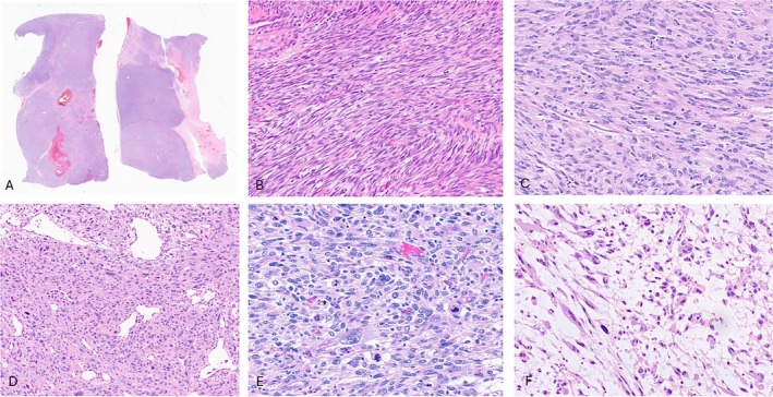
Histology of high‐grade sarcoma with myogenic differentiation andFGFRfusion. (A) Low power view showing well circumscribed borders and multilobular architecture with areas of geographic necrosis and hemorrhage (case 3). (B, C) The spindle cells are arranged in fascicular growth (cases 6 and 3; ×200 magnification). (D) Some tumors have overt cytologic atypia with marked nuclear pleomorphism (case 2; ×200 magnification) (E) and frequent mitoses (case 1; ×400 magnification). (F) Areas of myxoid stroma are present (case 2; ×400 magnification).
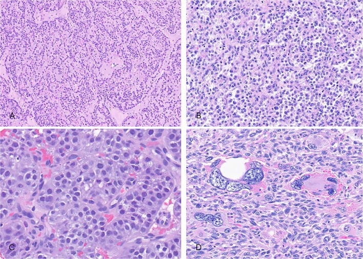
Morphologic appearance of one of theFGFRfusion high‐grade sarcomas with myogenic differentiation. (A–C) Epithelioid cells arranged in trabeculae, nests, cords, and alveolar‐like structures with relatively uniform cytology (case 4). (D) Multinucleated giant cells are seen in one case (case 1).
Immunohistochemically, all cases showed positivity for at least one smooth muscle marker, in keeping with myogenic differentiation. All except one case were positive for desmin, with an additional three cases being positive for h‐caldesmon (3/3) and SMA (3/5) positivity (Figure3). One case (case 6) was positive for all three markers. Detailed immunohistochemical results were summarized in Table2.
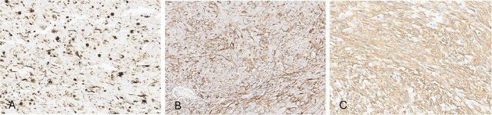
Immunohistochemical staining results of (A) desmin (case 1), (B) h‐caldesmon (case 3), and (D) smooth muscle actin (case 6).
Table: Key immunohistochemical and molecular findings of the study group
Genomic findings
The molecular features were summarized in Table2and Figure4. Half of the cases (3/6) harboredFGFR1fusions, with one case showingFGFR2and two casesFGFR4fusions. All the breakpoints inFGFR1/FGFR2/FGFR4genes were located at exon 17 except one at exon 13, with all retaining the kinase domain in the predicted fusion oncoprotein (Figure5). In four cases, the fusion transcript was detected by both IMPACT and Archer. In two cases, theFGFR4fusions were detected by IMPACT only but not by Archer. In both cases, the fusion was predicted by IMPACT to be in‐frame and included the kinase domain of FGFR4. Specifically, in case 5, theFGFR4::SH3PXD2Bfusion resulted from an inversion, withFGFR4exons 1–17 being fused toSH3PXD2Bexons 2–13. Similarly, the IMPACT analysis in case 6 detected anFGFR4rearrangement with breakpoint within exon 13 and including the kinase domain. In a retrospective review, the primer set for the Archer gene panel does not cover exons 17 and 13 ofFGFR4gene, respectively, thus leading to a false‐negative result. Therefore, these two cases are expected to produce a functional fusion transcript.
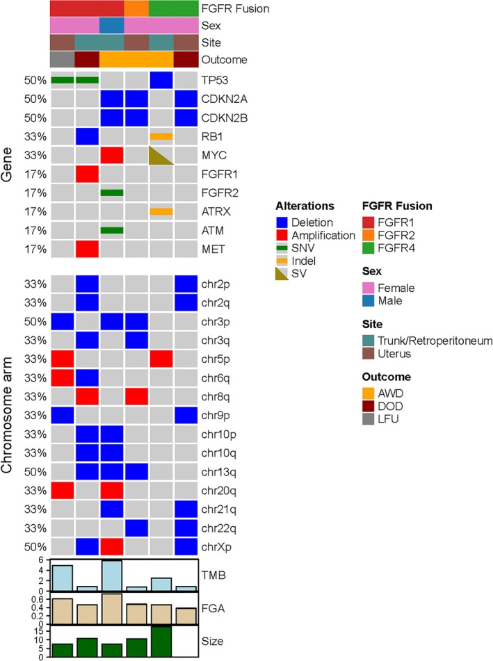
Oncoplot of molecular and clinicopathological findings in sarcoma with FGFR fusion, showing genomic alterations in selected genes and recurrent arm‐level copy number changes.
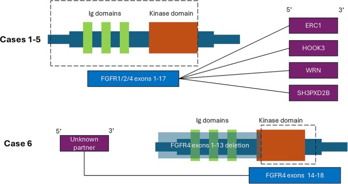
Schematic diagram of FGFR fusions in our cohort. In cases 1–5, FGFR genes were included as 5′ partner fused with different genes includingERC1,HOOK3,WRN, andSH3PXD2B. The breakpoint was consistently located after exon 17, thus retaining the kinase domain. In case 6, theFGFR4gene was present as 3′ partner, also retaining the kinase domain at exons 14–18.
By MSK‐IMPACT, additional secondary events were identified, includingTP53alterations (n= 3),CDKN2Ahomozygous deletions (n= 3),RB1alterations (n= 2), andMYCalterations (n= 2) (Table2, Figure4). The presence ofTP53alterations andCDKN2Adeletions was mutually exclusive, whileRB1gene alterations co‐occurred withTP53mutations. In addition, all cases had multiple copy number alterations, with a high FGA (median: 47.53%). However, the tumor mutation burden (TMB) was low in all cases (<10 mt/mb), with a median of 1.7 mt/mb. Recurrent arm‐level copy number changes included amplifications in 5p, 8q, and 20q, and deletions in 2p, 2q, 3p, 3q, 9p, 10p, 10q, 13q, 21q, and 22q.
Clinical management and outcome
All except one patient presented with localized disease and underwent definitive resection with negative margins, either a wide local excision or a total hysterectomy with bilateral salpingo‐oophorectomy. The remaining patient received palliative liposomal doxorubicin chemotherapy due to surgically unresectable disease with widespread distant metastases, including lung and pleura (case 2). Follow‐up was available in five patients with a median of 10.5 months (range 7–132 months). Most patients followed an aggressive clinical course. All five patients with available follow‐up developed local recurrence. In addition to the patient presenting with metastatic disease, one additional patient developed distant metastasis to bilateral lungs, 7 months after diagnosis (case 4). This patient was currently on chemotherapy with doxorubicin and trabectedin. At last follow up, two patients succumbed of disease, and three patients were alive with disease. None of the patients were given FGFR inhibitors.
FGFRfusions as secondary genetic alterations in common sarcoma types with known driver events
We identified 12 cases withFGFRfusions across a diverse spectrum of established sarcoma subtypes, as detailed in Table3. These included osteosarcomas (n= 3), intimal sarcomas (n= 2), and single cases of dedifferentiated liposarcoma, myxoinflammatory fibroblastic sarcoma, spindle cell/sclerosing rhabdomyosarcoma, embryonal rhabdomyosarcoma with anaplasia, undifferentiated pleomorphic sarcoma (UPS), and synovial sarcoma. These cases were characterized by either typical morphologic features, immunoprofile and/or displayed their pathognomonic genomic alterations (i.e.,MDM2/CDK4amplification,SS18fusion, etc.) for their respective diagnosis.FGFR1fusions were the most common (7/12), followed byFGFR3(n= 3) andFGFR2(n= 2), with all fusions predicted to be in‐frame and retaining the kinase domain, suggesting functional significance. The breakpoints were located at either exon 17 (n= 5) or exon 18 (n= 5) if theFGFRgenes were the 5′ partner and located at exon 9 (n= 1) or exon 4 (n= 1) if they were the 3′ partner. In 10 cases, fusion transcripts were identified by Archer. In two cases,FGFRfusions were detected by MSK‐IMPACT with no confirmation by Archer. Specifically, one case of dedifferentiated liposarcoma harboredFGFR1::TACC1fusion and one case of synovial sarcoma foundFGFR2::CCDC46fusion by MSK‐IMPACT, which were both predicted to be in‐frame with retained kinase domain. None of these patients were treated with FGFR inhibitors.
Table: FGFR fusion as likely secondary genetic alterations in specific sarcoma entities found in institutional database
Discussion
Constitutive activation of various kinases through oncogenic gene fusions plays an important role in the pathogenesis of human neoplasia, spanning many different lineages and risks of malignancies. In one large pan‐cancer study, FGFRs alterations were identified in 7.1% of tumors [15], with gene amplifications and mutations being more common than gene fusions. MostFGFRfusion genes have been reported in cholangiocarcinoma [16], glioblastomas [17], and urothelial carcinoma [18]. In soft tissue tumors, recurrentFGFRfusions often involving theFN1gene partner were first described in phosphaturic mesenchymal tumors [6], soft tissue chondroma [1], and calcified CCMN [1,2,3,4,5]. More recently,FGFRfusions have also been described in three cases of pediatric mesenchymal tumors sharing histologic overlap with IFS and ‘NTRK‐rearranged’ spindle cell neoplasms [8]. Additionally,FGFRfusions have been reported in quadruple wild‐type GIST [7], uterine sarcomas [19,20], rhabdomyosarcoma [21], and a high‐grade undifferentiated spindle cell sarcoma [22]. Mesenchymal tumors featuringFGFRfusions are summarized in Table4.
Table: Mesenchymal neoplasms featuring FGFR fusion
The FGFR family consists of four highly conserved single transmembrane tyrosine kinase receptors (FGFR1‐4) that are essential to physiologic processes such as cell proliferation, survival, and differentiation [23]. Similar to most other receptor tyrosine kinases (RTKs), FGFR comprises an extracellular ligand‐binding domain and a cytoplasmic conserved tyrosine kinase domain. However, in contrast to most other RTKs, there are two mechanisms generating oncogenic FGFR fusions, both resulting in ligand‐independent dimerization and constitutive activation of the FGFR kinase signaling pathway (Figure6). The first includes the FGFR as a 5′ partner, in which the N‐terminal domain, including the kinase domain of FGFR1‐4 (exons 11–18), is fused to a 3′ partner containing a constitutive dimerization/oligomerization motif [24,25]. This fusion transcript is encountered in pediatric kinase fusion‐positive mesenchymal tumors [8], uterine sarcoma [20], GIST [7], and rhabdomyosarcoma [21]. This fusion structure was also found in all cases of the current cohort of high‐grade sarcomas with myogenic differentiation, includingFGFR1::ERC1,FGFR1::HOOK3,FGFR2::WRN, andFGFR4::SH3PXD2B. Among these fusion partners, the coiled‐coil domains of HOOK3 and the oligomerization motifs of SH3PXD2B may facilitate spontaneous dimerization or clustering of the fusion proteins, promoting autophosphorylation and sustained kinase activity. This ligand‐independent activation of FGFR kinases results in the persistent activation of downstream signaling pathways, such as MAPK/ERK and PI3K/AKT, which are critical regulators of cell proliferation, survival, and migration. For the second mechanism, the C‐terminal of the FGFR protein is retained in the fusion protein, which is fused to the N‐terminal of FN1. The FN1 partner functions as a strong promoter, with the resulting fusion protein facilitating secretion and multimerization, and constitutive activation of the FGFR kinase signaling pathway. This alternative mechanism of fusion is detected within the spectrum of benign mesenchymal neoplasms, such as soft tissue chondroma, CCMN, and PMT [6]. In these cases, the 3′ end of the FGFR gene (breakpoint typically occurs at exons 5–9) includes the transmembrane domain (exon 10) and the entire tyrosine kinase domain (exons 11–18) [2].
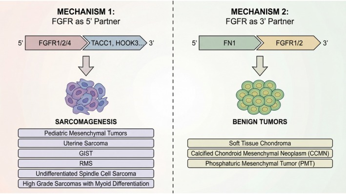
Clinical dichotomy ofFGFRfusions based on gene topology. The position of theFGFRgene within the fusion transcript correlates with biologic potential. Fusions featuringFGFRas the 5′ partner drive malignant sarcomagenesis, whereas fusions withFGFRas the 3′ partner define a spectrum of benign mesenchymal neoplasms.
Strikingly, the topologic orientation of theFGFRgene within the fusion construct strongly correlates with the histotype and clinical behavior, as 5′‐FGFRfusions predominantly drive sarcomas, whereas 3′‐FGFRfusions characterize benign neoplasms. This profound clinical divergence likely stems from differences in the spatial distribution and amplitude of their kinase signaling. WhenFGFRacts as the 5′ partner, fusion to 3′ coiled‐coil domains (e.g.,HOOK3,TACC1) drives robust, ligand‐independent oligomerization. This intense constitutive activation – potentially exacerbated by the fusion protein evading normal lysosomal degradation via cytoplasmic mislocalization – overwhelms intrinsic feedback loops and drives relentless downstream signaling, culminating in cellular dedifferentiation and sarcomagenesis [26]. Conversely, in 5′‐FN1fusions, fibronectin likely tethers the kinase to the secretory pathway or the extracellular matrix [27]. We postulate that this spatial confinement restricts the kinase from accessing critical intracellular substrates, resulting in a lower‐amplitude signal. Furthermore, while nativeFGFRpromoters (active in 5′ fusions) may link to cellular plasticity, the strongFN1promoter primarily drives localized matrix deposition and paracrine/endocrine signaling (e.g., FGF23). Thus, rather than cell‐autonomous malignant transformation, FN1‐driven fusions create a localized, hyperactive matrix‐modulating loop, leading to histologically benign neoplasms. Ultimately, fusion topology does not merely act as an ‘on‐switch’ forFGFR, but fundamentally dictates the spatial context and oncogenic trajectory of the signal [28].
Kinase fusion‐positive spindle cell neoplasms represent an emerging, molecularly defined subset, sharing a spectrum of morphologies and immunoprofiles [29]. Recent studies have implicated a wide range of kinases, includingNTRK1,NTRK2,NTRK3[30,31,32,33],BRAF[34,35,36],RAF1[37,38],RET[39], andLTK[40]. The phenotypic diversity includes mostly three architectural patterns, such as lipofibromatosis‐like neural tumors, IFS‐like and MPNST‐like [41], with common, but inconsistent, co‐expression of CD34 and S100 markers. In a recent study,FGFR1fusions have been found in three infants with IFS‐like tumors, arising in the gluteal and pelvic deep soft tissues [8]. Additionally, two cases of uterine sarcomas withFGFR1fusion were reported sharing a FS‐like phenotype with moderate nuclear atypia and co‐expression of CD34 and S100, likely fitting among the spectrum of kinase fusion sarcomas with high‐grade morphology [19,20]. In contrast, the three uterine tumors in the current series, harboredFGFR1, FGFR2, andFGFR4fusions, diverse histologic features and expression of smooth muscle markers, suggesting distinct pathogenesis. Another case report of anFGFR1‐rearranged high‐grade sarcoma occurring in the thigh of an 83‐year‐old man showed an FS‐like morphology and expression of S100, while CD34 and all three muscle markers were negative [22].
The histologic features seen in the current series did not fit with any well‐defined pathologic entities, including the morphologic spectrum of kinase fusion positive spindle cell neoplasms. Instead, tumors showed a high‐grade mostly spindle cell morphology with variable nuclear pleomorphism, increased mitotic activity and necrosis. Notably, all tumors showed expression of smooth muscle markers, including desmin in all except one case, in keeping with a high‐grade sarcoma with myogenic differentiation. Of note, none of the cases fulfilled the diagnostic criteria for leiomyosarcoma, lacking both the distinctive architectural pattern of perpendicularly intersecting long fascicles, as well as the typical cytologic features, such as fibrillary eosinophilic cytoplasm and elongated, cigar‐shaped nuclei. We have considered the possibility of a dedifferentiated leiomyosarcoma; however, no well‐differentiated component was identified in any of the extensively sampled cases. Moreover, no skeletal muscle markers were expressed to suggest a rhabdomyosarcoma diagnosis. Especially for the uterine location, another consideration was that of a malignant PEComa, but no expression of melanocytic markers, such as HMB45 or Melan‐A, was detected. In the retroperitoneum, another major differential diagnosis was dedifferentiated liposarcoma; however, there was no well‐differentiated liposarcoma component norMDM2andCDK4gene amplifications. The possibility of a high‐grade UPS was also considered, but the relatively monomorphic phenotype and consistent smooth muscle expression argued against.
Our findings suggest thatFGFRfusions may represent the driver alteration in a subset of high‐grade sarcomas with myogenic differentiation, adding this phenotype to the growing list of tumor types led by this oncogenic fusion.FGFR1fusions were the most common variant, but regardless of the gene involved, the kinase domain retained in the fusion was provided by the N‐terminal of the FGFR protein. The same mechanism is shared by all the other soft tissue sarcomas and kinase fusion spindle cell neoplasms harboring this fusion [8], in contrast to the opposite orientation ofFN1::FGFR1fusions found in the benign mesenchymal neoplasms [2]. In addition, our study sheds light on the incidence and sarcoma histotypes in whichFGFRfusions were detected likely as passengers or secondary events in otherwise well‐characterized sarcoma entities. The morphologic spectrum of this latter group was quite broad, but appeared enriched in genomically complex entities, such as osteosarcoma, UPS, etc. The most common fusion involvedFGFR1, as the 5′ partner. Notably, theFGFR3::TACC3fusion identified in two cases of osteosarcoma aligns with recent bioinformatics and functional studies confirming their role in enhancing invasiveness and metastasis [42,43]. The functional significance was further validated by anin vivostudy that demonstrated the success of induced pluripotent stem cell‐generated cytotoxic T lymphocytes targetingFGFR3::TACC3to inhibit growth and metastasis in a nude mouse osteosarcoma orthotopic transplantation model [44].
Finally, the identification ofFGFRfusions does not only impact the diagnosis and classification of these tumors, but may also represent an important druggable target. FGFR selective inhibitors targeting FGFR fusions, such as erdafitinib, pemigatinib, and futibatinib [45], have been approved by the U.S. Food and Drug Administration (FDA) to treat patients with urothelial cancer and cholangiocarcinoma. Our findings underscore the importance of integrating molecular testing into the diagnostic workup of unclassified high‐grade sarcomas and provide new insights into the biology of this emerging sarcoma subset.
In summary, we describe the clinical, histopathologic, and molecular features of six cases of high‐grade sarcoma with myogenic differentiation harboringFGFRfusions. Patients followed an aggressive clinical course, with local recurrences and distant metastases. As these tumors were initially deemed unclassified, not fitting any known pathologic categories, it is likely that theFGFRfusions may define a novel subset of high‐grade sarcomas with myogenic lineage. In addition, the presence ofFGFRfusions highlights their potential as therapeutic targets, in using FGFR inhibitors as a promising strategy for these patients. Future larger studies with integrated genomic alterations may further support the accurate classification of these lesions. Additional functional studies are also warranted to elucidate the precise mechanisms by which these fusions contribute to sarcomagenesis and to explore their therapeutic implications.
Author contributions statement
MCFY performed data retrieval and genomic analysis, reviewed histology and wrote the manuscript. CS reviewed the genomic data. CRA conceived and supervised the study, reviewed the histology and wrote the manuscript. All authors reviewed and approved the manuscript.
References
- AmaryF, Perez‐CasanovaL, YeH, et al. Synovial chondromatosis and soft tissue chondroma: extraosseous cartilaginous tumor defined by FN1 gene rearrangement. Mod Pathol2019; 32: 1762–1771. doi.org/10.1038/s41379-019-0315-8
- LiuYJ, WangW, YehJ, et al. Calcified chondroid mesenchymal neoplasms with FN1‐receptor tyrosine kinase gene fusions including FGFR2, FGFR1, MERTK, NTRK1, and TEK: a molecular and clinicopathologic analysis. Mod Pathol2021; 34: 1373–1383. doi.org/10.1038/s41379-021-00786-x
- BenardC, Le LoarerF, Gomez‐MascardA, et al. Comprehensive molecular characterization of a large series of calcified chondroid mesenchymal neoplasms widening their morphologic spectrum. Am J Surg Pathol2024; 48: 991–1004. doi.org/10.1097/PAS.0000000000002260
- FengX, WangS, WeiJ, et al. Calcified chondroid mesenchymal neoplasm: a clinicopathological and molecular analysis. J Clin Pathol2025: jcp‐2024‐209806. . doi.org/10.1136/jcp-2024-209806
- GeorgantzoglouN, ShenG, JourG, et al. A case of FN1‐fused calcified chondroid mesenchymal neoplasm of the hand with novel FGFR3 partner gene. Genes Chromosomes Cancer2023; 62: 237–241. doi.org/10.1002/gcc.23115
- LeeJC, JengYM, SuSY, et al. Identification of a novel FN1‐FGFR1 genetic fusion as a frequent event in phosphaturic mesenchymal tumour. J Pathol2015; 235: 539–545. doi.org/10.1002/path.4465
- ShiE, ChmieleckiJ, TangCM, et al. FGFR1 and NTRK3 actionable alterations in ‘wild‐type’ gastrointestinal stromal tumors. J Transl Med2016; 14: 339. doi.org/10.1186/s12967-016-1075-6
- WarmkeLM, Al‐IbraheemiA, WangL, et al. FGFR1 gene fusions in a subset of pediatric mesenchymal tumors: expanding the genetic spectrum of tumors sharing histologic overlap with infantile fibrosarcoma and ‘NTRK‐rearranged’ spindle cell neoplasms. Genes Chromosomes Cancer2023; 62: 641–647. doi.org/10.1002/gcc.23179
- ChengDT, MitchellTN, ZehirA, et al. Memorial Sloan Kettering‐integrated mutation profiling of actionable cancer targets (MSK‐IMPACT): a hybridization capture‐based next‐generation sequencing clinical assay for solid tumor molecular oncology. J Mol Diagn2015; 17: 251–264. doi.org/10.1016/j.jmoldx.2014.12.006
- WangK, LiM, HakonarsonH. ANNOVAR: functional annotation of genetic variants from high‐throughput sequencing data. Nucleic Acids Res2010; 38: e164. doi.org/10.1093/nar/gkq603
- ShenR, SeshanVE. FACETS: allele‐specific copy number and clonal heterogeneity analysis tool for high‐throughput DNA sequencing. Nucleic Acids Res2016; 44: e131. doi.org/10.1093/nar/gkw520
- GuZ, EilsR, SchlesnerM. Complex heatmaps reveal patterns and correlations in multidimensional genomic data. Bioinformatics2016; 32: 2847–2849. doi.org/10.1093/bioinformatics/btw313
- SpurrLF, TouatM, TaylorAM, et al. Quantification of aneuploidy in targeted sequencing data using ASCETS. Bioinformatics2021; 37: 2461–2463. doi.org/10.1093/bioinformatics/btaa980
- ZhengZ, LiebersM, ZhelyazkovaB, et al. Anchored multiplex PCR for targeted next‐generation sequencing. Nat Med2014; 20: 1479–1484. doi.org/10.1038/nm.3729
- HelstenT, ElkinS, ArthurE, et al. The FGFR landscape in cancer: analysis of 4, 853 tumors by next‐generation sequencing. Clin Cancer Res2016; 22: 259–267. doi.org/10.1158/1078-0432.CCR-14-3212
- NeumannO, BurnTC, AllgauerM, et al. Genomic architecture of FGFR2 fusions in cholangiocarcinoma and its implication for molecular testing. Br J Cancer2022; 127: 1540–1549. doi.org/10.1038/s41416-022-01908-1
- LiuJ, WangZ. The landscape of FGFR‐TACC fusion in adult glioblastoma: from bench to bedside. Mutat Res Rev Mutat Res2025; 795: 108536. doi.org/10.1016/j.mrrev.2025.108536
- De LucaA, Esposito AbateR, RachiglioAM, et al. FGFR fusions in cancer: from diagnostic approaches to therapeutic intervention. Int J Mol Sci2020; 21. . doi.org/10.3390/ijms21186856
- DevereauxKA, WeielJJ, MillsAM, et al. Neurofibrosarcoma revisited: an institutional case series of uterine sarcomas harboring kinase‐related fusions with report of a novel FGFR1‐TACC1 fusion. Am J Surg Pathol2021; 45: 638–652. doi.org/10.1097/PAS.0000000000001644
- ZylaRE, GoebelEA, JangJH, et al. Uterine sarcoma with FGFR1‐TACC1 gene fusion: a case report and review of the literature. Int J Gynecol Pathol2022; 41: 588–592. doi.org/10.1097/PGP.0000000000000846
- de Traux De WardinH, CyrtaJ, DermawanJK, et al. FGFR1 fusions as a novel molecular driver in rhabdomyosarcoma. Genes Chromosomes Cancer2024; 63: e23232. doi.org/10.1002/gcc.23232
- YauDT, LacambraMD, ChowC, et al. The novel finding of an FGFR1: : TACC1 fusion in an undifferentiated spindle cell sarcoma of soft tissue with aggressive clinical course. Genes Chromosomes Cancer2022; 61: 206–211. doi.org/10.1002/gcc.23024
- DaiS, ZhouZ, ChenZ, et al. Fibroblast growth factor receptors (FGFRs): structures and small molecule inhibitors. Cells2019; 8. . doi.org/10.3390/cells8060614
- FrattiniV, PagnottaSM, Tala, et al. A metabolic function of FGFR3‐TACC3 gene fusions in cancer. Nature2018; 553: 222–227. doi.org/10.1038/nature25171
- SinghD, ChanJM, ZoppoliP, et al. Transforming fusions of FGFR and TACC genes in human glioblastoma. Science2012; 337: 1231–1235. doi.org/10.1126/science.1220834
- LatyshevaNS, BabuMM. Discovering and understanding oncogenic gene fusions through data intensive computational approaches. Nucleic Acids Res2016; 44: 4487–4503. doi.org/10.1093/nar/gkw282
- DallasSL, SivakumarP, JonesCJ, et al. Fibronectin regulates latent transforming growth factor‐beta (TGF beta) by controlling matrix assembly of latent TGF beta‐binding protein‐1. J Biol Chem2005; 280: 18871–18880. doi.org/10.1074/jbc.M410762200
- CasalettoJB, McClatcheyAI. Spatial regulation of receptor tyrosine kinases in development and cancer. Nat Rev Cancer2012; 12: 387–400. doi.org/10.1038/nrc3277
- SuurmeijerAJH, KaoYC, AntonescuCR. New advances in the molecular classification of pediatric mesenchymal tumors. Genes Chromosomes Cancer2019; 58: 100–110. doi.org/10.1002/gcc.22681
- BrcicI, GodschachnerTM, BergovecM, et al. Broadening the spectrum of NTRK rearranged mesenchymal tumors and usefulness of pan‐TRK immunohistochemistry for identification of NTRK fusions. Mod Pathol2021; 34: 396–407. doi.org/10.1038/s41379-020-00657-x
- CoccoE, ScaltritiM, DrilonA. NTRK fusion‐positive cancers and TRK inhibitor therapy. Nat Rev Clin Oncol2018; 15: 731–747. doi.org/10.1038/s41571-018-0113-0
- CostiganDC, NucciMR, DicksonBC, et al. NTRK ‐rearranged uterine sarcomas: clinicopathologic features of 15 cases, literature review, and risk stratification. Am J Surg Pathol2022; 46: 1415–1429. doi.org/10.1097/PAS.0000000000001929
- KaoYC, SuurmeijerAJH, ArganiP, et al. Soft tissue tumors characterized by a wide spectrum of kinase fusions share a lipofibromatosis‐like neural tumor pattern. Genes Chromosomes Cancer2020; 59: 575–583. doi.org/10.1002/gcc.22877
- KaoYC, FletcherCDM, AlaggioR, et al. Recurrent BRAF gene fusions in a subset of pediatric spindle cell sarcomas: expanding the genetic spectrum of tumors with overlapping features with infantile fibrosarcoma. Am J Surg Pathol2018; 42: 28–38. doi.org/10.1097/PAS.0000000000000938
- KobayashiH, ZhangL, OkajimaK, et al. BRAF mutations and concurrent alterations in patients with soft tissue sarcoma. Genes Chromosomes Cancer2023; 62: 648–654. doi.org/10.1002/gcc.23182
- PenningAJ, Al‐IbraheemiA, MichalM, et al. Novel BRAF gene fusions and activating point mutations in spindle cell sarcomas with histologic overlap with infantile fibrosarcoma. Mod Pathol2021; 34: 1530–1540. doi.org/10.1038/s41379-021-00806-w
- YeungMCF, LamAYL, ShekTWH. Novel MAP4: : RAF1 fusion in a primary bone sarcoma: expanding the spectrum of RAF1 fusion sarcoma. Int J Surg Pathol2022; 30: 682–688. doi.org/10.1177/10668969221080059
- ZhuP, ZengX, YangL, et al. RAF1‐rearranged spindle cell neoplasm: a clinicopathological and molecular genetic study of six cases with review of the literature. Histopathology2025; 87: 388–396. doi.org/10.1111/his.15445
- AntonescuCR, DicksonBC, SwansonD, et al. Spindle cell tumors with RET gene fusions exhibit a morphologic spectrum akin to tumors with NTRK gene fusions. Am J Surg Pathol2019; 43: 1384–1391. doi.org/10.1097/PAS.0000000000001297
- YeungMCF, DermawanJK, LiuAPY, et al. Spindle cell neoplasms with novel LTK fusion – expanding the spectrum of kinase fusion‐positive soft tissue tumors. Genes Chromosomes Cancer2024; 63: e23227. doi.org/10.1002/gcc.23227
- AntonescuCR. Emerging soft tissue tumors with kinase fusions: an overview of the recent literature with an emphasis on diagnostic criteria. Genes Chromosomes Cancer2020; 59: 437–444. doi.org/10.1002/gcc.22846
- WangX, JiaW, WangM, et al. Human papillomavirus integration perspective in small cell cervical carcinoma. Nat Commun2022; 13: 5968. doi.org/10.1038/s41467-022-33359-w
- ZhouWY, ZhengH, DuXL, et al. Characterization of FGFR signaling pathway as therapeutic targets for sarcoma patients. Cancer Biol Med2016; 13: 260–268. doi.org/10.20892/j.issn.2095-3941.2015.0102
- ZhanH, XiaoJ, ShiS, et al. Pluripotent stem cell‐derived CTLs targeting FGFR3‐TACC3 fusion gene in osteosarcoma. Int Immunopharmacol2024; 142: 112862. doi.org/10.1016/j.intimp.2024.112862
- GoyalL, Meric‐BernstamF, HollebecqueA, et al. Futibatinib for FGFR2‐rearranged intrahepatic cholangiocarcinoma. N Engl J Med2023; 388: 228–239. doi.org/10.1056/NEJMoa2206834
Republished from the open web under CC-BY. Authors: Yeung MC, Saoud C, Chiang S, Hameed M, Antonescu CR. Read the original.