Age- and Sex-Dependent Dynamics in Pituitary Thyrotroph and Thyroid Hormones in Dromedary Camels (Camelus dromedarius): A Histochemical Approach.
Background The hypothalamic-pituitary-thyroid axis plays a crucial role in regulating metabolism and reproduction, yet its age- and sex-related dynamics in dromedary camels remain poorly understood. Objective This study aimed to investigate age- and sex-dependent variations in pituitary thyrotroph morphometry and thyroid hormone profiles in dromedary camels. Methods A total of 90 clinically healthy Brela breed camels of both sexes (aged 2 to ≥11 years) were grouped based on age and physiological status (lactating and non-lactating). Pituitary tissues (n = 60) were subjected to immunohistochemical analysis for thyroid stimulating hormone (TSH)-producing thyrotrophs. Morphometric parameters, including cell count, size, and nuclear dimensions, were measured. Plasma concentrations of TSH, triiodothyronine (T 3 ), and thyroxine (T 4 ) were determined using ELISA. Results Thyrotroph cell counts did not differ significantly among groups. However, older camels (≥11 years) showed significantly increased cell and nuclear dimensions (p 3 concentrations varied significantly with age and sex, with higher levels in younger males. T 4 levels declined with age but were not significantly influenced by sex (p > 0.05). Conclusion Thyrotroph morphology and thyroid hormone dynamics in dromedary camels are significantly influenced by age and sex, indicating adaptive modulation of the hypothalamic-pituitary-thyroid axis in response to metabolic and reproductive demands.
Introduction
Recent advancements in animal research have highlighted the critical role of animal models in both biomedical and veterinary sciences, particularly in understanding physiological mechanisms and supporting surgical and implant‐based innovations (Choudhary2025). Animal models not only facilitate translational research but also enhance anatomical and histophysiological investigations across species, while also serving as effective tools in veterinary education and anatomical training (Choudhary and Sarkar2025). Furthermore, emerging technologies such as artificial intelligence (AI) are increasingly being integrated into animal anatomy and diagnostic sciences, offering novel opportunities for data analysis, imaging interpretation, and predictive modelling (Choudhary2026). Incorporating these modern approaches strengthens the scientific framework for investigating endocrine dynamics, such as pituitary–thyroid interactions, in species like the dromedary camel.
The dromedary camel (Camelus dromedarius) plays a crucial role in sustaining livelihoods in arid and semi‐arid regions, where it provides milk, meat, transport, and economic stability (Babar and Ashraf2023; Tharwat et al.2025). Unlike other domestic livestock, camels are uniquely adapted to withstand environmental challenges such as heat stress, water scarcity, and feed limitation. These adaptive traits are underpinned by physiological, behavioural, and endocrine regulatory mechanisms, enabling survival and productivity under desert conditions (Hassan et al.2025; Mohammed et al.2025).
Among the endocrine systems, the hypothalamic–pituitary–thyroid (HPT) axis is central to metabolic regulation and thermoregulation in mammals. Thyrotrophs of the adenohypophysis secrete thyroid stimulating hormone (TSH), which drives thyroid hormone synthesis and modulates energy metabolism, reproduction, and lactation (Al‐Suhaimi and Khan2022). Morphological changes in thyrotrophs often reflect functional alterations in hormone production during different physiological states such as growth, aging, and lactation (Duntas2021).
Lactation, a physiologically demanding stage, requires major endocrine adjustments to support milk synthesis and energy partitioning. Studies in dairy animals demonstrate structural and functional modifications in pituitary cells during lactation (Gul et al.2024). However, such adaptations remain largely unexplored in camels, despite the nutritional and therapeutic value of their milk. Similarly, advancing age is known to alter pituitary cell morphology and endocrine capacity, potentially reducing reproductive efficiency and milk yield in camels (Mostafa et al.2022).
Morphometric evaluation of pituitary thyrotrophs provides an indirect indicator of their functional state, with increased cell size, nuclear volume, and cytoplasmic area often associated with heightened activity. Immunocytochemical techniques allow precise characterisation of these parameters. While such approaches have been applied in ruminants and poultry (Gnanadevi et al.2025; Goralsky et al.2021; Jaspal et al.2019; Khan et al.2013), equivalent studies in camels are lacking. Previous camel research has focused mainly on circulating thyroid hormones or thyroid gland histology (Ahmed et al.2016; Rejeb et al.2011), leaving the pituitary contribution to thyroid regulation poorly defined. No study has compared adenohypophyseal thyrotroph morphology in lactating and non‐lactating she‐camels across different age groups. Therefore, this study was undertaken to evaluate the morphometric characteristics of adenohypophyseal thyrotrophs in lactating and non‐lactatingCamelus dromedariusacross three age groups (2–4, 5–10, and ≥11 years). This approach provides new insights into the endocrine adaptations of the dromedary camel to physiological state and age, contributing to a better understanding of reproductive and metabolic regulation in this desert‐adapted species.
Materials and Methods
Experimental Animals
A total of 90 apparently healthy dromedary camels (Camelus dromedarius) were examined and sampled during the peak breeding season. All animals were sourced from a single commercial camel farm that supplies the municipal abattoirs, ensuring a uniform feeding and management background. They were categorised into three age groups: 2–4 years, 5–10 years, and ≥ 11 years, with 15 males and 15 females per group. Age was determined by the management records from the farm staff for each animal. The rationale for this age‐based classification is that 2–4 years generally encompasses the peri‐pubertal to early‐reproductive stage, 5–10 years corresponds to prime reproductive maturity, and ≥ 11 years represents late maturity or senescence. Previous work indicates that female dromedaries typically attain puberty between 3 and 4 years, while males reach full sexual maturity at approximately 4–6 years but can exhibit rutting behaviour earlier under favourable management.
Because all animals originated from the same farm with a consistent feeding regimen (grazing supplemented with concentrate and mineral mix), we minimised potential confounding from nutritional variation. To further reduce misclassification related to reproductive status, we incorporated direct reproductive assessments (e.g., ovarian and testicular examination at slaughter) in addition to age. This combined approach supports the validity of our three‐tier grouping while allowing meaningful comparison with earlier endocrine and morphometric studies that used similar age cut‐offs.
Assessment of Reproductive Status
To minimise sampling error related to reproductive physiology, each animal underwent a brief clinical and reproductive tract evaluation immediately after slaughter. In females, the ovaries and uterus were inspected to confirm the presence of follicles, corpora lutea, or other evidence of cyclic activity. Males were examined for testicular size, consistency, and the presence of characteristic rutting signs (e.g., poll gland enlargement, neck muscle hypertrophy). Only animals with no gross pathological lesions and with normal reproductive organs were included. This combined clinical examination and age verification ensured that the categorised animals were indeed reproductively representative of their assigned groups, addressing concerns about potential misclassification and strengthening the biological basis for age‐group comparisons.
Sample Collection
Pituitary glands were collected from 60 clinically normal camels using the extirpation technique described by Jaspal et al. (2019). To prevent hormone diffusion and loss, samples were harvested within 4 h post‐slaughter.
Tissue Preparation and Immunohistochemistry
Pituitary glands were fixed in Bouin's solution, dehydrated using standard protocols, and embedded in freshly melted paraffin wax. Midsagittal sections (4 µm thick) were prepared using a rotary microtome (Leica RM‐2235) and mounted on Poly‐L‐Lysine–coated slides (Sigma, St. Louis, MO, USA). Slides were stored in dust‐proof boxes for 7 days prior to analysis. For immunohistochemistry, slides were immersed in Lugol's iodine (5 min), followed by 2.5% (w/v) sodium thiosulphate solution (1 min) to remove residual colour. Sections were treated with 3% H2O2for 15 min to block endogenous peroxidase activity. After deparaffinisation in xylene, sections were incubated in 0.3% H2O2in methanol for 30 min. Non‐specific binding was blocked using normal goat serum (30 min, room temperature, humidity chamber). Sections were incubated at 37°C for 120 min with guinea pig–raised anti‐porcine TSHβ antibody (dilution 1:50), generously provided by Dr. A.F. Parlow (National Hormone and Peptide Program, NIDDK, USA). After washing with Tris‐buffered saline (TBS, pH 7.4) three times (5 min each), slides were incubated for 30 min with biotinylated goat anti‐guinea pig IgG (KPL, Cat. No. 71‐00‐30). Following another three TBS washes, sections were incubated with a streptavidin–phosphatase complex (KPL, Cat. No. 71‐00‐45) for 30 min. Immunoreactivity was visualised using Histomark Red solution (KPL, Cat. No. 55‐69‐00). Finally, sections were rinsed with wash buffer, and excess streptavidin–phosphatase was removed by additional rinsing (5 min). Following the washing steps, tissue sections were treated for 10 min with a working solution of chromogen and DAB substrate (dilution 1:50). The slides were subsequently rinsed, air‐dried, and coverslipped using DPX mounting medium. Primary antibodies were applied at a dilution of 1:300. To verify the specificity of the immunostaining, negative control slides were prepared by replacing the primary antibody with 10% normal goat serum. Additional controls were carried out by incubating sections with secondary antibodies raised in a species other than guinea pig, which resulted in no detectable immunoreactivity (Figure1).
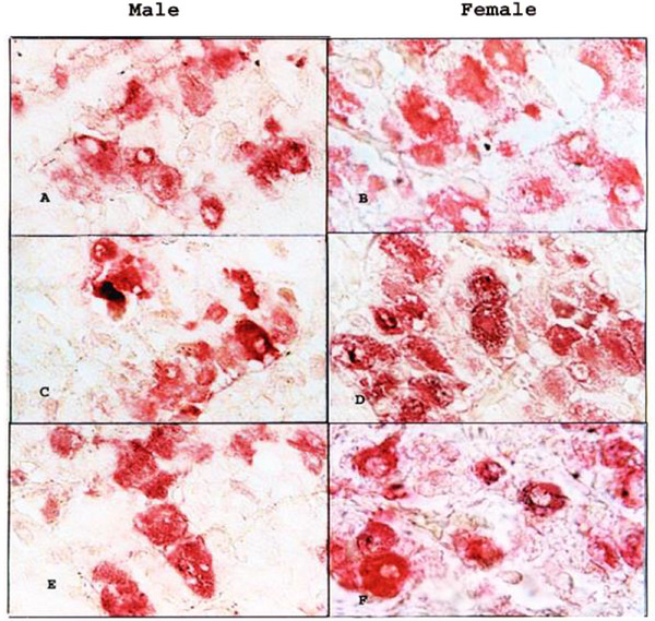
Thyroid stimulating hormone (TSH) immunoreactive cells in the adenohypophysis of camel (Camelus dromedarius). Streptavidin–alkaline phosphatase immunoreaction: X5000.(A)2–4 years male: immunoreactive cells small spherical rather than elongated with small nuclei.(B)2–4 years female: immunoreactive cells elongated or polygonal with large nuclei.(C)5–10 years male: immunoreactive cells spherical with spherical nuclei.(D)5–10 years female: immunoreactive cells polygonal with irregular nuclei.(E)11–onward years male: Immunoreactive cells polygonal with small irregular nuclei.(F)11–onward years female: immunoreactive cells large spherical with spherical nuclei in the centre.
Quantitative Morphometric Assessment
Morphometric analysis was carried out using a compound microscope, considering only thyrotrophs with complete cross‐sections and clear, non‐reactive nuclei. Six samples per group were examined, with five slides prepared from each sample and ten microscopic fields analysed per slide. Measurements included thyrotroph count, cell diameter, cell area, cell volume, nucleus diameter, nucleus area, and nucleus volume. Cell and nuclear diameters (µm) were recorded at two perpendicular points (widest axis and 90° angle) using ImageJ software (ImageJ 1.44P, NIH, Bethesda, MD, USA) and AutoCAD 2004. From these diameters, areas (A= πr2) and volumes (V= 4/3πr3) were calculated, following the method of Justin et al. (1999).
Blood Collection
Blood samples (10 mL) were collected via direct jugular venipuncture to minimise variability due to sampling site. Samples were drawn into heparinised tubes, centrifuged at 2500 rpm for 10 min, and plasma was stored at –20°C until hormone assays were conducted.
Hormonal Assays
Plasma thyroid stimulating hormone (TSH; μIU/mL) was measured using a TSH ELISA kit (Catalog No. DSL‐10‐5300; Diagnostic Systems Laboratories, Webster, TX, USA). Plasma triiodothyronine (T3; ng/mL) was quantified using an Enzyme Immunoassay kit (Lot No. RN15685; Cat. No. BC‐1005; BioCheck, Burlingame, CA, USA). Total thyroxine (T4; µg/dL) was measured using a T4Enzyme Immunoassay kit (Lot No. RN‐21055; Cat. No. BC‐1007; BioCheck, Burlingame, CA, USA). All procedures were performed according to the manufacturers’ instructions.
The sensitivity (minimum detectable concentration) of the assays was 0.05 μIU/mL for TSH, 0.2 ng/mL for T3, and 0.5 µg/dL for T4. The intra‐assay coefficients of variation (CVs) for three reference samples (low, medium, and high concentrations) were <6%, <5%, and <7% for TSH; <5%, <6%, and <8% for T3; and <6%, <5%, and <7% for T4. Inter‐assay CVs were consistently below 10% for all assays.
Statistical Analysis
Data were expressed as mean ± standard error of the mean (SEM). Differences among groups were analysed using one‐way analysis of variance (ANOVA), and when significant, means were compared using a post‐hoc multiple comparison test. Statistical significance was set atp< 0.05. Means sharing the same superscript letters were considered not significantly different.
Results
According to Table1, the mean cell count of adenohypophyseal thyrotrophs did not differ significantly across age groups or sexes. However, cell diameter, area, and volume were significantly greater in the oldest age group compared to the younger groups (p< 0.05). Similarly, nuclear dimensions (diameter, area, and volume) showed significant enlargement with advancing age, particularly in camels aged 11 years and above (p< 0.05). Across sexes, males generally exhibited larger nuclear dimensions than females, with significant differences observed in nucleus diameter, area, and volume (p< 0.05). Overall, the data indicate that thyrotroph morphometry is influenced by both age and sex, with the most pronounced changes evident in older camels.
Table: Mean adenohypophyseal thyrotroph parameters of camel (Camelus dromedarius) at different age groups (immunocytochemistry).
As shown in Figure2, plasma thyroid stimulating hormone (TSH) levels differed significantly among age groups (p< 0.05). The highest concentration was observed in females aged 5–10 years, followed by intermediate levels in the 2–4 year group. In contrast, the lowest TSH concentrations were recorded in camels aged 11 years and above. Overall, an age‐related decline in TSH was evident. Furthermore, females consistently exhibited higher values than males across all age groups.
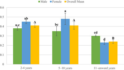
Mean plasma thyroid stimulating hormone in male and female camels (Camelus dromedarius).
Figure3shows the overall mean plasma TSH levels in male and female camels. Females exhibited higher mean TSH concentrations compared to males, although the difference was not statistically significant (p> 0.05).
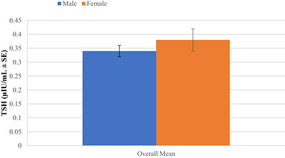
Overall mean plasma thyroid stimulating hormone in male and female camels (Camelus dromedarius).
Figure4shows that mean plasma triiodothyronine (T3) levels in camels varied significantly with age and sex. In the 2–4 years age group, males had higher T3concentrations than females. A similar trend was observed in the 5–10 years group, where males showed significantly higher values than females. However, in the 11–onward years group, both sexes exhibited reduced T3levels, with males showing the lowest values, while females maintained comparatively higher levels. Different superscripts indicate significant differences (p< 0.05) among age groups and between sexes.
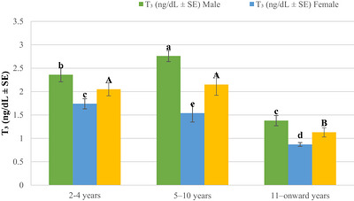
Mean plasma triiodothyronine in male and female camels (Camelus dromedarius).
Figure5shows the overall mean plasma triiodothyronine (T3) levels in male and female camels. Males had significantly higher T3concentrations compared to females, as indicated by different superscripts (p< 0.05).
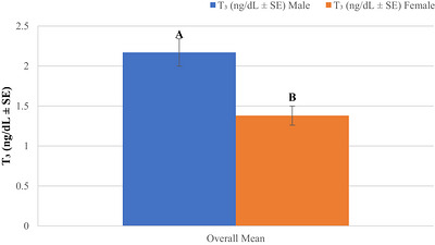
Overall mean plasma triiodothyronine in male and female camels (Camelus dromedarius).
Figure6shows that mean plasma thyroxin (T4) levels in camels were influenced by age but not markedly by sex. The highest T4concentrations were recorded in the 2–4 years age group, followed by a decline in the 5–10 years group and the lowest levels in the 11–onward years group. Different superscripts (A, B, C) indicate significant differences (p< 0.05) among age groups, whereas values between males and females within each age group did not differ significantly.
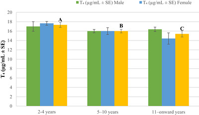
Mean plasma thyroxin in male and female camels (Camelus dromedarius).
Figure7illustrates the overall mean plasma thyroxin (T4) concentrations (µg/mL ± SE) in male and female camels. The mean T4level in male camels was slightly higher than in females, with males showing a concentration of approximately 16.4 µg/mL and females around 16.0 µg/mL. However, the standard error bars indicate that this difference is minimal and may not be statistically significant. Thus, the overall mean thyroxin levels in male and female camels appear to be relatively comparable.
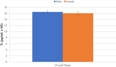
Overall mean plasma thyroxin in male and female camels (Camelus dromedarius).
Figure7illustrates the overall mean plasma thyroxin (T4) concentrations in male and female camels. Although the mean T4level in males appears slightly higher than in females, the difference was not statistically significant (p> 0.05).
Discussion
This study highlights how lactation, age, and sex influence thyrotroph morphology and thyroid hormone profiles in dromedary camels. The results demonstrate the adaptability of the hypothalamic–pituitary–thyroid (HPT) axis to physiological demands such as lactation and aging, while also revealing sex‐specific differences that add complexity to thyroid regulation in camels.
Lactation is one of the most metabolically demanding stages in mammals, requiring extensive endocrine adjustments to ensure adequate milk synthesis while maintaining maternal homeostasis (Khan et al.2025). The present findings indicate that lactating camels exhibited a higher number of pituitary thyrotrophs, whereas non‐lactating camels demonstrated larger thyrotroph cell and nuclear dimensions. Morphologically, an increase in cell count may reflect proliferative adaptation or enhanced turnover of TSH‐producing cells, while enlarged nuclear and cytoplasmic areas in non‐lactating animals may suggest storage or accumulation of secretory products. Similar adaptations have been reported in other species, where lactation is associated with structural remodelling of anterior pituitary cell populations (Capuco et al.2003; Lombardero et al.2020).
The endocrine correlates of these morphological changes provide further insights. Plasma TSH concentrations were lower in lactating compared with non‐lactating camels, yet immunocytochemical analysis revealed an abundance of thyrotrophs in lactating animals. This apparent paradox may be explained by increased secretory turnover of TSH during lactation, resulting in smaller amounts of hormone retained within cells and lower circulating levels. In lactating rats, Ni et al. (2021) reported an upregulation of hypothalamic pro‐TSH mRNA during early lactation, followed by a decline, suggesting temporal modulation of TSH synthesis (Ni et al.2021). These findings align with current observations, where lactation appears to be accompanied by an initial stimulatory drive of the HPT axis, followed by adjustments that prioritise efficient thyroid hormone utilisation rather than high systemic TSH levels.
Interestingly, lactating camels displayed higher circulating triiodothyronine (T3) levels despite reduced TSH and thyroxine (T4). This pattern strongly suggests an enhanced peripheral conversion of T4to T3via deiodinase activity, an adaptive mechanism widely described in mammals during lactation (Bianco and Kim2006). The conversion of T4into the metabolically active T3ensures adequate energy metabolism to sustain milk production, even when TSH and T4levels are relatively low. This lactation‐driven metabolic reprogramming may represent an evolutionary adaptation in camels, allowing them to maintain lactation under the energy constraints of desert environments.
Age‐related changes were also evident in thyrotroph morphology and thyroid hormone profiles. Younger camels (5–10 years) displayed higher T3concentrations compared to older animals, consistent with the general decline in thyroidal activity with age documented in humans and livestock (Monzani et al.1996;Rahman et al.2012). Morphologically, thyrotroph diameter, area, and nuclear dimensions were significantly greater in camels aged 11 years and above, suggesting a compensatory enlargement of cells despite lower functional output. This hypertrophy may reflect an attempt to sustain hormone production in the face of diminished secretory efficiency, a phenomenon also observed in aging pituitary tissues across mammalian species (Veldhuis2013).
The interaction between lactation and age highlights an important feature of endocrine adaptation. While lactation in younger camels was clearly associated with elevated T3and greater thyrotroph counts, this effect was less pronounced in older animals, where hormonal and morphological differences between lactating and non‐lactating individuals were attenuated. Such reduced plasticity of the HPT axis with advancing age may contribute to the diminished reproductive and lactational performance of older she‐camels, as has been reported in other domestic ruminants (Lucy2001).
The inclusion of sex‐based comparisons further revealed distinct patterns in thyroid regulation. Male camels demonstrated significantly larger thyrotroph cell and nuclear profiles at 11 years and above, suggesting heightened pituitary activity compared with females. These morphological traits were associated with higher plasma T3concentrations in males, particularly at younger ages. Elevated metabolic rates in males, especially during the breeding season, likely underlie these differences. The role of photoperiod and seasonal breeding patterns in camels may also influence thyrotroph activity, as seasonal modulation of pituitary function has been documented in other large herbivores.
Interestingly, while male camels exhibited significantly higher T3concentrations and larger thyrotroph morphometry, no significant sex‐related differences were observed in plasma T4concentrations (p> 0.05), as shown in Figure7. This supports the idea that thyroxine serves as a relatively stable reservoir of thyroid hormone, with sex‐related differences more likely reflected in T3production and conversion. These findings contrast with those of Tajik et al., who also reported no sex differences in T3levels in dromedaries (Tajik et al.2013). However, they are consistent with studies in other livestock species, such as Turkoman horses, sheep, and water buffaloes (Eshratkhah et al.2010;Nazifi et al.2003; Tajik et al.2011), where T4levels remained relatively constant across sexes.
Taken together, these findings suggest that thyrotroph morphology and thyroid hormone profiles are subject to dynamic regulation by lactation, age, and sex. The reduced TSH and T4but elevated T3in lactating camels underscores the importance of peripheral deiodinase‐mediated conversion of T4to T3as a key compensatory mechanism during milk production. Furthermore, the increased thyrotroph counts in lactating females may reflect heightened demand for thyroidal stimulation, even if circulating TSH is reduced, highlighting the complex feedback loops governing the HPT axis.
The enlarged thyrotrophs and nuclei observed in older camels suggest an age‐related decline in pituitary efficiency, whereby cellular hypertrophy attempts to compensate for reduced hormone output (Jaspal et al. 2025). Similarly, the heightened T3concentrations and increased thyrotroph dimensions in males likely reflect their greater metabolic activity and potential seasonal endocrine modulation. The lack of significant sex differences in T4levels reinforces its role as a stable endocrine reservoir, with T3production being more finely regulated to meet sex‐ and age‐specific metabolic demands.
The present study demonstrates that lactation in camels is associated with a functional reorganisation of the HPT axis, characterised by lower TSH and T4but enhanced T3availability. Age modifies these responses, with younger camels showing greater plasticity, while sex introduces additional variation, with males displaying heightened thyrotroph activity and T3output. These findings provide valuable insights into the endocrine strategies employed by camels to sustain lactation and adapt to age‐ and sex‐related metabolic demands, and they align with the broader understanding of pituitary–thyroid interactions in mammals.
Conclusion
This study provides comprehensive insights into the morphological and hormonal dynamics of the hypothalamic–pituitary–thyroid (HPT) axis in dromedary camels, emphasising the influence of lactation, age, and sex. The findings demonstrate that lactation is associated with increased thyrotroph cell numbers and elevated plasma T3concentrations, despite reduced TSH and T4levels, suggesting enhanced peripheral conversion of T4to T3as a key adaptive mechanism. Age‐related changes revealed increased thyrotroph cell and nuclear dimensions in older camels, likely reflecting compensatory hypertrophy in response to declining endocrine function. Sex differences were evident in T3levels and thyrotroph morphology, with males showing greater metabolic activity, while T4levels remained stable across sexes. Overall, the study highlights the plasticity of the HPT axis in camels and underscores its critical role in maintaining metabolic and reproductive homeostasis under varying physiological demands.
Author Contributions
Shabana Naz: writing – review and editing, writing – original draft.Ibrahim A. Alhidary: funding acquisition, resources.Robina Shaukat: investigation, validation, methodology.Shaukat Ali Shaukat Jaspal: conceptualisation, investigation, methodology, software, data curation.Muhammad Mubashar Shaukat: formal analysis, validation, visualisation.Tahmina Shaukat: formal analysis, software, data curation, resources.Antonella Perillo: writing – review and editing, writing – original draft.Rifat Ullah Khan: writing – review and editing, writing – original draft.Naseer Khan Momand: writing – review and editing, writing – original draft.
Funding
This research was funded by the King Saud University, Riyadh, Saudi Arabia, through the Ongoing Research Funding Program (ORF‐2026‐833)
Ethics Statement
The study was approved by the Ethical Committee of Faculty of Animal Husbandry & Veterinary Sciences, The University of Agriculture, Peshawar, Pakistan (Approval No. 12/FAH&VS/2021). Chatgpt has been used for English language
Conflicts of Interest
No potential conflict of interest was reported by the authors.
Contributor Information
Shabana Naz, Email: drshabananaz@gcuf.edu.pk.
Naseer Khan Momand, Email: naseerkhanm101@gmail.com.
Data Availability Statement
The relevant data are provided in the paper. The data of the current experiment can be obtained from corresponding author when needed.
Associated Data
Data Availability Statement
The relevant data are provided in the paper. The data of the current experiment can be obtained from corresponding author when needed.
References
- Ahmed, Y. F. , Mahmoud K. G. M., Kandiel M. M., et al. 2016. “Histomorphometry Aspect of Thyroid Gland and Biochemical Profile in Pregnant and Non‐Pregnant Dromedary Camels.” African Journal of Biotechnology 15, no. 10: 370–375. 10.5897/AJB2015.15029. doi.org/10.5897/AJB2015.15029
- Al‐Suhaimi, E. A. , and Khan F. A.. 2022. “The Pituitary Gland: Functional Relationship With the Hypothalamus, Structure, and Physiology.” In Emerging Concepts in Endocrine Structure and Functions, edited by E. A. Al‐Suhaimi, 73–131. Singapore: Springer Nature. 10.1007/978-981-19-2159-5_4. doi.org/10.1007/978-981-19-2159-5_4
- Babar, M. E. , and Ashraf M.. 2023. Biology and Breeding of Camels: Focus on Pakistan Camels. Boca Raton: CRC Press.
- Bianco, A. C. , and Kim B. W.. 2006. “Deiodinases: Implications of the Local Control of Thyroid Hormone Action.” Journal of Clinical Investigation 116, no. 10: 2571–2579. 10.1172/JCI29812. doi.org/10.1172/JCI29812
- Capuco, A. V. , Ellis S. E., Hale S. A., et al. 2003. “Lactation Persistency: Insights From Mammary Cell Proliferation Studies.” Journal of Animal Science 81, no. Suppl. 3: 18–31. 10.2527/2003.8118_suppl_3E18x. doi.org/10.2527/2003.8118_suppl_3E18x
- Choudhary, O. P. 2025. “Animal Models for Surgeries and Implants: A Vital Tool in Medical Research and Development.” Annals of Medicine and Surgery 87, no. 7: 4090–4095. doi.org/10.1097/MS9.0000000000003400
- Choudhary, O. P. 2026. “Artificial Intelligence in Animal Anatomy With Special Reference to Diagnostic Imaging.” Annals of Anatomy 265: 152810. doi.org/10.1016/j.aanat.2026.152810
- Choudhary, O. P. , and Sarkar R.. 2025. “Animal Anatomical Teaching Models for Enhanced Veterinary Anatomy Education and Learning.” Pakistan Veterinary Journal 45, no. 3: 961–982. 10.29261/pakvetj/2025.250. doi.org/10.29261/pakvetj/2025.250
- Duntas, L. H. 2021. “Aging and the Hypothalamic‐Pituitary‐Thyroid Axis.” Vitamins and Hormones 115: 1–14. 10.1016/bs.vh.2020.11.006. doi.org/10.1016/bs.vh.2020.11.006
- Eshratkhah, B. , Sadaghian M., Eshratkhah S., et al. 2010. “Relationship Between the Blood Thyroid Hormones and Lipid Profile in Moghani Sheep; Influence of Age and Sex.” Comparative Clinical Pathology 19, no. 1: 15–20. 10.1007/s00580-009-0881-4. doi.org/10.1007/s00580-009-0881-4
- Gnanadevi, R. , Basha S. H., Paramasivan S., et al. 2025. “Gross Morphological Studies on Hypothalamo‐Pituitary Axis of Male Coimbatore Sheep.” Journal of Advanced Biology and Biotechnology 28, no. 8: 425–432. 10.9734/jabb/2025/v28i81499. doi.org/10.9734/jabb/2025/v28i81499
- Goralsky, L. , Sokulsky I., Kolesnik N., et al. 2021. “Features of Histomorphology of the Pituitary Gland, Spinal Cord and Cerebral in Cattle.” Agrarian Bulletin of the Black Sea Littoral 87: 21–30.
- Gul, F. , Amin H., Naz S., et al. 2024. “Evaluation of Blood Minerals and Oxidative Stress Changing Pattern in Pre‐Partum and Post‐Partum Achai and Holstein Friesian Dairy Cows.” Reproduction in Domestic Animals 59: e14525. 10.1111/rda.14525. doi.org/10.1111/rda.14525
- Hassan, N. M. F. , Fouad R., Fouad H., et al. 2025. “Prevalence of Nasopharyngeal Myiasis Among Dromedary Camels and Influence of Cut Dates on Chemical Composition, Antioxidant and Larvicidal Efficacy ofMentha longifoliaL. Essential Oil Against Cephalopina Titillator.” International Journal of Veterinary Science 14, no. 5: 985–997. 10.47278/journal.ijvs/2025.054. doi.org/10.47278/journal.ijvs/2025.054
- Jaspal, S. A. , Shaukat M. M., Shaukat R., et al. 2025. “Interplay Between Thyrotroph Morphometry and Plasma Thyroid Hormones in Lactating and Non‐Lactating She‐Camels (Camelus dromedarius).” Veterinary Science 12, no. 9: 917. doi.org/10.3390/vetsci12090917
- Jaspal, S. A. , Zia‐ur‐Rahman, and Cheema A. M.. 2019. “Gross Morphological and Ontological Studies on Pituitary of Camel (Camelus dromedarius).” Pakistan Veterinary Journal 39, no. 4: 578. 10.29261/pakvetj/2019.116. doi.org/10.29261/pakvetj/2019.116
- Justin, H. S. , Kendall S. K., and Brinkmeier M. L., et al. 1999. “Thyroid Hormone Is Essential for Pituitary Somatotrophs and Lactotrophs.” Endocrinology 140: 1884–1892. 10.1210/endo.140.4.6622. doi.org/10.1210/endo.140.4.6622
- Khan, R. U. , Rahman Z. U., Javed I., et al. 2013. “Supplementation of Dietary Vitamins, Protein and Probiotics on Semen Traits and Immunohistochemical Study of Pituitary Hormones in Zinc‐Induced Molted Broiler Breeders.” Acta Histochemica 115: 698–704. 10.1016/j.acthis.2013.01.005. doi.org/10.1016/j.acthis.2013.01.005
- Khan, S. M. , Khan R. U., Naz S., et al. 2025. “Comparative Analysis of Metabolic, Reproductive, and Sub‐Clinical Mastitis in Pure Holstein‐Friesian and Sahiwal‐Crossbred Cows.” Archive of Animal Breeding 68: 89–100. 10.5194/aab-68-89-2025. doi.org/10.5194/aab-68-89-2025
- Lombardero, M. , Quintanar‐Stephano A., Rotondo F., et al. 2020. “Effect of Thyroxin on Cell Morphology and Hormone Secretion of Pituitary Grafts in Rats.” Annals of Anatomy 230: 151486. 10.1016/j.aanat.2020.151486. doi.org/10.1016/j.aanat.2020.151486
- Lucy, M. C. 2001. “Reproductive Loss in High‐Producing Dairy Cattle: Where Will It End?” Journal of Dairy Science 84, no. 6: 1277–1293. 10.3168/jds.S0022-0302(01)70158-0. doi.org/10.3168/jds.S0022-0302(01)70158-0
- Mohammed, A. A. , Almuyidi A., Almarri H., et al. 2025. “Unique Characteristics of Camel Body Systems: Adaptation to Harsh Conditions, Productive and Reproductive Performances: A Review.” Indian Journal of Animal Research 59: 12–21. 10.18805/IJAR.BF-1637. doi.org/10.18805/IJAR.BF-1637
- Monzani, F. , Del Guerra P., Caraccio N., et al. 1996. “Age‐Related Modifications in the Regulation of the Hypothalamic‐Pituitary‐Thyroid Axis.” Hormone Research in Paediatrics 46, no. 3: 107–112. 10.1159/000185998. doi.org/10.1159/000185998
- Mostafa, T. H. , Al‐Sagheer A. A., Arafa M. M., et al. 2022. “Changes in Milk Production, Hematology, Metabolites, Mineral and Hormonal Parameters of Primiparous and Multiparous Maghrebi Dairy She‐Camel During Nonbreeding Season.” Biological Rhythm Research 53, no. 2: 216–233. 10.1080/09291016.2020.1830031. doi.org/10.1080/09291016.2020.1830031
- Nazifi, S. , Saeb M., and Abedi M.. 2003. “Serum Lipid Profiles and Their Correlation With Thyroid Hormones in Clinically Healthy Turkoman Horses.” Comparative Clinical Pathology 12, no. 1: 49–52. 10.1007/s00580-003-0464-2. doi.org/10.1007/s00580-003-0464-2
- Ni, Y. , Chen Q., Cai J., et al. 2021. “Three Lactation‐Related Hormones: Regulation of Hypothalamus‐Pituitary Axis and Function on Lactation.” Molecular and Cellular Endocrinology 520: 111084. 10.1016/j.mce.2020.111084. doi.org/10.1016/j.mce.2020.111084
- Rahman, S. , Jahan N., and Sultana N.. 2012. “Age Related Change in Thyroid Function.” Journal of Bangladesh Society of Physiology 7, no. 2: 72–77. 10.3329/jbsp.v7i2.15025. doi.org/10.3329/jbsp.v7i2.15025
- Rejeb, A. , Amara A., Rekik M., et al. 2011. “Histomorphometry and Hormone Secretion of the Thyroid Gland of the Dromedary (Camelus dromedarius).” Journal of Camelid Science 4: 10–22.
- Tajik, J. , Rasooli A., and Sazmand A.. 2011. “Serum Thyroid Hormone Concentrations in Water Buffaloes.” Comparative Clinical Pathology 20: 359.
- Tajik, J. , Sazmand A., and Rasooli A.. 2013. “Serum Concentrations of Thyroid Hormones, Cholesterol and Triglyceride, and Their Correlations Together in Clinically Healthy Camels (Camelus dromedarius): Effects of Season, Sex and Age.” Veterinary Research Forum 4, no. 4: 239.https://www.ncbi.nlm.nih.gov/pmc/articles/PMC3919486.
- Tharwat, M. , Alkheraif A. A., Elmoghazy H. M. M., et al. 2025. “Disseminated Pyogranulomas in a Female Dromedary Camel: Hematobiochemical, Sonographic and Pathologic Investigations.” International Journal of Veterinary Science 14, no. 1: 120–124. 10.47278/journal.ijvs/2024.221. doi.org/10.47278/journal.ijvs/2024.221
- Veldhuis, J. D. 2013. “Changes in Pituitary Function With Ageing and Implications for Patient Care.” Nature Reviews Endocrinology 9, no. 4: 205–215. 10.1038/nrendo.2013.37. doi.org/10.1038/nrendo.2013.37
Republished from the open web under CC-BY. Authors: Jaspal SAS, Shaukat MM, Shaukat R, Shaukat T, Khan RU, Naz S, Perillo A, Momand NK, Alhidary IA. Read the original.