Application of PAX7 Gene Therapy for Muscle Wound Healing and Regeneration
Muscle injuries, ranging from minor exercise-induced tears to severe injuries, necessitate effective recovery strategies to restore tissue functions. This study integrates a comprehensive literature review with gene ontology-based analysis to identify the candidate genes involved in the muscle repair process. The key genes, including VEGF, TGF-β, and the IGF family, were identified for their roles in muscle cell regeneration during angiogenesis, myogenesis, and satellite cell activation. The overlapping analysis further highlighted NOTCH1, ENO3, MSTN, MYF6, and notably PAX7 as potential therapeutic targets for gene therapy. Focused cell experiments demonstrated that overexpression of PAX7 in human muscle cells using the DNA plasmid transfection significantly enhanced wound recovery. PAX7 induced 70% wound closure compared to 20% controls and provided significant protection against H₂O₂-induced oxidative stress (cell viability increased from 40% to 80%). Overall, these results indicate the promising potential of PAX7 gene therapy as a targeted intervention to activate muscle cell regeneration and protect from cellular damage in injured muscle tissues.
INTRODUCTION and GOAL
The term "muscle injury" encompasses a wide range of conditions, from severe trauma experienced by millions of athletes worldwide to minor post-workout soreness. 1 For instance, running, climbing stairs, or lifting weights can cause microscopic muscle tears, which are classified as muscle injuries. As a result, muscle recovery is a critical area of research. 2
Like most wounds, muscle injur
ies follow a structured, intricate three-phase healing process. The first phase, the inflammatory phase, alerts the body's systems, closes the wound, and removes bacteria and other debris, thereby limiting further damage. The next phase, the proliferative phase, facilitates the regeneration of lost tissue. This process is driven by the migration of cells from the wound’s peripheral areas and lasts significantly longer than the inflammatory phase. The final phase, remodeling, strengthens the affected area while breaking down excess collagen, ultimately enhancing the durability of the wound. Unfortunately, the remaining scar will never achieve total recovery. 3
Recent research has focused on developing innovative therapies for both mild and severe muscle injuries. Gene therapy and cell therapy are two promising approaches that attempt to accelerate recovery by modifying cellular activity. Gene therapy introduces nucleic acids into cells to alter gene expression, while cell therapy utilizes stem cells, such as mesenchymal stem cells and induced pluripotent stem cells, to stimulate regeneration. 4
However, challenges remain in identifying specific genes with high success rates due to the relatively recent emergence of these therapies. 5 There is also much further research to be done regarding the execution of gene therapy and its implications, specifically for treating muscle injuries, including determining the optimal methods for administering the target genes to patients.
One key gene involved in muscle regeneration is Paired Box 7, or PAX7. 6 It is a transcription factor crucial for muscle repair, regulating satellite cells: the stem cells responsible for skeletal muscle regeneration. It belongs to the Paired Box (PAX) family, which plays a fundamental role in tissue development and cellular differentiation. Following muscle injury, satellite cells proliferate and differentiate to replace damaged muscle fibers, a process heavily controlled by PAX7. 7 Proper PAX7 expression is essential for maintaining the satellite cell pool and ensuring effective regeneration. Research indicates that increased PAX7 expression enhances muscle repair, whereas a deficiency in PAX7 significantly impairs recovery.
Research Aim:
This study aimed to identify potential genetic targets for muscle wound healing by analyzing previous research in this field. Following a gene-ontology-based analysis, several promising genes were found to be crucial in the muscle recovery process. Ultimately, PAX7 was selected as the main candidate for further research. We hypothesized that overexpressing PAX7 through DNA plasmid transfection would improve muscle wound healing. To test this, PAX7 was overexpressed in human muscle cells, followed by a wound healing assay and a hydrogen peroxide stress test to evaluate its effects.
METHODS
PubMed database data search
A literature review was performed using the PubMed database to identify genes that are related to muscle wound healing and regeneration. The following keywords were used to search for information: “Muscle,” “Regeneration,” and “Wound Healing.” Peer-reviewed primary sources were selected to discuss the gene involvement in muscle injury recovery processes. Gene expression data were also collected for the wound-healing bs and for potential therapeutic targets.
Gene Ontology(GO)-based gene interaction analysis
An expanded Gene Ontology-based analysis was performed to identify candidate genes for gene therapy. The GO databases were used to retrieve gene and gene product annotations across multiple biological species, based on known biological processes, molecular functions, and cellular components. The genes that were annotated under the biological process were analyzed using the keywords muscle regeneration, muscle cell proliferation, and muscle cancer.
GO analysis integration with previous studies
The genes identified in the GO analysis were investigated using the gene lists collected from the literature review. This allowed for a comprehensive list of candidate genes. These integrations demonstrated cross-referencing of genes to identify overlapping genes. The known pathways and experimental results from the selected studies were used to validate the findings of the candidate genes for muscle wound healing.
Statistical methods for identifying significant candidate genes
Statistical analyses were conducted to assess the significance of the candidate genes in muscle wound healing. The overlap gene analysis was performed, and the Venn diagrams were used to visualize the shared genes between the muscle cancer, muscle cell proliferation, and muscle tissue regeneration categories. A statistical test was used to assess the significance of gene overlaps across datasets, with a p-value threshold of < 0.05.
Cell culture and maintenance
A human muscle cell line was obtained from the Korea Cell Line Bank and was used in the muscle wound-healing assays. DMEM cell culture media was used to culture these cells. A cell incubator at 37 °C with 5% CO2 was used for cell culture.
PAX transfection and wound healing assay
A scratched wound healing assay was performed to analyze the effect of PAX7 gene overexpression. The PAX7 gene was transfected with the Lipofectamine 2000 transfection reagents. After mixing PAX7 and the transfection reagent, the complex molecules were formed in 5 minutes. Then, the mixed solutions were added to the muscle cells. After 24 hours, a cross-shaped wound was formed with the micro pipette tips, and the wound healing area was analyzed with the ImageJ image analysis program.
H2O2 stress test
A hydrogen peroxide test was also performed. After PAX7 genes were overexpressed in muscle cells, hydrogen peroxide was added to the culture solutions. After incubating for 48 hours, the muscle cell viability was measured using the automated cell counter LUNA-FL device.
Statistical test
All data were collected and analyzed using the statistical test, the ANOVA test, and the post hoc test.
RESULTS
Table 1. List of candidate genes that can be used for gene therapy based on the previous studies collected from the PubMed database.
| Gene | How is this gene associated with muscle wound healing? | Wound healing phase | Association type | Reference |
| VEGF | Angiogenesis | Phase 3 | Mutation | 3 |
| TGF-β | Myogenesis | Phase 2 | N/A | 4 |
| IGF family | Satellite cell activation in injured muscle | Phase 2 | SNP | 8 |
| TIMP2 VEGF165 iNOS CARP SMAD FGF-2 | General wound healing genes | Multi-phase | N/A | 9 |
| Pax7 TNF-α TGF-β | Overlapping Genes of Wound Healing and Muscle Regeneration | Multi-phase | N/A | 4 |
| TP53 | May play a role in developing rhabdomyosarcoma (RMS) earlier in life | Phase 2 | Missense Mutation | 10 |
| FOXO1 | May play a role in the onset of RMS | Phase 2 | Mutations | 10 |
Several candidate genes for muscle wound healing were identified for potential gene therapy using the PubMed database. VEGF, associated with angiogenesis during Phase 3 of wound healing, indicates that the mutations of VEGF influence this process. TGF-β promotes myogenesis during phase 2 of wound healing. IGF family genes were involved in satellite cell activation in injured muscles during phase 2. The general wound healing genes (TIMP2, VEGF165, iNOS, CARP, SMAD, and FGF-2) are known to be involved in the multiple phases of wound healing. These overlapping genes contribute to wound healing and muscle regeneration, including PAX7, TNF-α, and TGF-β, which are involved in multiple processes. Additionally, mutations in TP53 (missense mutations) and FOXO1 are associated with the development of rhabdomyosarcoma (RMS) earlier in life during Phase 2.
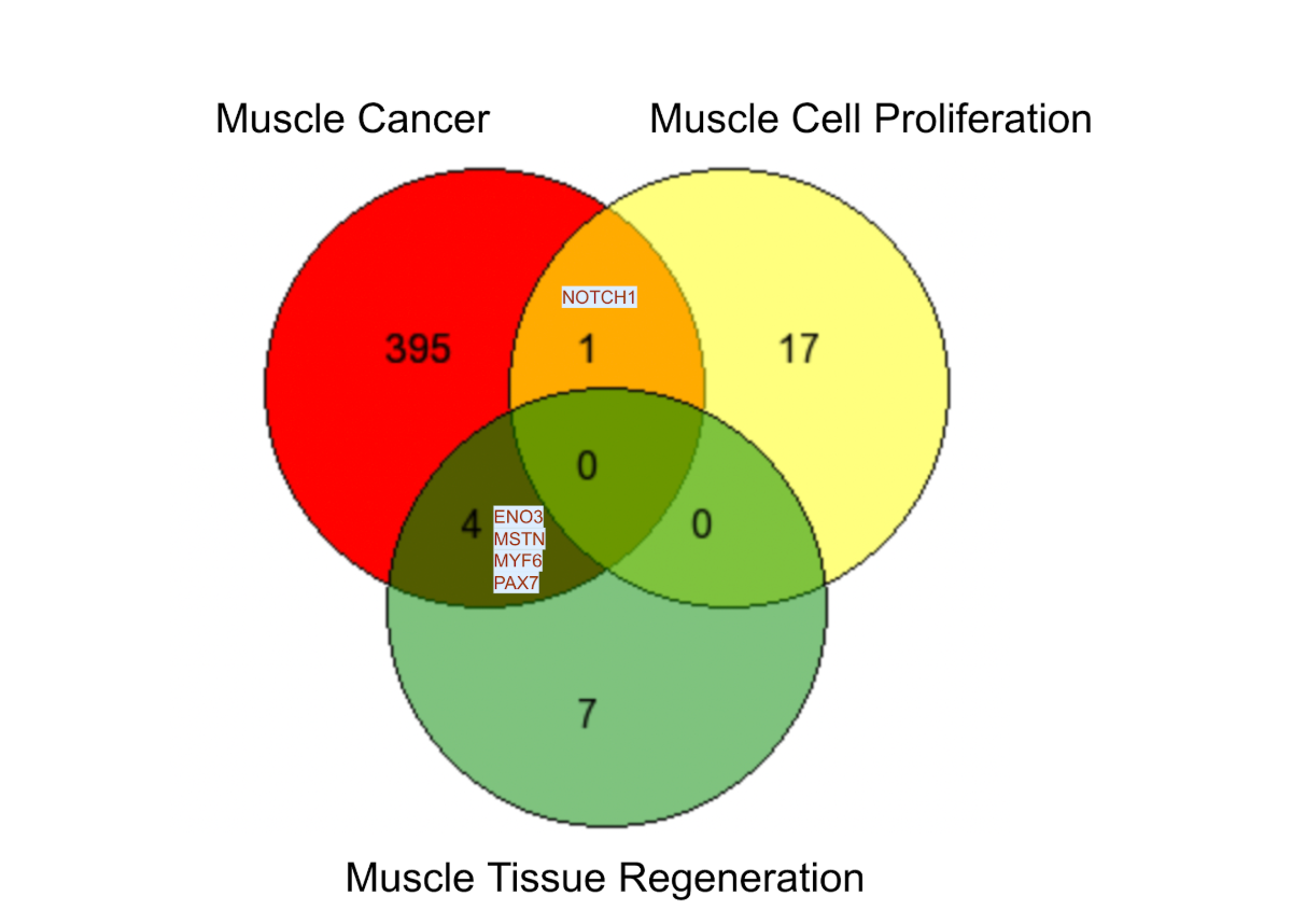
Figure 1. Overlapping genes of muscle cancer, muscle cell proliferation, and muscle tissue regeneration from Gene Ontology Biological Process Annotations Database.
The overlapping genes were analyzed using the keywords "muscle cancer," "muscle cell proliferation," and "muscle tissue regeneration" from the Gene Ontology Biological Process annotation database. Figure 1 shows the overlap genes associated with these keywords. A novel set of 395 genes was uniquely associated with the muscle cancer, while 17 genes are known to be involved in muscle cell proliferation. Also, seven genes were linked to muscle tissue regeneration. NOTCH1 is the only gene that is shared by the muscle cancer and muscle cell proliferation. The four genes (ENO3, MSTN, MYF6, and PAX7) are shared between muscle cancer and muscle tissue development. However, no genes overlap across all three categories. These results provide potential therapeutic candidate genes for muscle tissue recovery.
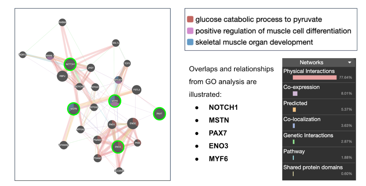
Figure 2. Gene network analysis identifying key functions associated with overlapping genes from GO analysis
This diagram presents a gene network analysis that identifies key functions of the overlapping genes identified through Gene Ontology analysis. On the left, a gene network diagram illustrates the connections and interactions among genes such as NOTCH1, MSTN, PAX7, ENO3, and MYF6. Each node is represented by various colored lines, which signify different types of interactions, including physical interactions, co-expression, and predicted relationships with surrounding genes.
From this analysis, three primary functional categories emerged. The first, Glucose Catabolic Process to Pyruvate (highlighted in red), is involved in glucose breakdown, with genes such as ENO3 playing a key role in this metabolic pathway. The second category, Positive Regulation of Muscle Cell Differentiation (shown in purple), includes genes such as MYF6 and PAX7, which are essential for muscle cell differentiation and regeneration. Lastly, Skeletal Muscle Organ Development (indicated in blue) encompasses the development and maintenance of muscle tissue, with NOTCH1 playing a crucial role in muscle organogenesis and satellite cell behavior. On the right, we have a breakdown of interaction types among these genes. The largest proportion of interactions is physical, indicating that these genes frequently function together within cellular processes.
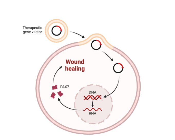
Figure 3. Overview of overexpressing the PAX7 gene using a therapeutic gene vector.
We hypothesized that a PAX7 gene-containing therapeutic gene vector may support the wound healing process in human muscle cells. The therapeutic gene vector introduces the PAX7 gene into human muscle cells (Figure 3). This leads to transcription of the PAX7 gene into RNA, which is then translated into PAX7 protein. Therefore, PAX7 protein supports the wound healing process in muscle tissue.
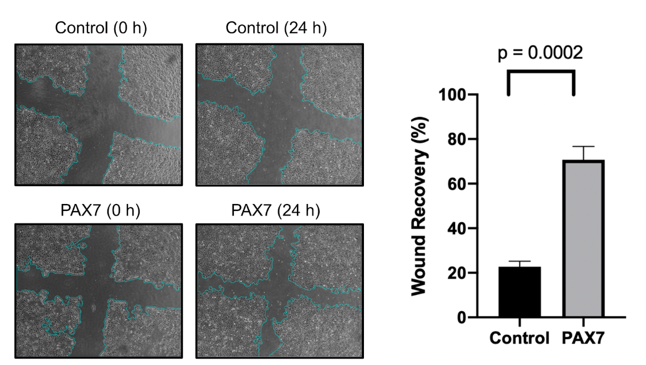
Figure 4. Wound healing test with the human muscle cells. The image shows the wound area at 0 hours and 24 hours for both control and PAX7 overexpressing cells. The graph shows the percentage of wound recovery after 24 hours.
Human muscle cells were grown and scratched with a cross shape. The wound region is indicated by the blue area. Compared to the control sample, overexpression of PAX7 significantly enhances wound healing in human muscle cells. The image shows the initial wound area at 0 hours, and the extent of wound closure is initiated after 24 hours. The result shows that PAX7 overexpressing cells displayed a significantly increased wound healing recovery. The quantitative analysis showed that PAX7 overexpression resulted in ~70% recovery compared to ~20% in the control group. This result demonstrates that PAX7 plays a crucial role in promoting cell migration and wound healing of human muscle cells.
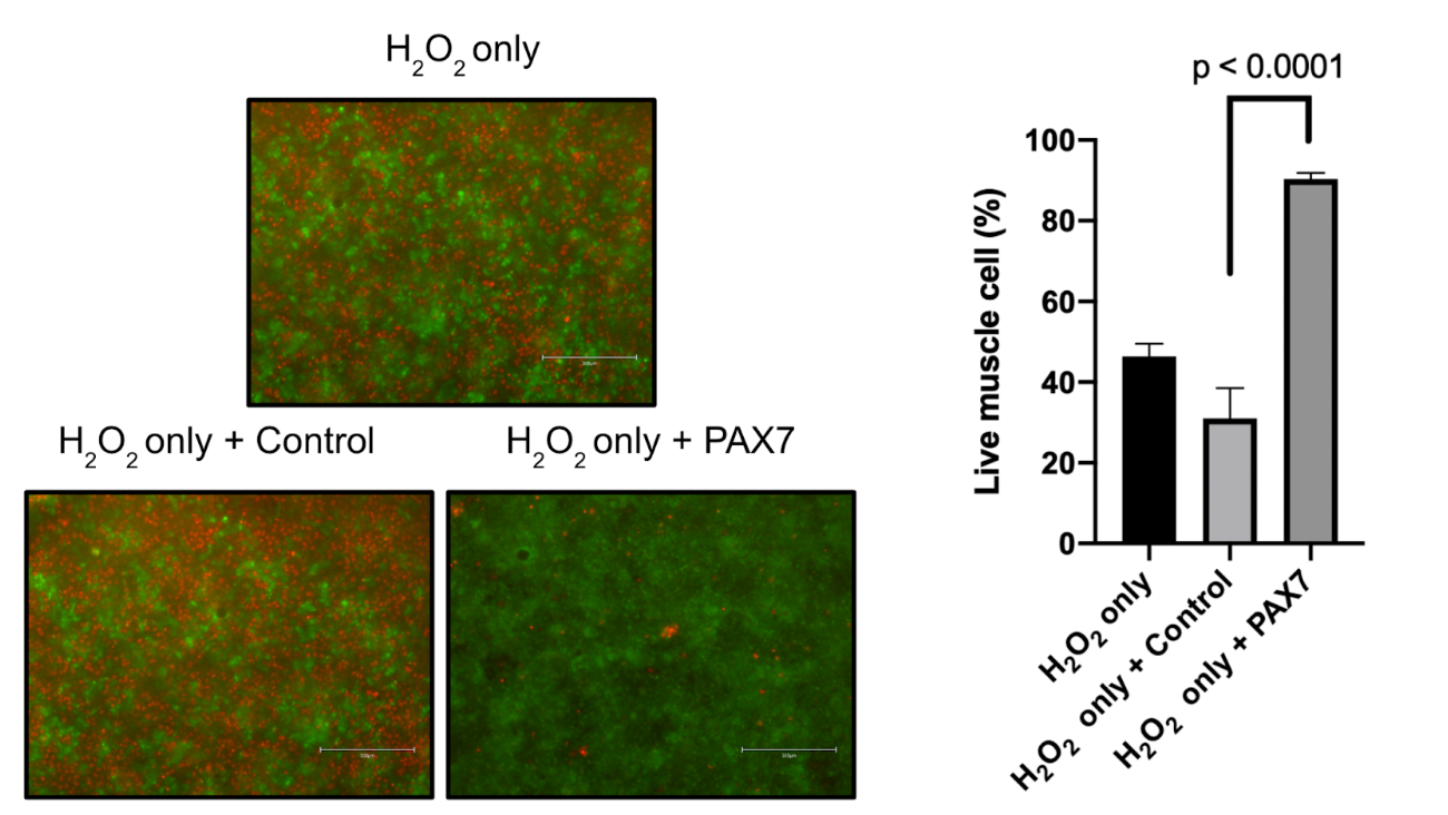
Figure 5. Effect of muscle protection of PAX7 against H2O2-induced cellular stress. PAX7 protected the muscle cells from H2O2 induced cell death. The bar graph quantifies the percentage of live cells, indicating a significant increase in cell viability with PAX7 overexpression compared to control groups.
When the muscle cells were exposed to H2O2, they showed a reduction in cell viability. However, when PAX7 was overexpressed, it protected the cells from H₂O₂-induced damage (Figure 5). Approximately 80% of the cells remained viable compared to only 40% in the control group in the H₂O₂-only group. These findings showed that PAX not only facilitates muscle cell regeneration but also activates protection against oxidative stress-induced cell death.
DISCUSSION
The results of this study indicate a promising approach to muscle wound healing and regeneration using PAX7 gene therapy. Using a comprehensive literature review and Gene Ontology-based analysis, this research identified candidate genes involved in multiple steps of muscle repair. Interestingly, PAX7, a key transcription factor that regulates satellite cell proliferation and differentiation, was evaluated as a strong candidate given its multifaceted role in the muscle stem cell pool. The cell experiments showed that PAX7-overexpressing cells exhibited 70% wound recovery, compared to only 20% in controls. This enhanced recovery indicates improved cell migration and proliferation during the proliferative phase of muscle recovery.
Additionally, this study examined oxidative stress using the H₂O₂ stress test. This result further highlights the therapeutic benefits of PAX7 overexpression. A significant increase was observed in cell viability when PAX7 was overexpressed in human muscle cells. These findings suggest that PAX7 gene therapy may address both regenerative deficits and increased vulnerability to stress-induced cell death observed in injured muscle tissue.
Limitations:
Although this research provides a solid foundation for the application of the PAX7 gene to muscle wound healing, long-term outcomes must be validated in in vivo models, such as a mouse model. Additionally, the side effects of PAX7 gene insertion must be addressed for future use of this gene therapy.
CONCLUSIONS
This study shows that overexpression of the PAX7 gene significantly enhances muscle wound healing and provides protection against oxidative stress. The improvement of cell migration, accelerated wound closure, and increased cell viability under H₂O₂ stress showed strong evidence of PAX7's potential for gene therapy. However, further research is required to optimize the gene delivery and evaluate the long-term efficacy in mouse models. These results provide a strong foundation for the future application of PAX7 gene therapy in human muscle cells.
REFERENCES
(1) Furrer, R.; Hawley, J. A.; Handschin, C. The Molecular Athlete: Exercise Physiology from Mechanisms to Medals. Physiol. Rev. 2023, 103 (3), 1693–1787. DOI: 10.1152/physrev.00017.2022.
(2) Owens, D. J.; Twist, C.; Cobley, J. N.; Howatson, G.; Close, G. L. Exercise-Induced Muscle Damage: What Is It, What Causes It and What Are the Nutritional Solutions? Eur. J. Sport Sci. 2019, 19 (1), 71–85. DOI: 10.1080/17461391.2018.1505957.
(3) Wallace, H. A.; Basehore, B. M.; Zito, P. M. Wound Healing Phases. In StatPearls; StatPearls Publishing: Treasure Island (FL), 2024.
(4) Stilhano, R. S.; Martins, L.; Ingham, S. J. M.; Pesquero, J. B.; Huard, J. Gene and Cell Therapy for Muscle Regeneration. Curr. Rev. Musculoskelet. Med. 2015, 8 (2), 182–187. DOI: 10.1007/s12178-015-9268-9.
(5) Kohn, D. B.; Chen, Y. Y.; Spencer, M. J. Successes and Challenges in Clinical Gene Therapy. Gene Ther. 2023, 30 (10–11), 738–746. DOI: 10.1038/s41434-023-00390-5.
(6) Rahman, N. I. A.; Lam, C. L.; Sulaiman, N.; Abdullah, N. A. H.; Nordin, F.; Ariffin, S. H. Z.; Yazid, M. D. PAX7, a Key for Myogenesis Modulation in Muscular Dystrophies through Multiple Signaling Pathways: A Systematic Review. Int. J. Mol. Sci. 2023, 24 (17). DOI: 10.3390/ijms241713051.
(7) Kaczmarek, A.; Kaczmarek, M.; Ciałowicz, M.; Clemente, F. M.; Wolański, P.; Badicu, G.; Murawska-Ciałowicz, E. The Role of Satellite Cells in Skeletal Muscle Regeneration-The Effect of Exercise and Age. Biology (Basel) 2021, 10 (10). DOI: 10.3390/biology10101056.
(8) Ahmad, S. S.; Ahmad, K.; Lee, E. J.; Lee, Y.-H.; Choi, I. Implications of Insulin-Like Growth Factor-1 in Skeletal Muscle and Various Diseases. Cells 2020, 9 (8). DOI: 10.3390/cells9081773.
(9) Farhangniya, M.; Samadikuchaksaraei, A. A Review of Genes Involved in Wound Healing. Med. J. Islam. Repub. Iran 2023, 37, 140. DOI: 10.47176/mjiri.37.140.
(10) Martin-Giacalone, B. A.; Weinstein, P. A.; Plon, S. E.; Lupo, P. J. Pediatric Rhabdomyosarcoma: Epidemiology and Genetic Susceptibility. J. Clin. Med. 2021, 10 (9). DOI: 10.3390/jcm10092028.