Combined Effects of Arsenic and Cadmium With Selenium on Oxidative Stress Indicators in Goldfish: Implications for Veterinary Toxicology.
Background The individual and combined effects of toxic heavy metals, including arsenic (As) and cadmium (Cd) and their interactions with selenium (Se), play a crucial role in modulating bioaccumulation patterns and oxidative stress responses in aquatic organisms. Objectives This study aimed to evaluate the effects of As, Cd and Se, individually and in combination, on tissue metal accumulation and oxidative stress biomarkers in goldfish (Carassius auratus). Methods A total of 210 fish were randomly assigned to seven experimental groups with three replicates each and exposed to single or combined concentrations of As, Cd and Se via water for 14 days. At the end of the exposure period, tissue concentrations of metals were determined using inductively coupled plasma mass spectrometry (ICP-MS). Oxidative stress biomarkers, including malondialdehyde (MDA), catalase (CAT) and superoxide dismutase (SOD), were assessed using spectrophotometric methods and commercial assay kits. Results and conclusions Body weight increased in all treatment groups compared to initial values. Co-exposure to Se significantly reduced As accumulation, with tissue concentrations decreasing from 0.16 ± 0.02 mg/kg in the As group to 0.10 ± 0.009 mg/kg in the combined exposure groups (approximately 62 % reduction; p 0.05). A significant positive correlation was observed between As concentration and MDA levels across all groups, including the control (p < 0.01). Conversely, Se exhibited a negative correlation with MDA and a positive correlation with SOD, indicating its protective role against oxidative damage. Overall, the results demonstrate that combined exposure to heavy metals can produce variable toxicological interactions, ranging from antagonistic to potentially synergistic effects, depending on the type of metal and exposure conditions. These interactions are closely associated with oxidative stress responses, highlighting potential ecological risks to aquatic organisms and public health concerns associated with the consumption of contaminated fish.
Introduction
The rapid development of industrial activities has led to the widespread release of heavy metals into aquatic and terrestrial ecosystems, resulting in significant environmental contamination, reduced food safety and both acute and chronic toxic effects in living organisms (Ali et al.2019). Heavy metals are generally defined as elements with high atomic weight and density, which are naturally present in the Earth's crust but are increasingly mobilized through anthropogenic activities such as mining, industrial discharge, agriculture and wastewater effluents.
Due to their persistence, non‐biodegradability and strong affinity for biological macromolecules, heavy metals readily enter food chains and accumulate in plant and animal tissues, ultimately reaching humans through contaminated water, food and air (Jaishankar et al.2014). Chronic exposure to these metals has been associated with a wide range of adverse health outcomes, including carcinogenicity, neurotoxicity, endocrine disruption and oxidative damage at the cellular level (Briffa et al.2020).
Among these elements, arsenic (As), cadmium (Cd), mercury (Hg) and lead (Pb) are considered highly toxic even at low concentrations (Genchi et al.2020). Cd exposure, primarily through food consumption, cigarette smoke and industrial emissions, leads to long biological half‐life and accumulation in vital organs such as the liver and kidneys, resulting in renal dysfunction, skeletal damage and increased cancer risk (Budi et al.2024). As exposure, mainly via contaminated drinking water and agricultural products, is associated with gastrointestinal disturbances in acute cases and skin lesions, cardiovascular disorders and various cancers under chronic exposure conditions (Barchielli et al.2022).
In contrast, selenium (Se) is an essential trace element required for antioxidant defence, immune function and thyroid hormone metabolism; however, its narrow margin between nutritional requirement and toxicity makes both deficiency and excess clinically relevant (Rayman2012). At physiological levels, Se exerts protective effects through selenoproteins involved in redox regulation, whereas excessive intake may lead to selenosis, characterized by gastrointestinal, hepatic and neurological disorders.
A key toxicological mechanism shared by many heavy metals, particularly Cd and As, is the induction of oxidative stress through excessive production of reactive oxygen species (Raeeszadeh et al.2024). This leads to lipid peroxidation, protein oxidation and DNA damage. Oxidative stress biomarkers such as malondialdehyde (MDA), along with antioxidant enzymes including superoxide dismutase (SOD), catalase (CAT) and glutathione peroxidase (GPx), are widely used to evaluate cellular redox imbalance in toxicological studies (Sies et al.2022; Ghafarifarsani et al.2024).
Fish are widely accepted as sensitive bioindicators of aquatic pollution due to their direct exposure to waterborne contaminants. In particular, goldfish (Carassius auratus) has been extensively used as a model organism in toxicology and environmental biology due to its physiological sensitivity, ease of maintenance and well‐characterized biochemical responses (Hong and Zha2019).
Although previous studies have examined single‐metal toxicity, there remains a significant gap in understanding the interactive effects of combined exposure to As, Cd and Se. These interactions may be antagonistic or synergistic depending on concentration and exposure conditions. Se, in particular, has been reported to modulate the toxicity of heavy metals by altering their bioavailability, promoting the formation of metal–Se complexes and regulating oxidative stress pathways (Zwolak2020; Uddin et al.2024).
Therefore, the main objective of this study was to investigate the individual and combined effects of As, Cd and Se on bioaccumulation patterns and oxidative stress responses in C. auratus. Specifically, this study aimed to evaluate tissue metal accumulation and key oxidative stress biomarkers to clarify the mechanistic interactions between these elements. This approach provides a clearer link between exposure conditions and physiological outcomes, contributing to a better understanding of metal toxicity interactions in aquatic organisms and their potential implications for environmental and human health.
Materials and Methods
The selection of metal concentrations was based on previously reported 96‐h median lethal concentration (LC50‐96 h) values for As, Cd and Se in freshwater fish species, which are commonly used to establish sublethal exposure levels in aquatic toxicology studies. Sublethal exposure is typically defined as a fraction (commonly 1/10 or lower) of the LC50 value to avoid acute mortality while inducing measurable physiological and biochemical responses (Sprague1973).
Based on a review of relevant toxicological literature, exposure concentrations equivalent to approximately 10% of reported LC50 values were selected to ensure biologically relevant but non‐lethal stress conditions. Accordingly, concentrations of 4 mg/L for As, 2.8 mg/L for Cd and 2 mg/L for Se were chosen to induce oxidative and biochemical responses without causing acute toxicity (Monteiro et al.2009; Kavitha et al.2010; Ghafari Farsani et al.2015).
This approach is consistent with established experimental designs in aquatic toxicology, where sublethal metal exposures are used to evaluate oxidative stress biomarkers, enzyme activity alterations and bioaccumulation patterns (Grosell et al.2006).
All biochemical substances and metal salts (Sodium arsenite, Sigma‐Aldrich, Lot No: 104266100; Cd chloride, Sigma‐Aldrich, Lot No: 210527; and sodium selenite, Sigma‐Aldrich, Lot No: 1203690) were obtained from certified analytical‐grade suppliers and prepared according to the manufacturers’ specifications to ensure consistency and experimental reliability.
Experimental Study Design
A total of 210 C. auratus with a mean body weight of 20 ± 8 g were obtained in spring 2024 from a commercial fish farm in Gilan Province, Iran and transported to the Laboratory of the Faculty of Veterinary Medicine, University of Kurdistan, Sanandaj. Upon arrival, fish were acclimated and randomly allocated to 21 glass aquaria (40 L each) under controlled laboratory conditions (20 ± 2°C; pH 7.5–8.5) with continuous aeration.
It should be noted that fish were evenly distributed among aquaria, with each treatment consisting of three independent replicates. Treatments were applied at the aquarium level; therefore, the aquarium was considered the experimental unit. At the end of the 14‐day exposure period, fish were randomly sampled from each aquarium, and measurements were averaged per tank to avoid pseudoreplication. These mean values were used as independent replicates in the statistical analysis (n= 3 per treatment).
Prior to the main experiment, the 96‐h median lethal concentrations (LC50) of As, Cd and Se were determined to define sublethal exposure levels. Fish were then exposed to 10% of the respective LC50 values for 14 days in separate treatment groups (three replicates per treatment). This sublethal exposure design is widely used in aquatic toxicology to assess physiological and biochemical responses without causing acute mortality (S. Eroglu et al.2015; M. A. Aldoghachi2016; J. Choi et al.2022; M. Rabbane et al.2022).
At the end of the exposure period, fish were collected, mortality was verified and biometric measurements were recorded. Muscle tissues were subsequently excised for biochemical and metal accumulation analyses.
Sample Collection and Preparation for Analysis
All solutions were prepared using analytical‐grade reagents. Throughout the study, ultrapure double‐distilled water was used. Nitric acid and hydrogen peroxide (H2O2) of supra‐pure quality were obtained from Merck (Darmstadt, Germany). Standard solutions of As, Cd and Se (1000 mg/L) were purchased from Sigma–Aldrich (St. Louis, USA) and diluted appropriately before use.
Muscle samples (∼10 g) were collected from the dorsal white muscle using a sterile surgical blade. Immediately after sampling, tissues were placed in sterile containers on dry ice to maintain the cold chain and then transported to the laboratory for further processing. All samples were analysed within one week of collection, well before their expiration date. Before analysis, samples were dried, crushed and ground, and 5 g of the resulting powder was used for measurements. Metal concentrations were expressed based on the dry weight of the samples.
Inductively Coupled Plasma Mass Spectrometry (ICP‐MS) was employed to quantify heavy metals. ICP‐MS is a sensitive analytical technique in which the sample is atomized and ionized by an inductively coupled plasma. The resulting atomic and small polyatomic ions are detected and quantified, allowing precise measurement of metals and certain non‐metals at trace levels. ICP‐MS can also differentiate isotopes of the same element, making it suitable for isotopic studies. Compared with atomic absorption spectroscopy, ICP‐MS offers higher sensitivity, precision and throughput.
For sample digestion, Pyrex glass tubes (Foss, USA) were used. Each tube was pre‐cleaned with 10% nitric acid for 48 h, followed by thorough rinsing with double‐distilled water to minimize external metal contamination. Approximately 0.2 g of each sample was digested with 3 mL of 70% nitric acid (Merck) and 1 mL of 30% H2O2at 160°C for 8 h, until complete tissue dissolution. The digestion temperature was gradually increased from 50°C to 160°C. After cooling, the digested solutions were filtered through Whatman No. 1 filter paper, transferred into labelled containers, and diluted to 10 mL with double‐distilled water. The prepared samples were then analysed using a SCIEX ELAN DRC II ICP‐MS (PerkinElmer).
Metals were calibrated using a multi‐element standard (IV‐ICPMS‐71A, Inorganic Ventures) diluted to 0.05 µg/mL in 19.6% (w/w) nitric acid, matching the acid content of the samples. Six‐point calibration curves were constructed for each analyte, covering concentrations from the respective LOD to 200 ng/g. Measurements were conducted in full quantitative mode, with all calibration curves showing excellent linearity (R2≥ 0.9999). Polyatomic interferences were evaluated by measuring multiple isotopes and verifying isotopic ratios in the digested samples (Rito et al.2022; Poopak et al.2024).
Preparation of Tissue Homogenates and Stress Oxidative Biomarkers
Approximately 0.5–1 g of dorsal white muscle tissue was excised, washed with cold phosphate‐buffered saline (PBS, pH 7.4), and immediately placed on ice. The tissue was then homogenized in 4–5 mL of cold PBS using a glass–Teflon homogenizer. The homogenate was centrifuged at 10,000 ×gfor 15 min at 4°C, and the supernatant was collected for biochemical analyses (Dias et al.2020).
Catalase Activity
CAT activity was determined spectrophotometrically by measuring the rate of H2O2decomposition at 240 nm. The decrease in absorbance over time was used to calculate enzyme activity following standard protocols (Hadwan and Kadhum Ali2018).
Superoxide Dismutase Activity
SOD activity was measured based on its ability to inhibit the photochemical reduction of nitroblue tetrazolium (NBT). The reaction was initiated under light exposure, and the reduction of NBT was monitored at 560 nm, where lower absorbance indicates higher SOD activity (Weydert and Cullen2010).
Malondialdehyde Level
Lipid peroxidation was assessed by measuring MDA levels using the thiobarbituric acid reactive substances (TBARS) assay. Samples were incubated at 95°C for 30 min, and the resulting chromogen was measured spectrophotometrically at 532 nm (De Leon and Borges2020).
Protein Normalization
All enzymatic activities and MDA levels were normalized to total protein concentration in tissue homogenates, which was determined using the Bradford colourimetric method (Raeeszadeh et al.2023).
Data Analysis
All statistical analyses were conducted with the aquarium considered as the experimental unit, and tank‐level clustering was incorporated into the study design. Measurements obtained from fish within each tank were averaged, and these mean values were used for all analyses (n= 3 per treatment) to avoid pseudoreplication.
Prior to analysis, data distribution and assumptions were evaluated. Normality was assessed using the Shapiro–Wilk and Kolmogorov–Smirnov tests, while homogeneity of variances was examined using Levene's test. When the assumptions for parametric analysis were satisfied, one‐way analysis of variance (ANOVA) was applied to compare quantitative variables among groups, followed by Tukey's post hoc test for multiple comparisons. Pairedt‐tests were used, where appropriate, to assess within‐group differences (e.g., pre‐ and post‐intervention weights). Pearson correlation coefficients were calculated based on tank‐level mean values to evaluate associations between variables. Data visualization was performed using box plots, scatter plots and line graphs. Statistical analyses were conducted using SPSS (v22), GraphPad Prism (v10) and R (v4.4.1), with the level of significance set atp< 0.05.
Results
In Table1, the survival rate of the different groups is presented. The highest survival rate was observed in the control group, while the lowest was recorded in the As group. However, the differences between groups were not statistically significant.
Table: Survival rate of the different groups during the experimental study.
Figure1presents the initial and final body weights of the experimental groups. The results indicate no significant differences in initial mean body weights among the groups (p> 0.05), confirming uniformity at the start of the experiment. In contrast, final body weights increased in all treatment groups compared to their respective initial values. Moreover, significant differences were observed among the groups in terms of final body weight (p< 0.001). The lowest mean final weight was recorded in the As+Se group, whereas the highest final body weight was observed in the Cd+Se group.
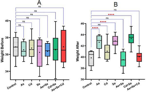
*Comparison of initial and final weights of animals in the different study groups. ns, Not significant; ***p< 0.0001.
The results regarding the concentrations of the toxic heavy metals As, Cd and Se in the experimental groups are presented in Figure2. Accordingly, the data indicate that muscle As accumulation was lowest in the control group and was significantly altered following exposure to Se and Cd (p< 0.001) (Figure2A). Cd concentrations were also lowest in the control group, while a reduction in Cd accumulation was observed in the As+Se treatment group (Figure2B). In addition, Se levels showed a significant decrease following exposure to both Cd and As compared with the control group (Figure2C).
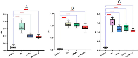
*Concentration of heavy metals arsenic, cadmium and selenium in the different experimental groups, ***p< 0.0001.
The levels of oxidative stress biomarkers in muscle tissue across the experimental groups are presented in Figure3. The results indicate significant variations among the groups.
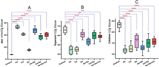
*Comparison of oxidative stress parameters across the studied groups. *p< 0.05, **p< 0.01, ***p< 0.001, ***p< 0.0001.
As shown in Figure3A, the mean MDA levels (nmol/g tissue) differed significantly among the experimental groups. The lowest MDA levels were observed in the control and Se groups. In contrast, a statistically significant increase in MDA concentration was observed in the As+Cd+Se group compared with the control group (p< 0.0001).
Figure3Bpresents the mean activity of SOD (U/g tissue) across the different groups, revealing a significant difference among treatments (p< 0.001). The highest SOD activity was recorded in the control and Se groups, whereas the lowest activity was observed in the As and Cd groups. However, Se supplementation in the As and Cd groups resulted in an increase in SOD antioxidant activity.
Similarly, the mean CAT activity (U/g tissue) showed significant differences among groups (p< 0.001). The highest CAT activity was observed in the control and Se groups, respectively, while the lowest values were recorded in the As and Cd groups. Notably, CAT activity increased in the combined As+Se and Cd+Se groups, and this difference was statistically significant (p< 0.01) (Figure3).
The correlations between heavy metal concentrations (As, Cd and Se) in the independent treatment groups and oxidative stress biomarkers are presented in Figure4. The results indicate distinct patterns of association between metal accumulation and biochemical responses.
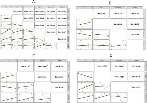
Correlation between heavy metal concentrations and oxidative stress markers in the control, As, Cd and Se groups.
Figure4Ashows that in the control group, a strong positive and statistically significant correlation was observed between As concentration and MDA (nmol/g tissue) (r= 0.910,p< 0.001). In contrast, the correlation between Se concentration and MDA (nmol/g tissue) was negative but not statistically significant (r= −0.348,p> 0.05). Similarly, correlations between Se and Cd concentrations were negative and not statistically significant (p> 0.05).
Figure4Billustrates that in the As‐exposed group, As concentration showed a positive correlation with MDA (nmol/g tissue) (r= 0.769,p< 0.05), while exhibiting a negative correlation with SOD activity (U/g tissue) (r= −0.740,p< 0.05). The correlation between As concentration and CAT activity (U/g tissue) was negative (r= −0.076), but not statistically significant (p> 0.05).
Figure4Cdemonstrates that in the Cd group, Cd concentration was positively correlated with MDA (nmol/g tissue) (r= 0.710,p< 0.05) and negatively correlated with SOD activity (U/g tissue) (r= −0.836,p< 0.05).
Finally, Figure4Dshows that Se concentration exhibited a significant positive correlation with SOD activity (U/g tissue) (r= 0.890,p< 0.05) and a significant negative correlation with MDA (nmol/g tissue) (r= −0.769,p< 0.05).
Figure5presents the correlations between oxidative stress biomarkers and heavy metal concentrations (As and Cd) in the presence of Se. The results indicate distinct interaction patterns among treatments.
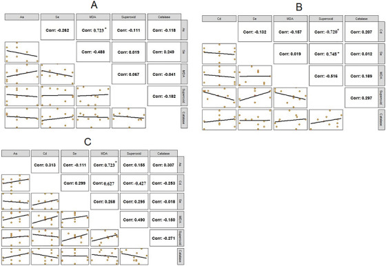
Correlation between heavy metal concentrations and oxidative stress markers in the As+Se, Cd+Se and As+Cd+Se groups.
Figure5Ashows that in the As+Se group, As concentration exhibited a negative correlation with CAT activity (U/g tissue) and SOD activity (U/g tissue), while showing a positive correlation with MDA (nmol/g tissue) (r= 0.723,p< 0.05), which was statistically significant. In addition, Se concentration showed a positive correlation with CAT activity (U/g tissue) (r= 0.249) and SOD activity (U/g tissue) (r= 0.019), and a negative correlation with MDA (nmol/g tissue) (r= −0.488); however, these correlations were not statistically significant (p> 0.05).
Figure5Bdemonstrates that in the Cd+Se group, Se concentration showed a significant positive correlation with CAT activity (U/g tissue) (r= 0.748,p< 0.05), while Cd concentration exhibited a significant negative correlation with SOD activity (U/g tissue) (r= −0.720,p< 0.05).
Figure5Cindicates that in the As+Cd+Se group, Cd concentration was negatively correlated with CAT activity (U/g tissue) (r= −0.253) and SOD activity (U/g tissue) (r= −0.427), while showing a positive correlation with MDA (nmol/g tissue) (r= 0.627). In addition, As concentration showed a positive and significant correlation with MDA (nmol/g tissue) (r= 0.723,p< 0.05).
Discussion
One of the major concerns associated with the consumption of aquatic organisms is the accumulation of heavy metals in their muscle tissues (Garai et al.2021). This accumulation may not only adversely affect aquatic organisms—potentially leading to behavioural alterations, impaired growth and increased mortality—but also pose risks to human health due to the persistence and bioaccumulative nature of these metals (Garai et al.2021). It has been suggested that metal accumulation in fish tissues is influenced by multiple factors, including metal concentrations in water as well as environmental parameters such as temperature, pH, salinity and hardness (Menon et al.2023). In this context, the present study aimed to investigate the potential interactive effects of toxic and essential metals on oxidative stress responses under controlled experimental conditions.
The findings of this study may indicate cumulative and relatively non‐degradable effects of metals such as As and Cd. The observed modulation following Se exposure could suggest a mitigating role, as reflected by the possible reduction in MDA levels and enhancement of antioxidant enzyme activities. The significant correlation between As concentration and MDA levels in the control group might reflect underlying environmental exposure, possibly linked to background contamination sources such as well water. This possibility is supported by previous reports indicating As contamination in water resources of western Iran (Raeeszadeh et al.2023; Poopak et al.2024).
Cd and As are recognized as important environmental toxicants, and organisms are often exposed to them in combination. However, their interactive toxicological effects remain incompletely understood. Metallothioneins (MTs), which are cysteine‐rich metal‐binding proteins, are known to play a key role in Cd detoxification, although their involvement in As toxicity may be more complex and less clear.
Previous reviews have emphasized that heavy metals such as Hg, Cd, Pb and As can bioaccumulate through aquatic environments and exert toxicity primarily via oxidative stress mechanisms, potentially leading to tissue damage in vital organs (Zahran et al.2025). Similarly, it has been suggested that stress‐related proteins, including heat shock proteins, antioxidant enzymes and MTs, may be involved in cellular defence responses and could serve as biomarkers of metal exposure (Gora et al.2025).
Some studies have reported increased MT levels following As exposure, whereas others suggest that pre‐exposure to Cd may not significantly alter As‐induced toxicity, indicating that metal–metal interactions may depend on exposure conditions and doses (Amalraj et al.2017). In line with this, it has been proposed that the toxicity of metal mixtures cannot be reliably predicted based on single‐metal exposure data alone, with interactions ranging from additive to synergistic effects (Yoo et al.2021). The reduced tissue accumulation observed in the present study under combined exposure (particularly As+Se) may therefore reflect possible interaction effects influencing uptake, distribution or detoxification processes (Magos et al.1980; Wu et al.2016).
The distribution of heavy metals in fish tissues may also be influenced by metabolic activity, with lower concentrations typically observed in muscle compared to organs such as the liver (Shahjahan et al.2022). This pattern could be related to the role of proteins such as MTs in metal binding and detoxification (Aalami et al.2022). In addition, changes in metal distribution during growth and adaptation may further contribute to these differences (Wu et al.2016).
The observed reduction in Se concentrations in combined treatments compared to Se alone may suggest possible interactions between Se and other metals affecting its bioavailability or incorporation into tissues. Previous studies have indicated that Se may modulate the toxicity of Cd and As through mechanisms related to redox balance and antioxidant defence (Naz et al.2023). It has also been proposed that different chemical forms of Se may exert distinct effects on antioxidant pathways (Jamwal2018; Raeeszadeh et al.2022). Furthermore, Se may interact with certain metals to reduce their toxicity, possibly through the enhancement of selenoprotein activities such as GPx (Liang et al.2023).
The increase in MDA levels observed in treated groups may reflect enhanced lipid peroxidation and oxidative stress, whereas the decrease in SOD and CAT activities could indicate impairment of antioxidant defence systems. The comparatively improved status in Se‐treated groups may suggest a protective or modulatory role of Se against oxidative damage. Combined exposure to Se with Cd and As appeared to partially counteract oxidative stress, which may be associated with improved antioxidant enzyme activity and reduced lipid peroxidation.
Antioxidant enzymes such as SOD and CAT are critical components of cellular defence mechanisms against reactive oxygen species. SOD catalyses the conversion of superoxide radicals into molecular oxygen, thereby limiting oxidative damage (Hermes‐Lima2004). MDA, as a marker of lipid peroxidation, is widely used to assess oxidative stress status. Changes in these biomarkers may therefore reflect the balance between pro‐oxidant and antioxidant processes (Martins et al.2022).
Overall, the largely non‐significant variations observed in some metal concentrations may suggest the presence of possible antagonistic or interactive effects among metals, which could influence their bioaccumulation and toxicity patterns in fish tissues.
Conclusion
This study demonstrates that simultaneous exposure to heavy metals, including As, Cd and Se, significantly influences tissue bioaccumulation patterns and biochemical responses in aquatic organisms. Se supplementation markedly reduced the tissue accumulation of both As and Cd, suggesting a modulatory and potentially antagonistic interaction. A consistent positive correlation was observed between As and Cd concentrations and MDA levels, whereas a negative correlation was found with antioxidant enzymes, particularly in the case of As, indicating pronounced oxidative stress induction.
The observed reduction in metal accumulation and oxidative damage in Se‐treated groups suggests a competitive and protective mechanism by which Se mitigates the toxic effects of As and Cd. However, the magnitude of these interactions appears to be influenced by several environmental and biological factors, including exposure dose, water quality parameters (e.g., pH and salinity) and species‐specific physiological characteristics, all of which can modulate metal uptake and toxicity.
Despite the valuable findings of this study, several limitations should be acknowledged. The present work is limited to biochemical endpoints in muscle tissue and does not include comprehensive molecular assessments. Future studies are recommended to incorporate gene expression analyses (e.g., RT‐PCR) of key antioxidant and stress‐related enzymes, as well as to evaluate additional target organs such as liver, kidneys and gills to provide a more complete toxicological profile. Moreover, extending the exposure duration is necessary to assess chronic toxicity and long‐term accumulation effects of heavy metals.
It is also suggested that future research include more comprehensive experimental designs with expanded treatment groups to better evaluate the interactive effects of As, Cd and Se under varying environmental conditions. Such integrated approaches would enhance the understanding of both individual and combined metal toxicity.
Overall, the findings of this study contribute to the ongoing scientific discussion on the interactive and cumulative effects of heavy metals and provide useful insights for environmental toxicology and aquatic risk assessment.
Author Contributions
Sarina Hosseinkhani: conceptualization, data curation, formal analysis, investigation, methodology.Mahdieh Raeeszadeh: conceptualization, data curation, formal analysis, investigation, software, supervision, validation, visualization, methodology, writing – original draft.Behnam Salimi: conceptualization, data curation, formal analysis, investigation, methodology, writing – review and editing.
Funding
The authors have nothing to report.
Ethics Statement
All procedures followed international animal care guidelines and were approved by the Ethics Committee of Islamic Azad University, Sanandaj, with the code IR.IAU.SDJ.REC.1403.001. The study commenced after a 10‐day acclimatization period during which water parameters were monitored daily.
Conflicts of Interest
The authors declare no conflicts of interest.
Acknowledgements
The authors would like to sincerely appreciate the Research Vice‐Chancellor at the Islamic Azad University, Sanandaj Branch, for his collaboration to facilitate research.
Data Availability Statement
The datasets used and/or analysed during the current study are available from the corresponding author upon reasonable request.
Associated Data
Data Availability Statement
The datasets used and/or analysed during the current study are available from the corresponding author upon reasonable request.
References
- Aalami, A. H. , Hoseinzadeh M., Hosseini Manesh P., Jiryai Sharahi A., and Kargar Aliabadi E.. 2022. “Carcinogenic Effects of Heavy Metals by Inducing Dysregulation of microRNAs: A Review.” Molecular Biology Reports 49, no. 12: 12227–12238. doi.org/10.1007/s11033-022-07897-x
- Aldoghachi, M. A. 2016. “Toxicity Assessment of Heavy Metals in Aquatic Organisms: LC50‐Based Experimental Approaches.” Toxicology Reports 3: 1–8.28959520
- Ali, H. , Khan E., and Ilahi I.. 2019. “Environmental Chemistry and Ecotoxicology of Hazardous Heavy Metals: Environmental Persistence, Toxicity, and Bioaccumulation.” Journal of Chemistry 1–14.
- Amalraj, A. , Pius A., Gopi S., and Gopi S.. 2017. “Biological Activities of Curcuminoids, Other Biomolecules From Turmeric and Their Derivatives–A Review.” Journal of Traditional and Complementary Medicine 7, no. 2: 205–233. doi.org/10.1016/j.jtcme.2016.05.005
- Barchielli, G. , Capperucci A., and Tanini D.. 2022. “The Role of Selenium in Pathologies: An Updated Review.” Antioxidants 11, no. 2: 251. doi.org/10.3390/antiox11020251
- Briffa, J. , Sinagra E., and Blundell R.. 2020. “Heavy Metal Pollution in the Environment and Their Toxicological Effects.” Heliyon 6, no. 9: e04691. doi.org/10.1016/j.heliyon.2020.e04691
- Budi, H. S. , Catalan Opulencia M. J., Afra A., et al. 2024. “Source, Toxicity and Carcinogenic Health Risk Assessment of Heavy Metals.” Reviews on Environmental Health 39, no. 1: 77–90. doi.org/10.1515/reveh-2022-0096
- Choi, J. , Li Y., et al. 2022. “Sublethal Exposure of Heavy Metals in Fish: Oxidative Stress and Biochemical Responses.” Aquatic Toxicology 244: 106077.35091369
- De Leon, J. A. D. , and Borges C. R.. 2020. “Evaluation of Oxidative Stress in Biological Samples Using the Thiobarbituric Acid Reactive Substances Assay.” Journal of Visualized Experiments: JoVE 159: 10–3791. 10.3791/61122. doi.org/10.3791/61122
- Dias, P. R. F. , Gandra P. G., Brenzikofer R., and Macedo D. V.. 2020. “Subcellular Fractionation of Frozen Skeletal Muscle Samples.” Biochemistry and Cell Biology 98, no. 2: 293–298. doi.org/10.1139/bcb-2019-0219
- Eroglu, S. , Dogan Z., et al. 2015. “Acute Toxicity of Heavy Metals in Freshwater Fish Species and Determination of LC50 Values.” Environmental Toxicology and Pharmacology 39, no. 2: 770–776.
- Garai, P. , Banerjee P., Mondal P., and Saha N.. 2021. “Effect of Heavy Metals on Fishes: Toxicity and Bioaccumulation.” Journal of Clinical Toxicology 11: 1–10.
- Genchi, G. , Sinicropi M. S., Lauria G., Carocci A., and Catalano A.. 2020. “The Effects of Heavy Metals on Human Health.” Metals 10, no. 10: 1–25.
- Ghafari Farsani, H. , Hedayati S., Mansouri Chorehi M., Rostamian N., and Baladehi E. C.. 2015. “Study on Liver Histopathological Changes of Gold Fish (Carassius auratus) During Exposure to Lethal and Sub Lethal Concentrations of Cadmium.” Experimental animal Biology 3, no. 4: 9–16.
- Ghafarifarsani, H. , Hoseinifar S. H., Raeeszadeh M., et al. 2024. “Comparative Effect of Chemical and Green Zinc Nanoparticles on the Growth, Hematology, Serum Biochemical, Antioxidant Parameters, and Immunity in Serum and Mucus of Goldfish,Carassius auratus(Linnaeus, 1758).” Biological Trace Element Research 202, no. 3: 1264–1278. doi.org/10.1007/s12011-023-03753-6
- Gora, A. H. , Sreeram M. P., Rehman S., Chakraborty K., Siriyappagouder P., and PS A.. 2025. “A Review on Metallothionein Research in Marine and Estuarine Realms: Past Paradigms and Future Vistas.” Frontiers in Marine Science 12: 1636760.
- Grosell, M. , Blanchard J., Brix K. V., and Gerdes R.. 2006. “Physiology and Toxicology of Metal Uptake in Fish.” Aquatic Toxicology 91: 302–311.
- Hadwan, M. H. , and kadhum Ali S.. 2018. “New Spectrophotometric Assay for Assessments of Catalase Activity in Biological Samples.” Analytical Biochemistry 542: 29–33. doi.org/10.1016/j.ab.2017.11.013
- Hermes‐Lima, M. 2004. “Oxygen in Biology and Biochemistry: Role of Free Radicals.” Functional Metabolism: Regulation and Adaptation 1: 319–366.
- Hong, X. , and Zha J.. 2019. “Fish Behavior: A Promising Model for Aquatic Toxicology Research.” Science of the Total Environment 686: 311–321. doi.org/10.1016/j.scitotenv.2019.06.028
- Jaishankar, M. , Tseten T., Anbalagan N., et al. 2014. “Toxicity, Mechanism and Health Effects of Heavy Metals.” Interdisciplinary Toxicology 7: 60. doi.org/10.2478/intox-2014-0009
- Jamwal, A. 2018. “Interactive Effects of Selenium on Arsenic and Cadmium Induced Toxicity in Rainbow Trout (Oncorhynchus mykiss).” PhD thesis, University of Saskatchewan.
- Kavitha, C. , Malarvizhi A., Kumaran S. S., and Ramesh M.. 2010. “Toxicological Effects of Arsenate Exposure on Hematological, Biochemical and Liver Transaminases Activity in an Indian Major Carp,Catla catla.” Food and Chemical Toxicology 48, no. 10: 2848–2854. doi.org/10.1016/j.fct.2010.07.017
- Liang, Y. , Wang J., Wang T., et al. 2023. “Moderate Selenium Mitigates Hand Grip Strength Impairment Associated With Elevated Blood Cadmium and Lead Levels in Middle‐Aged and Elderly Individuals: Insights From NHANES 2011–2014.” Frontiers in Pharmacology 14: 1324583. doi.org/10.3389/fphar.2023.1324583
- Magos, L. , Webb M., and Clarkson T. W.. 1980. “The Interactions of Selenium With Cadmium and Mercury.” CRC Critical Reviews in Toxicology 8, no. 1: 1–42. doi.org/10.3109/10408448009037490
- Martins, C. d. C. e. , Oliveira A. S. d. S. S., da Silva L. A. A., Primo M. G. S., and de Carvalho Lira V. B.. 2022. “Biological Indicators of Oxidative Stress [Malondialdehyde, Catalase, Glutathione Peroxidase, and Superoxide Dismutase] and Their Application in Nutrition.” Biomarkers in Nutrition, 1–25. Springer.
- Menon, S. V. , Kumar A., Middha S. K., et al. 2023. “Water Physicochemical Factors and Oxidative Stress Physiology in Fish, a Review.” Frontiers in Environmental Science 11: 1240813.
- Monteiro, D. A. , Rantin F. T., and Kalinin A. L.. 2009. “The Effects of Selenium on Oxidative Stress Biomarkers in the Freshwater Characid Fish Matrinxã,Brycon cephalus(Günther, 1869) Exposed to Organophosphate Insecticide Folisuper 600 BR®(Methyl Parathion).” Comparative Biochemistry and Physiology Part C: Toxicology & Pharmacology 149, no. 1: 40–49. doi.org/10.1016/j.cbpc.2008.06.012
- Naz, S. , Chatha A. M. M., Téllez‐Isaías G., et al. 2023. “A Comprehensive Review on Metallic Trace Elements Toxicity in Fishes and Potential Remedial Measures.” Water 15, no. 16: 3017.
- Poopak, H. , Raeeszadeh M., and Salimi B.. 2024. “Accumulation of Heavy Metals in Meat and Their Relationship With Water and Food Intake of Aquatic Animals in Kermanshah, Western Iran.” International Journal of Environmental Health Research 34, no. 3: 1453–1465. doi.org/10.1080/09603123.2023.2221640
- Rabbane, M. , Kabir H., et al. 2022. “Toxicological Evaluation of Heavy Metals in Fish Using LC50‐Based Exposure Design.” Environmental Science and Pollution Research 29: 1–16.
- Raeeszadeh, M. , Gravandi H., and Akbari A.. 2022. “Determination of Some Heavy Metals Levels in the Meat of Animal Species (Sheep, Beef, Turkey, and Ostrich) and Carcinogenic Health Risk Assessment in Kurdistan Province in the West of Iran.” Environmental Science and Pollution Research 29, no. 41: 62248–62258. doi.org/10.1007/s11356-022-19589-x
- Raeeszadeh, M. , Khoei A. J., Parhizkar S., Rad F. T., and Salimi B.. 2023. “Assessment of Some Heavy Metals and Their Relationship With Oxidative Stress and Immunological Parameters in Aquatic Animal Species.” Biological Trace Element Research 201, no. 9: 4547–4557. doi.org/10.1007/s12011-022-03507-w
- Raeeszadeh, M. , Parhizkar S., Rahmani R., and Mohammadzadeh M.. 2024. “Assessment of Some Heavy Metals Concentration in Turkey Meat in Kurdistan Province and Their Relationship With Oxidative Stress and Human Health Indicators.” Environmental Monitoring and Assessment 196, no. 12: 1268. doi.org/10.1007/s10661-024-13397-1
- Rayman, M. P. 2020. “Selenium and Human Health.” Lancet 379: 1256–1268. doi.org/10.1016/S0140-6736(11)61452-9
- Rito, B. , Almeida D., Coimbra C., et al. 2022. “Post‐Measurement Compressed Calibration for ICP‐MS‐Based Metal Quantification in Mine Residues Bioleaching.” Scientific Reports 12, no. 1: 16007. doi.org/10.1038/s41598-022-19620-8
- Shahjahan, M. , Taslima K., Rahman M. S., Al‐Emran M., Alam S. I., and Faggio C.. 2022. “Effects of Heavy Metals on Fish Physiology–A Review.” Chemosphere 300: 134519. doi.org/10.1016/j.chemosphere.2022.134519
- Sies, H. , Berndt C., and Jones D. P.. 2022. “Oxidative Stress.” Annual Review of Biochemistry 86: 715–748. doi.org/10.1146/annurev-biochem-061516-045037
- Sprague, J. B. 1973. “The ABCs of Pollutant Bioassay Using Fish.” In Biological Methods for the Assessment of Water Quality, edited by Cairns J. and Dickson K. L.. ASTM Special Technical Publication.
- Uddin, M. H. , Ritu J. R., Putnala S. K., Rachamalla M., Chivers D. P., and Niyogi S.. 2024. “Selenium Toxicity in Fishes: A Current Perspective.” Chemosphere 364: 143214. doi.org/10.1016/j.chemosphere.2024.143214
- Weydert, C. J. , and Cullen J. J.. 2010. “Measurement of Superoxide Dismutase, Catalase and Glutathione Peroxidase in Cultured Cells and Tissue.” Nature Protocols 5, no. 1: 51–66. doi.org/10.1038/nprot.2009.197
- Wu, X. , Cobbina S. J., Mao G., Xu H., Zhang Z., and Yang L.. 2016. “A Review of Toxicity and Mechanisms of Individual and Mixtures of Heavy Metals in the Environment.” Environmental Science and Pollution Research 23: 8244–8259. doi.org/10.1007/s11356-016-6333-x
- Yoo, J.‐W. , Cho H., Lee K.‐W., Won E.‐J., and Lee Y.‐M.. 2021. “Combined Effects of Heavy Metals (Cd, As, and Pb): Comparative Study Using Conceptual Models and the Antioxidant Responses in the Brackish Water Flea.” Comparative Biochemistry and Physiology Part C: Toxicology & Pharmacology 239: 108863. doi.org/10.1016/j.cbpc.2020.108863
- Zahran, E. , Mamdouh A.‐Z., Elbahnaswy S., et al. 2025. “The Impact of Heavy Metal Pollution: Bioaccumulation, Oxidative Stress, and Histopathological Alterations in Fish Across Diverse Habitats.” Aquaculture International 33, no. 5: 371.
- Zwolak, I. 2020. “The Role of Selenium in Arsenic and Cadmium Toxicity: An Updated Review of Scientific Literature.” Biological Trace Element Research 193, no. 1: 44–63. doi.org/10.1007/s12011-019-01691-w
Republished from the open web under CC-BY. Authors: Hosseinkhani S, Raeeszadeh M, Salimi B. Read the original.