Echocardiographic Findings in Small-Breed Dogs With Myxomatous Mitral Valve Disease and the Severity of Mitral Regurgitation, Heart Size and Clinical Signs Myxomatous Mitral Valve Disease in Dogs.
Background The main goal of this study was to identify various brightness, motion and Doppler echocardiographic variables in small-breed dogs with myxomatous mitral valve disease. Animals Sixty client-owned small-breed dogs with mitral regurgitation murmurs. Methods Echocardiography was performed in brightness, motion and Doppler modality from the right parasternal view. On the basis of thoracic radiographs, the dogs were categorized into two groups: those with cardiomegaly and those without cardiomegaly. Additionally, on the basis of the jet area signal to left atrium ratio in colour Doppler echocardiography, the canines were divided into three groups: mild, moderate and severe mitral regurgitation. Furthermore, the dogs were classified into preclinical and clinical groups on the basis of the presence or absence of clinical signs, and various variables were compared across these groups. Results This difference was statistically significant and more prevalent in intact dogs than in spayed/neutered dogs. Significant differences were observed in variables including LAmax, LA:Ao, LVIDd, LVIDs, VMA, VMG, VMV, GMV, VMA-GMVT, LVOT and GLVOT in dogs with myxomatous mitral valve disease of varying mitral regurgitation severity and between normal and enlarged hearts. Significant differences were also found in the FS, GMV, VLVOT and GLVOT variables between the clinical and preclinical groups. A moderate and statistically significant correlation was observed between LA:Ao and VMA, VMV and LVOT. Weak and mostly insignificant correlations were found between the Doppler and motion-mode variables. Conclusions Varying mitral regurgitation severity, heart size and the presence of clinical signs can significantly affect certain brightness, motion and Doppler echocardiographic variables in dogs with myxomatous mitral valve disease (valvular heart disease).
Introduction
Cardiovascular diseases are among the leading causes of mortality and disability worldwide. Myxomatous mitral valve disease (MMVD) is the most common acquired heart disease in dogs (Keene et al.2019; Mattin et al.2015; Svensson et al.2024), particularly in small‐breed dogs (Burchell and Schoeman2014; Chetboul and Tissier2012). Initially, this disease progresses silently without clinical signs, but over time, it leads to changes in heart size and, in severe cases, may result in death (Borgarelli and Buchanan2012; O'Brien et al.2021). For this reason, MMVD is considered the leading cause of heart disease‐related mortality in dogs (Häggström et al.2009) and one of the primary causes of heart failure and death in humans (Levine et al.2015).
This disease is characterized by MR, which results in left atrial and ventricular enlargement (Ljungvall2024) in advanced stages, volume overload, left atrial and left ventricular remodelling, chordal rupture and even congestive heart failure (Bagardi et al.2022).
Various tools are used to diagnose heart diseases, with echocardiography being one of the most effective methods (Chetboul and Tissier2012; Klein et al.2022). This technique allows for the evaluation of cardiac structures via different modalities, including brightness, colour and Doppler imaging (Serres et al.2008). Colour Doppler imaging can detect even mild MR (Ljungvall2024). Additionally, Doppler modalities provide objective haemodynamic data, such as velocity and pressure, which are essential for quantifying ventricular function and assessing the severity of valve lesions (Adham Esfahani et al.2019; Harris and Kuppurao2016).
However, despite numerous studies, an accurate understanding of the hemodynamic changes in the hearts of dogs with MMVD remains incomplete. This study was therefore conducted to identify various echocardiographic variables in these patients and to determine the severity of these changes across different clinical, radiographic and MR severity classifications in small‐breed dogs. Additionally, this study included an epidemiological evaluation of the age, sex, weight and breed of the dogs.
Materials and Methods
Animals
This study was conducted on small‐breed dogs with owners referred to XXXXXX veterinary hospital in XXX, XXX, between 2022 and 2024. The dogs were examined by a small animal internal medicine specialist via a Littmann stethoscope, and those with mitral regurgitation murmurs were selected for the study. Dogs that were receiving cardiac treatment or who were diagnosed with conditions such as neoplasia, anaemia, kidney failure, respiratory disorders or endocrine diseases were excluded.
After animal selection, echocardiography was performed by a board‐certified radiologist to confirm MMVD. Disease was confirmed if a mosaic mitral valve pattern was observed via colour Doppler imaging (Figure1). Dogs diagnosed with other cardiac diseases were excluded. Thoracic radiographs were then obtained via a digital x‐ray machine (Konica, Japan) in right lateral recumbency and ventrodorsal views. On the basis of the criteria outlined by Bahr (2018), the hearts of the dogs were classified as either having cardiomegaly or being normal (Figure2).
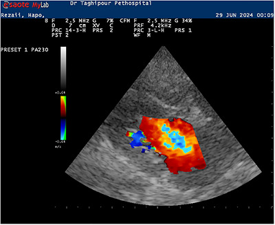
Echocardiographic image of a small‐breed dog with myxomatous mitral valve disease (MMVD). Right parasternal long‐axis four‐chamber view using Doppler modality, demonstrating mitral regurgitation (MR).
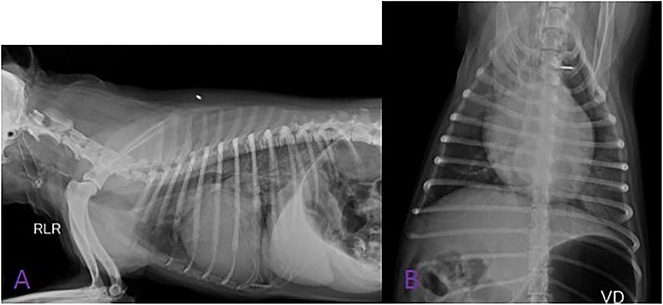
Radiographic images of a small‐breed dog with myxomatous mitral valve disease (MMVD) showing cardiomegaly. (A) Right lateral recumbency. (B) Left lateral recumbency.
Echocardiographic Measurements
Echocardiography was performed via a MyLab 40 device (Esaote, Italy) with cardiac phased‐array transducers (frequency 3.5 MHz) by a board‐certified radiologist, following the standard protocol described by Bonagura and Schober (2009).
In the right parasternal, long‐axis at the view four‐chamber level, with the Doppler sample volume positioned at the anterior aspect of the mitral valve leaflets, mitral inflow velocity (atrium side) (VMA) and the corresponding pressure gradient (GMA) were measured. Subsequently, the Doppler sample volume was repositioned to the posterior aspect of the mitral valve, and mitral inflow velocity (ventricle side) (VMV) and pressure gradient (GMV) were recorded at this location as well (Figure3A,B) (Boon2011).
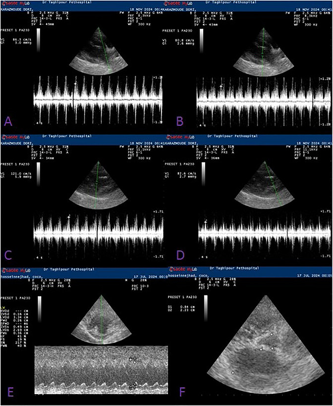
Echocardiographic images of a small‐breed dog with myxomatous mitral valve disease (MMVD). (A) Right parasternal long‐axis four‐chamber view, Doppler modality, showing peak velocity of mitral inflow at the atrial side. (B) Right parasternal long‐axis four‐chamber view, Doppler modality, showing peak velocity of mitral inflow at the ventricular side. (C) Right parasternal long‐axis five‐chamber view, Doppler modality, showing peak velocity of left ventricular outflow tract (LVOT). (D) Right parasternal long‐axis five‐chamber view, Doppler modality, showing peak velocity of the aorta. (E) Right parasternal short‐axis mushroom‐level view, motion (M‐mode) modality. (F) Right parasternal short‐axis aortic‐root view, brightness (B‐mode) modality, showing left atrium (LA) and aorta (Ao) diameters.
In the right parasternal, long‐axis view at the five‐chamber level, the Doppler sample volume was positioned at the level of the left ventricular outflow tract (LVOT) and LVOT velocity (VLVOT), along with the corresponding pressure gradient (GLVOT) were measured. Subsequently, in the same view, the sample volume was repositioned at the proximal portion of the aorta, where aortic flow velocity (VA) and the associated pressure gradient (GA) were recorded (Figure3C,D) (Boon2011).
In the right parasternal, short‐axis view at the level of the papillary muscles (mushroom level) using M‐mode echocardiography, measurements were obtained according to standard guidelines. First, the interventricular septal thickness in diastole (IVSd) was measured. Subsequently, the left ventricular internal diameter in diastole (LVIDd) was recorded in the same alignment, followed by measurement of the left ventricular free wall thickness in diastole (LVPWd). The cursor was then evaluated during systole to measure the interventricular septal thickness in systole (IVSs), the left ventricular internal diameter in systole (LVIDs) and finally the left ventricular free wall thickness in systole (LVPWs). Fractional shortening (FS) and ejection fraction (EF) were subsequently calculated using the Teichholz method (Figure3E) (Boon2011).
Using the short‐axis aortic root view in B‐mode, LAmax, Ao and the LA:Ao ratio were measured. For this method, the aortic diameter was measured at the level of the commissure between the non‐coronary and right coronary cusps. Subsequently, the left atrial diameter was obtained by drawing a linear measurement from the junction of the non‐coronary cusp to the left coronary cusp of the aortic valve and recording the maximal left atrial dimension (Figure3F) (Boon2011).
Classification of the Enrolled Patients
Mitral Regurgitation Severity: Using a Semiquantitative Assessment
Mitral regurgitation severity was assessed using a semiquantitative approach similar to that described by Muzzi et al. (2003). The area of the regurgitant jet (ARJ) was measured at the site of the mosaic colour flow pattern extending from the mitral valve into the left atrium during systole using colour Doppler imaging. The left atrial area (LLA) was also determined. All measurements were obtained from the right parasternal long‐axis view at the four‐chamber level. Subsequently, the ARJ/LLA ratio was calculated. Based on the ARJ/LLA ratio, dogs were classified as having mild (<–30%), moderate (–30% to 70%) or severe (> 70%) mitral regurgitation
Heart Size
Based on the radiographic measurements, the dogs were divided into two groups: those with cardiomegaly and those without cardiomegaly.
Clinical Signs
Dogs were grouped into preclinical and clinical categories on the basis of clinical signs, including difficulty breathing, exercise intolerance, weakness and coughing.
Statistical Analysis
Statistical analysis was performed via SPSS software (version 24). The Kolmogorov–Smirnov test was used to evaluate data normality. Chi‐square tests were applied for frequency comparisons, ANOVA with Tukey post hoc tests was used for comparing means across three groups, and independentt‐tests were applied for comparing means between two groups. Correlations between echocardiographic variables were analysed via scatter plots and Pearson's correlation coefficient. A significance level ofp< 0.05 was considered statistically significant.
Results
Assessment of Data Distribution
The results of the Kolmogorov‒Smirnov test indicated that the data were normally distributed (p> 0.05). Therefore, parametric tests were used for subsequent analyses.
Animal Characteristics
The findings revealed that MMVD was significantly more prevalent in intact dogs than in spayed/neutered dogs (p= 0.02). The Terrier, Pomeranian, Shih Tzu and Terrier Mix breeds had the highest prevalence of MMVD (p= 0.00). Notably, these breeds are among the most popular in Iran and, therefore, represent the majority of referrals. No differences in MMVD incidence were observed based on age (p= 0.34), weight (p= 0.28) or sex (p= 0.76) (Table1).
Table: Signalment characteristics of 60 small‐breed dogs diagnosed with myxomatous mitral valve disease (MMVD).
Group Characteristics
As shown in Figure4, in the mild MR group, the highest frequency was observed in dogs without cardiomegaly and in the preclinical group. In the moderate MR group, the highest frequency was observed in dogs with cardiomegaly, followed by those in the preclinical group. In the severe MR group, all dogs had cardiomegaly, with the highest frequency belonging to the clinical group. The highest frequency of clinical cases was observed in the severe MR group.
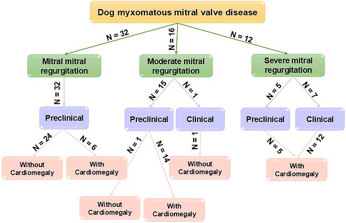
Integration of classifications in 60 small‐breed dogs with myxomatous mitral valve disease (MMVD) based on the severity of mitral regurgitation (MR), cardiac enlargement and the presence of clinical signs. N: number of cases.
Echocardiographic Variables
As shown in Table2, the mean values for LAmax, LVIDd, VMA‐VMV and VLVOT in the severe MR group were significantly greater than those in the mild and moderate groups. However, no differences were observed between the mild and moderate groups. The mean values for LA:Ao and VMA showed a decreasing trend from the severe to the moderate to the mild groups, with statistically significant differences. The mean values for LVIDs, GMA, VMV, GMV and GLVOT were significantly greater in the severe MR group than in the mild MR group, but no differences were observed between the other groups.
Table: Echocardiographic variables in 60 small‐breed dogs with myxomatous mitral valve disease (MMVD), grouped by mitral regurgitation severity and cardiac enlargement.
The mean values for LAmax, LA:Ao, LVIDd, LVIDs, VMA, GMA, GMV, VMA‐GMA, VLVOT and GLVOT were significantly greater in dogs with mitral regurgitation and cardiomegaly than in those with mitral regurgitation but without cardiomegaly (p≤ 0.05). No statistically significant differences were observed in the variables Ao, IVSd, LVPWd, IVSs, LVPWs, EF, FS, VA and GA among the groups with varying severities of mitral regurgitation and different heart sizes (p> 0.05).
Table3indicates that there are statistically significant differences in the variables FS, GMV, VLVOT and GLVOT between the clinical and preclinical groups.
Table: Echocardiographic variables in 60 small‐breed dogs with myxomatous mitral valve disease (MMVD), grouped by clinical signs.
Correlation Analyses
As shown in Figure5, a moderate positive statistical correlation was observed between LA:Ao and three Doppler echocardiographic variables: VMA (p= 0.00,r= 0.49), VMV (p= 0.008,r= 0.33) and LVOT (p= 0.002,r= 0.39). Additionally, a weak but statistically significant correlation was found between VMA‐VMV and LAmax (p= 0.02,r= 0.29). However, no differences in correlation were observed between these factors and motion‐mode echocardiographic variables.
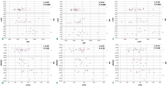
Correlations between echocardiographic measurements in 60 small‐breed dogs with mitral regurgitation (MR) of varying severity: mild (1), moderate (2) and severe (3). All measurements were obtained using brightness (B‐mode), motion (M‐mode) and Doppler echocardiography. Red spots indicate female dogs; blue spots indicate male dogs. (A) LA/Ao ratio versus VMA. (B) LA/Ao ratio versus VMV. (C) LA/Ao ratio versus LVOT. (D) VMA/VMV ratio versus LAmax. (E) VMA/VMV ratio versus LVIDd. (F) VMA/VMV ratio versus LVIDs. Ao, aorta; LA, left atrium; LAmax, maximum left atrium diameter; LVIDd, left ventricular internal diameter in diastole; LVIDs, left ventricular internal diameter in systole; LVOT, left ventricular outflow tract; MR, mitral regurgitation; VMA, mitral annular velocity at atrial side; VMV, mitral annular velocity at ventricular side.r= Pearson correlation coefficient.p= significance level for correlation (calculated using Pearson correlation test).
Discussion
This study revealed a statistically significant correlation between Doppler hemodynamic variables and MR severity. As MR severity increased, the velocity and gradient variables at the mitral valve and left ventricular outflow tract also increased. One of the earliest studies on echocardiography and MR in dogs was conducted by Braunwald et al. (1957) and their findings align with those of the present study. According to Vezzosi et al. (2021) and Morgan et al. (2020), the mitral valve velocity increases with the severity of MMVD. Similarly, Larouche‐Lebel et al. (2019) demonstrated significant correlations between Doppler echocardiographic variables and different MR severity levels in dogs with MMVD. Hezzell et al. (2012) also reported higher velocities in dogs with severe (fatal) MR than in nonfatal MR cases. Studies by Suzuki et al. (2013) and Thomas et al. (1998) Thomas et al. (1998) support these findings. Additionally, Muzzi et al. (2003) reported a significant correlation between colour flow mapping of the regurgitant jet and Doppler variables in dogs with congestive heart failure.
This study also revealed a significant correlation between LVID in systole and diastole and MR severity. Similarly, Lord et al. (2010), in their study on Cavalier King Charles Spaniels, reported a direct relationship between increased LVIDs, MR severity and the occurrence of congestive heart failure.
The results of this study demonstrated a significant increase in LA:Ao with increasing MR severity. Gouni et al. (2007) conducted a similar study on dogs with varying MR severities and reported significant differences in LA:Ao between groups. Serres et al. (2008) and Larouche‐Lebel et al. (2019) reported similar findings.
According to studies such as Höllmer et al. (2017), left atrial size increases in response to MR. Olsen et al. (2003) reported that in Dachshund dogs, mitral valve prolapse severity, regurgitant jet size and murmur intensity positively correlated with increased LA size over time. These findings align with human studies (Kihara et al.1988; Yellin et al.1979). However, some studies, such as Larouche‐Lebel et al. (2019), did not find significant correlations between B‐mode variables and MR severity.
The present study revealed differences in FS, LVOT and GLVOT between clinical and preclinical MMVD patients. Schober et al. (2010) reported that the LA:Ao ratio and velocity flow were significantly greater in clinical MR groups than in preclinical groups. Chetboul and Tissier. (2012) proposed Doppler echocardiographic measurements as valuable indices for evaluating MMVD in preclinical dogs and predicting disease progression. Similarly, studies in humans have shown that velocity variables are significantly greater in clinical MR patients (Al‐Wakeel et al.2015).
The results of this study indicated that LA size can be used to estimate the severity of the blood jet, but it is not an ideal criterion for distinguishing between clinical and preclinical MMVD patients. Dickson et al. (2017) reported similar findings.
No differences in FS or EF were observed among dogs with varying MMVD severity. However, Gouni et al. (2007) and Suzuki et al. (2013) reported that FS is correlated with disease severity. Vezzosi et al. (2021) measured FS in dogs with late‐stage, severe, moderate and mild MMVD as 51%, 50%, 46% and 39%, respectively, and reported statistically significant differences between each group and the mild/moderate groups. Serres et al. (2008) reported that FS and EF increased with disease severity. Differences in grouping, disease duration and sample size may explain the discrepancies between this study and others. Factors such as preload, afterload and the creation of a pathological low‐resistance pathway by mitral valve lesions can influence these variables, as noted by Boon (2011).
This study revealed that heart size was greater in dogs with severe MR and clinical signs. Similarly, Mihara et al. (2021) reported that the mitral valve annulus was larger and more circular in dogs with MMVD than in control dogs. However, Sargent et al. (2015) argued that MR severity provides prognostic value independent of heart size in MMVD patients.
The findings also revealed that MMVD incidence was greater in intact dogs. No differences were observed in terms of age, weight or sex. Similarly, Suzuki et al. (2013) reported no correlation between age, weight or MMVD incidence. However, Atkins et al. (2009) reported a higher MMVD incidence in male dogs than in female dogs, contrary to the findings of this study. This finding was consistent with the study by Elyasi et al. (2023) in cats. In human studies, postmenopausal estrogen reduction has been linked to adverse cardiovascular effects, including increased blood pressure, sympathetic tone and collagen/elastin deposition in the heart, potentially accelerating mitral degeneration and calcification (Huxley2007).
Limitations
In the present study, echocardiographic and clinical evaluations were conducted only at the initial visit. As a result, it was not possible to differentiate cases on the basis of disease duration (i.e., acute vs. chronic). Furthermore, the sample size was limited, highlighting the need for larger population studies in the future.
Conclusions
The findings of this study indicate that echocardiography, particularly Doppler echocardiography, is a valuable tool for diagnosing and grading MMVD. This study demonstrated that velocity variables in affected patients vary according to the severity of MR, changes in heart size and the presence of clinical signs. Therefore, evaluating these variables allows for more precise treatment strategies and follow‐up care. Additionally, the higher prevalence of this disease in intact dogs highlights the potential impact of neutering on cardiac health and the development of heart disease. Thus, the results of this study can serve as a reference for managing MMVD cases.
Author Contributions
Boshra Elyasi: conceptualization, investigation, writing – original draft, funding acquisition, methodology, validation, visualization, writing – review and editing, software, formal analysis, project administration, data curation, supervision, resources.
Funding
The author has nothing to report.
Ethics Statement
This study was conducted on client‐owned animals referred to Dr. Taghipour Veterinary Hospital, Tehran, Iran. All procedures were performed as part of routine clinical practice, and no additional diagnostic or therapeutic interventions were carried out for research purposes. Therefore, formal ethical committee approval was not required. Informed consent was obtained from all pet owners prior to inclusion in the study. Owners were informed that anonymized clinical data would be used for research purposes and may be published as part of a scientific article.
Conflicts of Interest
The author declares no conflicts of interest.
References
- Adham Esfahani, S. , K. Hassani, andD. M. Espino. 2019. “Fluid‐Structure Interaction Assessment of Blood Flow Hemodynamics and Leaflet Stress During Mitral Regurgitation. ”Computer Methods in Biomechanics and Biomedical Engineering22: 288–303. doi.org/10.1080/10255842.2018.1552683
- Al‐Wakeel, N. , J. F. Fernandes, A. Amiri, et al. 2015. “Hemodynamic and Energetic Aspects of the Left Ventricle in Patients With Mitral Regurgitation Before and After Mitral Valve Surgery. ”Journal of Magnetic Resonance Imaging42: 1705–1712. doi.org/10.1002/jmri.24926
- Atkins, C. , J. Bonagura, S. Ettinger, et al. 2009. “Guidelines for the Diagnosis and Treatment of Canine Chronic Valvular Heart Disease. ”Journal of Veterinary Internal Medicine23: 1142–1150. doi.org/10.1111/j.1939-1676.2009.0392.x
- Bagardi, M. , V. Zamboni, C. Locatelli, A. Galizzi, S. Ghilardi, andP. G. Brambilla. 2022. “Management of Chronic Congestive Heart Failure Caused by Myxomatous Mitral Valve Disease in Dogs: A Narrative Review From 1970 to 2020. ”Animals12: 209. doi.org/10.3390/ani12020209
- Bahr, R. 2018. “Canine and Feline Cardiovascular System. ” In: Textbook of Veterinary Diagnostic Radiology, 684–709. Elsevier.
- Bonagura, J. , andK. Schober. 2009. “Can Ventricular Function be Assessed by Echocardiography in Chronic Canine Mitral Valve Disease?”Journal of Small Animal Practice50: 12–24. doi.org/10.1111/j.1748-5827.2009.00803.x
- Boon, J. A. 2011. Veterinary Echocardiography. John Wiley & Sons.
- Borgarelli, M. , andJ. W. Buchanan. 2012. “Historical Review, Epidemiology and Natural History of Degenerative Mitral Valve Disease. ”Journal of Veterinary Cardiology14: 93–101. doi.org/10.1016/j.jvc.2012.01.011
- Braunwald, E. , G. H. WelchJr. , andS. J. Sarnoff. 1957. “Hemodynamic Effects of Quantitatively Varied Experimental Mitral Regurgitation. ”Circulation Research5: 539–545. doi.org/10.1161/01.res.5.5.539
- Burchell, R. K. , andJ. Schoeman. 2014. “Advances in the Understanding of the Pathogenesis, Progression and Diagnosis of Myxomatous Mitral Valve Disease in Dogs. ”Journal of the South African Veterinary Association85: 1–5. doi.org/10.4102/jsava.v85i1.1101
- Chetboul, V. , andR. Tissier. 2012. “Echocardiographic Assessment of Canine Degenerative Mitral Valve Disease. ”Journal of Veterinary Cardiology14: 127–148. doi.org/10.1016/j.jvc.2011.11.005
- Dickson, D. , D. Caivano, J. N. Matos, N. Summerfield, andM. Rishniw. 2017. “Two‐Dimensional Echocardiographic Estimates of Left Atrial Function in Healthy Dogs and Dogs With Myxomatous Mitral Valve Disease. ”Journal of Veterinary Cardiology19: 469–479. doi.org/10.1016/j.jvc.2017.09.003
- Elyasi, B. , M. N. Masouleh, andA. Jahandideh. 2023. “Effect of Ovariohysterectomy on Queen Myocardial Function: Echocardiographic Evidence. ”Veterinary Research Forum14, no. 9: 495. doi.org/10.30466/vrf.2022.552942.3456
- Gouni, V. , F. J. Serres, J. ‐L. Pouchelon, et al. 2007. “Quantification of Mitral Valve Regurgitation in Dogs With Degenerative Mitral Valve Disease by Use of the Proximal Isovelocity Surface Area Method. ”Journal of the American Veterinary Medical Association231: 399–406. doi.org/10.2460/javma.231.3.399
- Häggström, J. , K. Höglund, andM. Borgarelli. 2009. “An Update on Treatment and Prognostic Indicators in Canine Myxomatous Mitral Valve Disease. ”Journal of Small Animal Practice50: 25–33. doi.org/10.1111/j.1748-5827.2009.00800.x
- Harris, P. , andL. Kuppurao. 2016. “Quantitative Doppler Echocardiography. ”BJA Education16: 46–52.
- Hezzell, M. J. , A. Boswood, W. Moonarmart, andJ. Elliott. 2012. “Selected Echocardiographic Variables Change More Rapidly in Dogs That Die From Myxomatous Mitral Valve Disease. ”Journal of Veterinary Cardiology14: 269–279. doi.org/10.1016/j.jvc.2012.01.009
- Höllmer, M. , J. Willesen, A. Tolver, andJ. Koch. 2017. “Left Atrial Volume and Function in Dogs With Naturally Occurring Myxomatous Mitral Valve Disease. ”Journal of Veterinary Cardiology19: 24–34. doi.org/10.1016/j.jvc.2016.08.006
- Huxley, V. H. 2007. “Sex and the Cardiovascular System: the Intriguing Tale of How Women and Men Regulate Cardiovascular Function Differently. ”Advances in Physiology Education31: 17–22. doi.org/10.1152/advan.00099.2006
- Keene, B. W. , C. E. Atkins, J. D. Bonagura, et al. 2019. “ACVIM Consensus Guidelines for the Diagnosis and Treatment of Myxomatous Mitral Valve Disease in Dogs. ”Journal of Veterinary Internal Medicine33: 1127–1140. doi.org/10.1111/jvim.15488
- Kihara, Y. , S. Sasayama, S. Miyazaki, et al. 1988. “Role of the Left Atrium in Adaptation of the Heart to Chronic Mitral Regurgitation in Conscious Dogs. ”Circulation Research62: 543–553. doi.org/10.1161/01.res.62.3.543
- Klein, S. , I. Nolte, J. L. Granados‐Soler, et al. 2022. “Evaluation of New and Old Biomarkers in Dogs With Degenerative Mitral Valve Disease. ”BMC Veterinary Research18: 256. doi.org/10.1186/s12917-022-03343-z
- Larouche‐Lebel, É. , K. A. Loughran, andM. A. Oyama. 2019. “Echocardiographic Indices and Severity of Mitral Regurgitation in Dogs With Preclinical Degenerative Mitral Valve Disease. ”Journal of Veterinary Internal Medicine33: 489–498. doi.org/10.1111/jvim.15461
- Levine, R. A. , A. A. Hagége, D. P. Judge, et al. 2015. “Mitral Valve Disease—Morphology and Mechanisms. ”Nature Reviews Cardiology12: 689–710. doi.org/10.1038/nrcardio.2015.161
- Ljungvall, I. 2024. “Cardiovascular Disease. ” In: Ettinger's Textbook of Veterinary Internal Medicine. 9th ed. Elsevier, 110.
- Lord, P. , K. Hansson, C. Kvart, andJ. Häggström. 2010. “Rate of Change of Heart Size Before Congestive Heart Failure in Dogs With Mitral Regurgitation. ”Journal of Small Animal Practice51: 210–218. doi.org/10.1111/j.1748-5827.2010.00910.x
- Mattin, M. , A. Boswood, D. Church, et al. 2015. “Prevalence of and Risk Factors for Degenerative Mitral Valve Disease in Dogs Attending Primary‐Care Veterinary Practices in England. ”Journal of Veterinary Internal Medicine29: 847–854. doi.org/10.1111/jvim.12591
- Mihara, K. , I. Kanemoto, K. Sato, Y. Yasuhira, I. Watanabe, andK. Misumi. 2021. “Echocardiographic Evaluation of Deformity and Enlargement of the Canine Mitral Valve Annulus Associated With Myxomatous Degenerative Mitral Valve Disease. ”Journal of Veterinary Cardiology37: 8–17. doi.org/10.1016/j.jvc.2021.08.002
- Morgan, K. , G. Monteith, S. Raheb, M. Colpitts, andS. Fonfara. 2020. “Echocardiographic Parameters for the Assessment of Congestive Heart Failure in Dogs With Myxomatous Mitral Valve Disease and Moderate to Severe Mitral Regurgitation. ”Veterinary Journal263: 105518. doi.org/10.1016/j.tvjl.2020.105518
- Muzzi, R. A. , R. B. de Araújo, L. A. Muzzi, J. L. Pena, andE. F. Silva. 2003. “Regurgitant Jet Area by Doppler Color Flow Mapping: Quantitative Assessment of Mitral Regurgitation Severity in Dogs. ”Journal of Veterinary Cardiology5: 33–38. doi.org/10.1016/S1760-2734(06)70050-9
- O'Brien, M. J. , N. Beijerink, andC. Wade. 2021. “Genetics of Canine Myxomatous Mitral Valve Disease. ”Animal Genetics52: 409–421. doi.org/10.1111/age.13082
- Olsen, L. , T. Martinussen, andH. Pedersen. 2003. “Early Echocardiographic Predictors of Myxcomatous Mitral Valve Disease in Dachshunds. ”Veterinary Record152: 293–297. doi.org/10.1136/vr.152.10.293
- Sargent, J. , R. Muzzi, R. Mukherjee, et al. 2015. “Echocardiographic Predictors of Survival in Dogs With Myxomatous Mitral Valve Disease. ”Journal of Veterinary Cardiology17: 1–12. doi.org/10.1016/j.jvc.2014.11.001
- Schober, K. , T. Hart, J. Stern, et al. 2010. “Detection of Congestive Heart Failure in Dogs by Doppler Echocardiography. ”Journal of Veterinary Internal Medicine24: 1358–1368. doi.org/10.1111/j.1939-1676.2010.0592.x
- Serres, F. , V. Chetboul, R. Tissier, et al. 2008. “Comparison of 3 Ultrasound Methods for Quantifying Left Ventricular Systolic Function: Correlation With Disease Severity and Prognostic Value in Dogs With Mitral Valve Disease. ”Journal of Veterinary Internal Medicine22: 566–577. doi.org/10.1111/j.1939-1676.2008.0097.x
- Suzuki, R. , H. Matsumoto, T. Teshima, Y. Mochizuki, andH. Koyama. 2013. “Left Ventricular Geometrical Differences in Dogs With Various Stages of Myxomatous Mitral Valve Disease. ”Journal of Small Animal Practice54: 234–239. doi.org/10.1111/jsap.12058
- Svensson, M. , J. Selling, andM. Dirven. 2024. “Myxomatous Mitral Valve Disease in Large Breed Dogs: Survival Characteristics and Prognostic Variables. ”Veterinary Sciences11: 136. doi.org/10.3390/vetsci11030136
- Thomas, L. , E. Foster, andN. B. Schiller. 1998. “Peak Mitral Inflow Velocity Predicts Mitral Regurgitation Severity. ”Journal of the American College of Cardiology31: 174–179. doi.org/10.1016/s0735-1097(97)00454-3
- Vezzosi, T. , G. Grosso, R. Tognetti, et al. 2021. “The Mitral Insufficiency Echocardiographic Score: A Severity Classification of Myxomatous Mitral Valve Disease in Dogs. ”Journal of Veterinary Internal Medicine35: 1238–1244. doi.org/10.1111/jvim.16131
- Yellin, E. L. , C. Yoran, E. H. Sonnenblick, S. Gabbay, andR. Frater. 1979. “Dynamic Changes in the Canine Mitral Regurgitant Orifice Area During Ventricular Ejection. ”Circulation Research45: 677–683. doi.org/10.1161/01.res.45.5.677
Republished from the open web under CC-BY. Authors: Elyasi B. Read the original.