Enzymatic 1,4-addition of 2-hydroxy-3-keto-glucal for β-selective aryl-C-glycosylation of polyphenols.
3-Ketoglycals are versatile Michael acceptors widely used in chemical C -glycosylation. Here, we report the enzymatic equivalent of this transformation, catalysed by a 3-keto- C -glycoside lyase, enabling selective C -glycosylation of polyphenolic natural products. The reaction proceeds with remarkable chemo- and stereo-selectivity, affording aryl- C -β-glycosides.
Aryl-C-glycosyl compounds, henceforth aryl-C-glycosides (Fig. S1), represent key structural motifs in numerous bioactive natural products, notably flavonoids.1–4Owing to their resistance to hydrolytic cleavage,C-glycosides are metabolically stable analogues ofO-glycosides.1,5Synthetic approaches typically introduce the sugar moiety on an aryl precursor,4,6–13often employing 1,2-unsaturated sugar derivatives (glycals).4,6,14Among these, 3-keto-glycals – featuring an α,β-unsaturated ketone system with an electron-donating oxygen at the β-carbon15–20– promote aryl-C-glycosylation through 1,4-addition to yield α-anomers (Fig. 1a).21–23An enzymatic equivalent to this transformation has not been reported. From a synthetic perspective, a β-selective glycosylation is desirable, since natural aryl-C-glycosides invariably adopt this configuration.1,2Metal-catalysed coupling of 3-ketoglycals has been shown for 2-deoxy-C-glycoside synthesis,21,23–28but stereo-control of these transformations is challenging.22,28–30
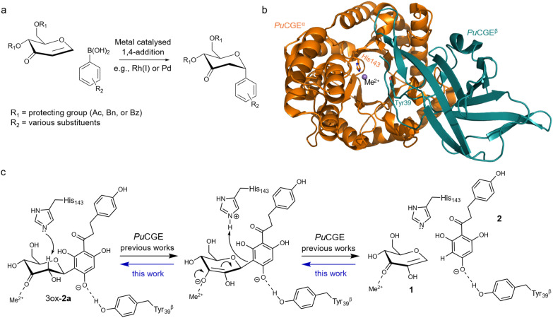
1,4-Addition of 3-keto-glycal for arylC-glycoside synthesis and enzymatic equivalent of the transformation catalysed by 3-keto-C-glycoside lyase. (a) Metal-catalysed chemical reaction. (b) Overall fold of the lyase heterodimer (PuCGE; PDB code: 7EXZ), showing the α-subunit with the divalent metal ion and catalytic histidine. The β-subunit provides a tyrosine to activate the aglycone. (c) Proposed mechanism for enzymatic C–C bond cleavage of 3″-keto-nothofagin (3ox-2a). The reverse reaction of the enzyme is characterized in this study.
Here, we demonstrated the formation of flavonoidC-glycosides through the reverse reaction of 3-keto-C-glycoside lyase (Fig. 1b and c). The enzyme is involved in non-hydrolytic deglycosylation of aryl-C-glycosides through a four-step biochemical pathway31–35(Fig. S3). The pathway starts with C3′ oxidation of the glycoside substrate. The C–C bond is then cleaved by the lyaseviaβ-elimination of the intermediary 3-keto-glycoside (Fig. 1c). The 1,2-unsaturated elimination product (see compound 1 inFig. 1c) is hydrated by the same or another lyase, and the resulting 3-keto-sugar is reduced to the final non-oxidised monosaccharide product.
For this study we used the 3-keto-β-glucoside lyase from the human intestinal bacterium strain PUE, henceforthPuCGE. The enzyme adopts a heterodimer fold depicted inFig. 1b.32The main α-subunit coordinates a divalent metal ion (Mn2+) in the active site and provides a catalytic histidine residue (His143α), proposed to function as general acid–base for the β-elimination (Fig. 1c).33The β-subunit contributes a conserved tyrosine (Tyr39β) to the binding pocket for the aglycone, providing interactions found to be mechanistically critical for the C–C bond cleavage.33PuCGE was shown in earlier work33to release 2-hydroxy-3-keto-glucal (1,5-anhydro-d-erythro-hex-1-en-3-ulose, 1,Fig. 1c) upon elimination of the 3″-keto derivative (3ox-2a;Fig. 1c) of nothofagin (phloretin 3′-C-β-glucoside; 2a; Fig. S1). The extent to which thePuCGE reaction is reversible was not known before this work. However, we noted the possible relevance for biocatalytic synthesis of such an enzymatic transformation.
Compound 1 was exploited here as an electrophilic substrate for 1,4-addition of flavonoids such as phloretin (2;Fig. 1c) or apigenin (3; Fig. S1). It was generated enzymaticallyviaC3′-oxidation of 4-nitrophenyl-α-d-glucoside (4) or sucrose (5),33followed byin situeliminative cleavage of the 3-keto-glycoside (Fig. S4). The elimination of 4-nitrophenyl-3′-keto-α-d-glucoside (3ox-4) was catalysed at pH 6.5 by a lyase variant (H275N-3-keto-O-glycoside-eliminating lyase fromBacteroides thetaiotaomicron) deficient in hydration activity toward 1.35Alternatively, 3′-keto-sucrose (3ox-5) afforded 1 chemically under alkaline conditions (1.0 M NaOH; Fig. S5 and S6).15The structure of isolated compound 1 was confirmed by NMR spectroscopy (Fig. S7).
We now show thatPuCGE catalyses the addition of 1 to 2 in the absence of any other promoting reagent in aqueous solution at ambient conditions (pH 7.5; 37 °C), forming exclusively theC-β-glycosidic product at the C3′ of 2 (Fig. 2a). Reaction progress was monitored by HPLC, and MS fragmentation confirmedC- rather thanO-glycoside formation (Fig. S8–S10). The major product was isolated and characterised by NMR (Fig. S11–S17, Table S1). NoO-glycoside was detected in thePuCGE reaction (Fig. S18) which is interesting considering the evidence33that the enzyme is active toward elimination of the 3″-keto derivative (3ox-2b; Fig. S1) of phlorizin (phloretin 3′-O-β-glucoside, 2b; Fig. S1), releasing 1 and 2. Another lyase (fromAgrobacterium tumefaciens) specific for 3-keto-O-glucosides showed no activity towards 1 and 2 (Fig. S19), confirming strict chemo-selectivity of the enzymatic C–C coupling. Comparable specific activities for elimination of 3″-keto-nothofagin (3ox-2a; 0.64 U mg−1)33and 3″-keto-phlorizin (3ox-2b; 1.25 ± 0.05 U mg−1)33suggest that the selectivity in the synthetic direction reflects the higher thermodynamic stability ofC-versus O-glycosides36–38rather than kinetic effects of the enzyme.
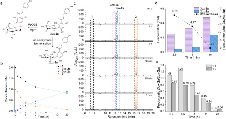
Kinetic analysis of phloretinC-glycoside formation byPuCGE-catalysed 1,4-addition of compounds 1 and 2. (a) Scheme of the enzymatic reaction at the C3′ of 2 (orange arrow) followed by spontaneous isomerisation (3ox-2a → 2ox-2a). (b) Time course of the reaction of 1 (2.0 mM) and 2 (1.0 mM). (c) Stacked HPLC traces showing the composition of the reaction mixture at different reaction times. (d) Column plot of product formation in the initial 10 min of reaction. Change in product ratio (3ox-2a/2ox-2a) over time is indicated with a dashed black line. (e) Column plot of the product ratio (3ox-2a/2ox-2a) over time for reactions using equimolar or two-fold molar excess of 2 over 1 (1.0 mM).N= 1 individual experiment. For experimental details, see the Methods section in the SI.
Experiments conducted with the isolatedPuCGE α-subunit showed no addition of 1 to 2 (Fig. S20), indicating that the fully formed enzyme α/β heterodimer is required for the activity. The result is consistent with Bitteret al.33who found the α-subunit to be completely devoid of activity toward 3ox-2a while a low level of 3ox-2b eliminating activity was retained.
Incubation ofPuCGE with 1 in two-fold excess of 2 (1.0 mM) revealed the kinetics ofC-glycoside formation (Fig. 2a–c). The initial enzymatic product 3ox-2a, underwent non-enzymatic isomerisation to 2″-keto-nothofagin (2ox-2a;Fig. 2a and d). Isomerisation to a 2″-keto glycoside has previously been observed for related 3″-keto aryl-C-glycosides.31,39Nagorski and Richard demonstrated that non-enzymatic sugar isomerisation can proceedviasolvent-assisted proton transfer catalysed by Brønsted bases.40In contrast, the presence of Zn2+was shown to accelerate an alternative mechanistic pathway involving direct hydride transfer.40Yiet al.also reported that addition of Ca2+shifted the carbonyl migration toward a pathway dominated by 1,2-hydride transfer.41In light of these studies, our observation of isomerisation is consistent with an inherent, metal-modulated reactivity of the glycoside scaffold rather than a requirement for specific catalytic assistance to the conversion of 3-keto- into 2-keto-glycoside. The effectively irreversible isomerisation under the conditions used, likely drives the reaction towards near-quantitative conversion of phloretin 2.
The isolated 2ox-2a product structure was confirmed by NMR and shown to contain a fully hydrated keto-group (2diol-2a; Fig. S11–S17, Table S1). The observed H3″–H4″ coupling constant (3.4 Hz) is significantly lower than the typical 8–10 Hz expected for an unconstrained glucosyl ring.42This localized reduction in vicinal coupling indicates that the 2″-diol sugar ring probably populates a distorted conformational state in solution. Reactions with varying molar ratios of 1 : 2 (1 : 1, 1 : 2) likewise yielded 2ox-2a stoichiometrically relative to the limiting substrate (Fig. 2eand Fig. S21, S22). The product 2ox-2a remained stable in solution over a prolonged incubation (up to 20 h) and was not utilised as a substrate for reverse enzymatic cleavage. The lack of reactivity for 2ox-2a can be explained by the requirements for the positioning in thePuCGE active site. Metal coordination of the substrate 3-keto group was suggested to be crucial for productive binding and catalytic activation.33
Extension of the analysis ofPuCGE reaction to apigenin 3 (0.4 mM;Fig. 3a) revealed twoC-glycosylation products when 1 was reacted in 2.5-fold excess (Fig. 3band Fig. S23). Both were identified asC-glycosides by MS (Fig. S24–S26), but degraded upon prolonged incubation (>4 h), even in the presence of tris(2-carboxyethyl)phosphine (2.0 mM) as a reducing agent (Fig. S27). NMR identification of the products from partially purified mixture was complicated by the effect of Mn2+on spectral line broadening. However, NaBH4reduction of the reaction mixture generated two new HPLC peaks (Fig. 3b), one co-eluting with authentic vitexin (apigenin 8-C-β-glucoside, 3a), suggesting glycosylation at C8. The second product, putatively the C6-glycoside (isovitexin, 3b), was assigned based onPuCGE substrate specificity.33
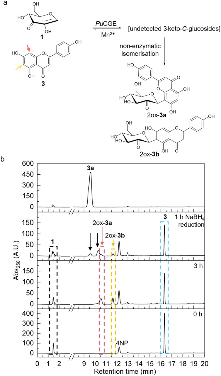
Product identification of thePuCGE-catalysed addition of 1 (1.0 mM) to apigenin (3; 0.4 mM). (a) Proposed reaction products following non-enzymatic isomerisation are 2″-keto-vitexin (2ox-3a) and 2″-keto-isovitexin (2ox-3b). Arrows indicate the glycosylation site (C6, yellow; C8, red). (b) HPLC traces of samples from the addition reaction, before and after reduction with NaBH4(2.0 mM). A vitexin (3a) standard is also shown. Black arrows indicate newly formed peaks.N= 1 individual experiment. For experimental details, see the Methods section in the SI.
Incubation of the reaction mixture with an NADPH-dependent reductase specific for 3-keto group reduction did not alter the initialC-glycosylation products (Fig. S28), supporting their identity as 2-keto-C-glycosides (2″-keto-vitexin, 2ox-3a; 2″-keto-isovitexin, 2ox-3b). The absence of 3-keto-isomers implies rapid 3,2-isomerisation, favouring the 2-keto species.
Incubation of 1 with daidzein (4; Fig. S1) in the presence ofPuCGE failed to yield a glycosylation product under a broad range of conditions (Fig. S29a). The daidzein (6) A ring is electronically less activated than those of phloretin (2) and apigenin (3).43,44Additionally, it offers only a single glycosylation site at C8, which arguably can make it more difficult for the enzyme to bring the aglycone into a reactive position (Fig. S29b).45Ionisation of the daidzein acidic hydroxy group at C7 (pKa= ∼7.3)46might additionally impair productive binding. The 3-keto derivative (3ox-4a; Fig. S1) of puerarin (daidzein 8-C-β-glucoside, 4a, Fig. S1) is, however, a 30-fold better substrate forPuCGE cleavage than 3ox-2a.33Therefore the accumulation of 3-keto-puerarin (3ox-4a) is highly unlikely. The specific activity ratio of addition and elimination of 1 to and from 2 is 1 : 9.7,33indicating that elimination is strongly favoured.
In summary, this study establishes an unprecedented aryl-C-glycosylation achieved through enzymatic 1,4-addition of 1 to flavonoid acceptors. The biocatalytic transformation exploits the reverse reaction catalysed by a 3-keto-C-glycoside lyase. The reaction proceeds with strict chemo-selectivity forC- overO-glycosylation and affords exclusively the β-configuredC-glycoside. The primary addition product is a 3-keto-β-C-glycoside, which undergoes spontaneous isomerisation to the corresponding 2-keto form under the applied reaction conditions. The addition of 1 to 2 proceeds to completion, followed by near quantitative isomerisation of 3ox-2a to 2ox-2a. In contrast, the reaction of 1 with 3 reaches partial conversion only. However the quantitative isomerisation occurs rapidly. Collectively, these findings constitute proof-of-principle of a novel enzymatic reaction and underscore the promising synthetic potential of enzymaticC-glycosylation for polyphenolic natural products. The 3-keto-C-glycoside lyase reaction expands the repertoire of enzymatic C–C couplings for biocatalytic transformations.47–52Further studies will show the role of spontaneous keto-group isomerisation for the enzymatic C–C bond formation to proceed.
Author contributions
Klara Kastner: investigation, writing – review & editing. Martin Pfeiffer: writing – review & editing. Bernd Nidetzky: conceptualization, writing – original draft, review & editing, and funding acquisition.
Conflicts of interest
The authors declare no conflict of interest.
References
- ZhangY. -Q. ZhangM. WangZ. -L. QiaoX. YeM. Biotechnol. Adv. 202260108030 doi.org/10.1016/j.biotechadv.2022.108030
- LiuC. -F. Molecules2022277439 doi.org/10.3390/molecules27217439
- BililignT. GriffithB. R. ThorsonJ. S. Nat. Prod. Rep. 200522742760 doi.org/10.1039/B407364A
- KitamuraK. AndoY. MatsumotoT. SuzukiK. Chem. Rev. 201811814951598 doi.org/10.1021/acs.chemrev.7b00380
- XiaoJ. CapanogluE. JassbiA. R. MironA. Crit. Rev. Food Sci. Nutr. 201656S29S45 doi.org/10.1080/10408398.2015.1067595
- YangY. YuB. Chem. Rev. 20171171228112356 doi.org/10.1021/acs.chemrev.7b00234
- LiaoH. MaJ. YaoH. LiuX. -W. Org. Biomol. Chem. 20181617911806 doi.org/10.1039/C8OB00032H
- ParidaS. P. DasT. AhemadM. A. PatiT. MohapatraS. NayakS. Carbohydr. Res. 2023530108856 doi.org/10.1016/j.carres.2023.108856
- Mejia-OtalvaroF. LaxB. M. KırtelO. WelnerD. H. ChemSusChem202518e202501094 doi.org/10.1002/cssc.202501094
- XuS. PingY. XuM. WuG. KeY. MiaoR. QiX. KongW. Nat. Chem. 20241620542065 doi.org/10.1038/s41557-024-01629-3
- ZhangC. XuS. -Y. ZuoH. ZhangX. DangQ. -D. NiuD. Nat. Synth. 20232251260 doi.org/10.1038/s44160-022-00214-1
- ShangW. HuY. HeY. NiuD. LiW. Sci. Adv. 202511eady2175 doi.org/10.1126/sciadv.ady2175
- BokorÉ. KunS. GoyardD. TóthM. PralyJ. -P. VidalS. SomsákL. Chem. Rev. 201711716871764 doi.org/10.1021/acs.chemrev.6b00475
- YadavY. SagarR. Chem. – Asian J. 202520e202401773 doi.org/10.1002/asia.202401773
- PietschM. WalterM. BuchholzK. Carbohydr. Res. 1994254183194 doi.org/10.1016/0008-6215(94)84251-5
- SridharP. R. AliI. LakshmiM. V. K. J. Org. Chem. 20228789398955 doi.org/10.1021/acs.joc.2c00663
- SharmaM. K. PandeyA. K. HussainN. ChemSelect202510e04180
- DasP. ThakurR. Carbohydr. Res. 2024536109032 doi.org/10.1016/j.carres.2024.109032
- CzerneckiS. VijayakumaranK. VilleG. J. Org. Chem. 19865154725475 doi.org/10.1021/jo00376a094
- DubbuS. Carbohydr. Res. 2024536109016 doi.org/10.1016/j.carres.2023.109016
- BellostaV. CzerneckiS. Carbohydr. Res. 1987171279288 doi.org/10.1016/S0008-6215(00)90892-9
- SinghA. K. VenkateshR. KandasamyJ. Synthesis202411671174
- RamnauthJ. PoulinO. BratovanovS. S. RakhitS. MaddafordS. P. Org. Lett. 2001325712573 doi.org/10.1021/ol016245d
- KirschningA. HardersJ. Tetrahedron19975378677876 doi.org/10.1016/S0040-4020(97)00475-4
- HayashiM. KawabataH. ShimonoS. KakehiA. Tetrahedron Lett. 20004125912594 doi.org/10.1016/S0040-4039(00)00212-4
- Kumar SinghA. VenkateshR. Kumar KanaujiyaV. TiwariV. KandasamyJ. Eur. J. Org. Chem. 2022e202200023 doi.org/10.1002/ejoc.202200023
- BenhaddouR. CzerneckiS. VilleG. J. Org. Chem. 19925746124616 doi.org/10.1021/jo00043a016
- SinghA. K. KanaujiyaV. K. TiwariV. SabiahS. KandasamyJ. Org. Lett. 20202276507655 doi.org/10.1021/acs.orglett.0c02843
- GoodwinT. E. CrowderC. M. WhiteR. B. SwansonJ. S. EvansF. E. MeyerW. L. J. Org. Chem. 198348376380 doi.org/10.1021/jo00151a019
- YunkerM. B. PlaumannD. E. Fraser-ReidB. Can. J. Chem. 19775540024009 doi.org/10.1139/v77-568
- NakamuraK. ZhuS. KomatsuK. HattoriM. IwashimaM. Appl. Environ. Microbiol. 202086e0060720 doi.org/10.1128/AEM.00607-20
- MoriT. KumanoT. HeH. WatanabeS. SendaM. MoriyaT. AdachiN. HoriS. TerashitaY. KawasakiM. HashimotoY. AwakawaT. SendaT. AbeI. KobayashiM. Nat. Commun. 2021126294 doi.org/10.1038/s41467-021-26585-1
- BitterJ. PfeifferM. BorgA. J. E. KuhlmannK. Pavkov-KellerT. Sánchez-MurciaP. A. NidetzkyB. Nat. Commun. 2023147123 doi.org/10.1038/s41467-023-42750-0
- BainsR. K. NasseriS. A. WardmanJ. F. WithersS. G. Curr. Opin. Chem. Biol. 202480102457 doi.org/10.1016/j.cbpa.2024.102457
- KastnerK. BitterJ. PfeifferM. GriningerC. OberdorferG. Pavkov-KellerT. WeberH. NidetzkyB. Angew. Chem. , Int. Ed. 202463e202410681 doi.org/10.1002/anie.202410681
- LiM. YanQ. WangY. QiuR. GaoL. ChenT. WangJ. Green Synth. Catal. 2025 doi.org/10.1016/j.gresc.2025.12.011
- GutmannA. KrumpC. BungaruangL. NidetzkyB. Chem. Commun. 20145054655468 doi.org/10.1039/C4CC00536H
- AshikariY. YaoY. KudoT. TakumiM. NagakiA. Eur. J. Org. Chem. 2025e202500038 doi.org/10.1002/ejoc.202500038
- KimH. MiH. T. N. AhnJ. -H. LeeJ. S. EserB. E. ChoiJ. HanJ. Front. Bioeng. Biotechnol. 2024121413854 doi.org/10.3389/fbioe.2024.1413854
- NagorskiR. W. RichardJ. P. J. Am. Chem. Soc. 2001123794802 doi.org/10.1021/ja003433a
- YiR. MojicaM. FahrenbachA. C. James CleavesH. KrishnamurthyR. LiottaC. L. JACS Au2023325222535 doi.org/10.1021/jacsau.3c00299
- LemieuxR. U. KullnigR. K. BernsteinH. J. SchneiderW. G. J. Am. Chem. Soc. 19588060986105 doi.org/10.1021/ja01555a051
- WangH. JianL. WangZ. JiaoY. WangY. MaF. LiP. Plant, Cell Environ. 20244743984415 doi.org/10.1111/pce.15031
- Chiorcea-PaquimA. -M. Int. J. Mol. Sci. 20232415667 doi.org/10.3390/ijms242115667
- XiaoJ. CaoH. WangY. ZhaoJ. WeiX. J. Agric. Food Chem. 20095766426648 doi.org/10.1021/jf901456u
- NanG. ShiJ. HuangY. SunJ. LvJ. YangG. LiY. J. Chem. Eng. Data20145913041311 doi.org/10.1021/je4010905
- SchmidtN. G. EgerE. KroutilW. ACS Catal. 2016642864311 doi.org/10.1021/acscatal.6b00758
- MiaoY. RahimiM. GeertsemaE. M. PoelarendsG. J. Curr. Opin. Chem. Biol. 201525115123 doi.org/10.1016/j.cbpa.2014.12.020
- ZetzscheL. E. NarayanA. R. H. Nat. Rev. Chem. 20204334346 doi.org/10.1038/s41570-020-0191-2
- HélaineV. GastaldiC. LemaireM. ClapésP. Guérard-HélaineC. ACS Catal. 202212733761 doi.org/10.1021/acscatal.1c04273
- XuG. PoelarendsG. J. Angew. Chem. , Int. Ed. 202261e202203613 doi.org/10.1002/anie.202203613
- ZhaoH. RSC Adv. 2024142593225974GasteigerE. , HooglandC. , GattikerA. , DuvaudS. , WilkinsM. R. , AppelR. D. andBairochA. , The Proteomics Protocols Handbook, ed. J. M. Walker, Humana Press, Totowa, NJ, 2005, pp. 571–607 doi.org/10.1039/D4RA03885A
- NasseriS. A. LazarskiA. C. LemmerI. L. ZhangC. Y. BrencherE. ChenH. -M. SimL. PanwarD. BetschartL. WorrallL. J. BrumerH. StrynadkaN. C. J. WithersS. G. Nature2024631199206 doi.org/10.1038/s41586-024-07574-y
- KachlickiP. PiaseckaA. StobieckiM. MarczakŁ. Molecules2016211494 doi.org/10.3390/molecules21111494
Republished from the open web under CC-BY. Authors: Kastner K, Pfeiffer M, Nidetzky B. Read the original.