Granulosa Cell Tumour in a Spayed Cat With Ovarian Remnant Syndrome and Hyperestrogenaemia Without Estrus Signs.
Background Granulosa cell tumour (GCT) is the most common primary ovarian neoplasm in cats and may arise from residual ovarian tissue following incomplete spay procedures. Objectives To describe the surgical management of a GCT identified in the setting of Ovarian Remnant Syndrome (ORS) in a spayed cat with hyperestrogenaemia without estrus signs, and to assess the potential utility of serum estradiol as an adjunct for diagnosis and postoperative monitoring. Methods A 6-year-old female domestic shorthair cat, reportedly spayed, presented with a 2-week history of weight gain and abdominal distension. Diagnostic imaging and cytology suggested a large neuroendocrine tumour occupying the abdominal cavity. Exploratory laparotomy was performed, and the abdominal mass along with one hepatic nodule was resected. Results Histopathology confirmed GCT in the abdominal mass with hepatic metastasis. A preoperative serum sample collected at initial presentation and assayed after receipt of the histopathology report showed markedly increased estradiol. Although clinical signs improved after surgery, the cat re-presented 4 weeks later with abdominal distension and severe anaemia; imaging revealed progression of hepatic lesions, and the patient died 5 weeks after surgery. Conclusions This case highlights that GCT can occur in the setting of ORS without estrus signs, despite marked hyperestrogenaemia. Ovarian remnants may undergo neoplastic transformation and cause life-threatening disease, even in the absence of estrus signs. Serum estradiol may be a valuable marker for diagnosis, treatment response, and recurrence monitoring in feline GCT.
Introduction
Ovarian remnant syndrome (ORS) is defined as the presence of functional ovarian tissue in a previously spayed animal, resulting in recurrence of estrus signs after ovariectomy or ovariohysterectomy. ORS is reported more frequently in queens than in bitches (Miller1995; Wallace1991). The most common cause is incomplete surgical excision of ovarian tissue (Miller1995; Wallace1991). Other proposed mechanisms include implantation of ovarian tissue fragments within the abdominal cavity during surgery, the presence of ectopic or accessory ovarian tissue located outside the normal anatomic position, and supernumerary ovaries (DeNardo et al. 2001; Kustritz and Rudolph2001; Miller1995; Wallace1991). Accessory ovarian tissue within the proper ligament of the ovary has been documented in humans, cats, and cows (MacLachlan and Kennedy2008). Nevertheless, most remnants are identified near the ovarian pedicles or within omental fat adjacent to the pedicle region rather than in ectopic locations (Ball et al.2010; Miller1995; Wallace1991). The principal clinical concern in ORS is recurrence of estrus signs due to functionally active tissue (DeNardo et al. 2001; Kustritz and Rudolph2001; Miller1995; Wallace1991). In some cases, however, residual tissue may undergo neoplastic transformation without overt estrus signs, delaying diagnosis and predisposing to life‐threatening disease.
Granulosa cell tumour (GCT) is a sex cord–stromal tumour arising from ovarian granulosa cells and is the most common primary ovarian neoplasm in cats, accounting for approximately half of reported feline ovarian tumours (Aliakbrai and Ivoghli1979; Heaps et al.2017; Norris et al.1969; Saba and Lawrence2013). The metastatic rate in cats has been reported to approach 50% (Heaps et al.2017; Norris et al.1969), with common sites including the peritoneum, regional lymph nodes, omentum, diaphragm, liver, kidney, spleen, and lungs (Heaps et al.2017). GCT is also the neoplasm most frequently reported in association with functionally active residual ovarian tissue (Ball et al.2010).
Some feline GCTs are known to produce hormones, such as estrogen, progesterone, testosterone, inhibin, insulin‐like growth factor‐1, glucocorticoids and anti‐müllerian hormone (AMH) (Flock et al.2022; MacLachlan and Kennedy2008; Uçmak et al.2018). In human medicine, these hormones are utilised to diagnose and to monitor remission in GCT (Chen et al.2020; Pectasides et al. 2008; Schumer and Cannistra2003). Several veterinary reports have stated the potential of serum hormonal assays as diagnostic tool for this disease, although validation of hormonal biomarkers for diagnosis and monitoring feline GCT remains limited (Heaps et al.2017; Holzworth1987; Shille et al. 1979). Estradiol is considered as useful biomarker for human GCT (Ciucci et al.2018; Pectasides et al.2008; Schumer and Cannistra2003). However, there is yet minimal information of its use in veterinary medicine.
The objectives of this case report were to describe the surgical management of a GCT identified in the setting of ORS in a spayed cat with hyperestrogenaemia without estrus signs, and to assess the potential utility of serum estradiol as an adjunct for diagnosis and postoperative monitoring.
Materials and Methods
Case Description
A 6‐year‐old female domestic shorthair cat, reportedly spayed, weighing 3.96 kg (body condition score 4/9), was presented with a 2‐week history of weight gain and abdominal distension, along with mild decreases in appetite and activity. Physical examination revealed a large abdominal mass. Complete blood count (CBC) showed moderate anaemia, with packed cell volume of 19.1% (reference range: 30.3–52.3%) and haemoglobin concentration of 6.7 g/dL (reference range: 9.8–16.2 g/dL). Serum amyloid A was mildly elevated at 5.93 µg/mL (reference range: < 5.49 µg/mL). Serum biochemistry and urinalysis were unremarkable. Abdominal radiographs of right lateral view and ventrodorsal view revealed a radiopaque mass occupying most of the abdominal cavity (Figure1). Ultrasonography examination (ARIETTA 70, Fujifilm Corporation, Tokyo, Japan) using a convex array transducer, 10 MHz demonstrated a hypoechoic solid mass with a cystic component of unknown origin.
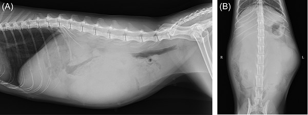
Preoperative radiographic images: (A) right lateral view; (B) ventrodorsal view. A large radiopaque mass occupying most of the abdominal cavity is observed.
Contrast‐enhanced computed tomography (CT) angiography (Aquilion 16, Canon Medical Systems Corporation, Tochigi, Japan) was performed before and at 25, 40, and 120 s after intravenous administration of iohexol (Omnipaque 300, GE Healthcare Pharma, Tokyo, Japan) at a dose of 2 mL/kg (600 mgI/kg). Images were acquired in helical mode with a slice thickness of 0.5 mm. CT revealed a large hypoattenuating mass (97 × 106 × 65 mm) with heterogeneous enhancement, most prominent during the arterial phase. The mass was in contact with the right ovarian vessel and the descending colon. Multiple hypoattenuating nodules were present in the liver, the largest measuring 20 × 26 × 25 mm. A 22‐mm hypoattenuating mass was also noted in the right kidney (Figure2). Both the hepatic and renal lesions showed contrast patterns similar to the abdominal mass. Mild ascites was present. Ultrasound‐guided (ARIETTA 70, Fujifilm Corporation, Tokyo, Japan) fine needle biopsy with a convex array transducer, 10 MHz and 23‐gauge hypodermal needle (Nipro Medical Industries LTD, Gunma, Japan) of the mass was performed under general anaesthesia following CT angiography. Cytology revealed loosely cohesive epithelial cells, some arranged in acinar‐like or rosette‐like structures with intercellular eosinophilic stroma (Figure3). The cells had moderately amphophilic to pale basophilic, finely vacuolated cytoplasm, dusty chromatin, and ovoid nuclei, with mild anisokaryosis and variable nuclear‐to‐cytoplasmic ratios. Findings suggested an epithelial tumour, such as neuroendocrine or carcinoid tumour.
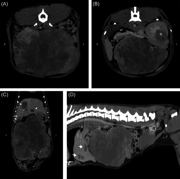
Preoperative contrast‐enhanced computed tomography images of the abdominal mass. (A) transverse view of the abdominal mass; (B) transverse view showing the right kidney mass (asterisk); (C) coronal view of the abdominal mass and multiple hepatic lesions (arrows); (D) sagittal view of the abdominal mass and multiple hepatic lesions (arrows). A heterogeneously contrast‐enhancing large mass of unknown origin is seen, along with hypoattenuating masses in the liver and right kidney parenchyma.
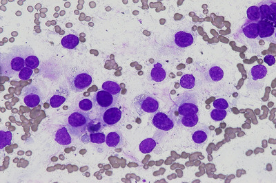
Cytological findings. Epithelial cells are loosely cohesive and partially arranged in acinar‐like or rosette‐like structures with intercellular eosinophilic stroma. The cells exhibit moderately amphophilic to pale basophilic, finely vacuolated cytoplasm, a dusty chromatin pattern, and ovoid nuclei. Mild anisokaryosis and variable nucleus‐to‐cytoplasm ratios are observed.
Additional evaluations included electrocardiography, blood pressure measurement, echocardiography, and serum catecholamine assays (Fujifilm VET Systems Corporation, Tokyo, Japan). Electrocardiography (Veterinary Automatic Electrocardiogram Analyzer. Fukuda ME Kogyo corporation, Tokyo, Japan) was unremarkable (mean heart rate 162 bpm). Mean systolic blood pressure were 156 mmHg and 148 mmHg by Doppler method (Bi‐directional Doppler ES‐100V3, Hadeco, Inc., Kanagawa, Japan) and oscillometric method (pettust NIBP, BioCare corporation, Taoyuan City, Taiwan), respectively. Echocardiography (Verifia V, Canon Medical Systems Corporation, Tochigi, Japan) using a sector array transducer 6 MHz revealed a left atrium‐to‐aortic root ratio of 1.29, interventricular septal thickness of 3.7 mm, left ventricular wall thickness of 3.7 mm, and fractional shortening of 54.8%, all within reference ranges. Serum catecholamines were: epinephrine, 1.68 nmol/L; norepinephrine, 16.23 nmol/mL; dopamine, 1.07 nmol/L, indicating elevated norepinephrine.
Surgery
In addition to the findings from these examinations, considering patient's spayed history and absence of estrus signs, the abdominal mass was suspected to be a neuroendocrine tumour. Given progressive enlargement of the abdominal mass and clinical decline, exploratory laparotomy was performed 10 days after the initial evaluation. The cat was premedicated intravenously with 0.25 mg/kg midazolam (Sandoz K.K., Tokyo, Japan) and 2 µg/kg fentanyl (Daiichi Sankyo, Tokyo, Japan). Anaesthesia was induced with 2 mg/kg propofol (Pfizer Inc., NY, USA) to effect, followed by endotracheal intubation (4.5‐mm tube). Anaesthesia was maintained with isoflurane (1.5–2.0% in oxygen, 1 L/min; DS Pharma, Osaka, Japan) and a fentanyl infusion (2–7 µg/kg/h). Amoxicillin (Fujita Pharma, Tokyo, Japan) was given intravenously at 20 mg/kg 30 min before surgery, then every 90 min intraoperatively and every 8 h for 48 h postoperatively. Lactated Ringer's solution was administered perioperatively at 3–5 mL/kg/h. Phentolamine and esmolol were prepared but not required.
A median celiotomy from the xiphoid to the pubis was performed. The intra‐abdominal mass occupied the cranial abdomen and was extensively adherent, precluding gross identification of the tissue of origin. The omentum adherent to the mass was carefully separated, and several vessels, including a branch from the caudal vena cava, were ligated with monofilament polyglyconate. One hepatic nodule in the right medial lobe was excised using the guillotine technique with vessel sealing device (LigaSure, Covidien Japan, Tokyo, Japan). The abdominal cavity was inspected for haemorrhage and other abnormalities before lavage. An active suction drain (OKID corporation, Shizuoka, Japan) was placed, and the abdomen was closed routinely with monofilament polyglyconate. Resected tissues were submitted for histopathology. Abdominal wound healed uneventfully and skin suture was removed at postoperative day (POD) 14.
Histopathology
The samples obtained from the resected tissues were immersed in 10% neutral buffered formalin for 48 h and embedded in paraffin. After paraffin sections (4 µm thick) were deparaffinised with xylene and immersed in ethanol, slides were stained with haematoxylin and eosin, and immunohistochemical staining was performed. An automated immunohistochemistry system (Histostainer, Nichirei Biosciences Inc., Tokyo, Japan) was used to process the deparaffinised and antigen‐retrieval tissues. The immunohistochemical antibodies were cytokeratin 19 (CK19; 1:150, Leica Biosystems, Newcastle, UK), vimentin (Histofine SAB‐PO(M) anti‐vimentin, Nichirei Biosciences Inc.) and inhibin (MCA951ST anti‐inhibin alpha, Bio‐Rad Laboratories Inc., Hercules, CA, USA). Peroxidase activity was demonstrated using diaminobenzidine solution, and slides were counterstained with haematoxylin.
Serum Collection and Sex Hormone Assays
At initial presentation (day 0), blood was drawn, and serum was separated and stored at –80°C until analysis. After histopathology confirmed GCT, sex hormones (estradiol and progesterone) were quantified on the stored preoperative serum and on a second serum sample obtained on postoperative day (POD) 30. Hormone assays were performed using the enzyme immunoassay method by a commercial laboratory (Fujifilm VET Systems Corporation, Tokyo, Japan).
Results
The patient recovered uneventfully from anaesthesia. Postoperative analgesia consisted of fentanyl constant rate infusion (1 µg/kg/h) maintained until the following day, followed by buprenorphine administered intravenously at 0.01 mg/kg every 8 h for 72 h. Drain output remained below 1 mL/kg/day from the day of surgery until POD 3, when the drain was removed. The patient resumed voluntary food intake the day after surgery and was discharged POD 5.
Histopathology
Histopathological findings are shown in Figure4. No morphologically recognisable non‐neoplastic ovarian tissue (e.g., follicles or corpora lutea) was identified in the submitted sections of the abdominal mass. The abdominal mass consisted of highly undifferentiated atypical spindle to round cells arranged in cord‐like, alveolar‐like, and microcystic patterns. Mucin retention was observed between tumour cells. Neoplastic cells had abundant eosinophilic cytoplasm and round nuclei with small, distinct nucleoli. Moderate nuclear atypia was present, and the mitotic index was 13 per 10 high‐power fields.
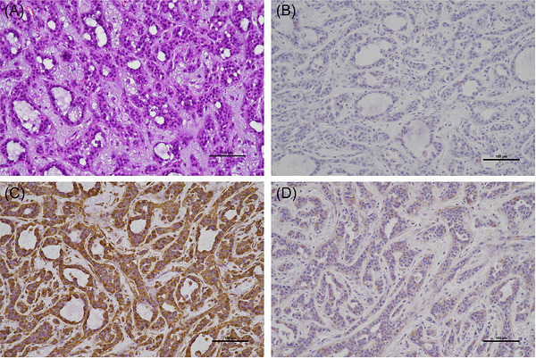
Histopathological findings. Findings are consistent with a granulosa cell tumour with hepatic metastasis. (A) Haematoxylin and eosin stain of the abdominal mass; immunohistochemistry shows neoplastic cells; (B) negative for cytokeratin; (C) positive for vimentin; (D) partially positive for inhibin.
The hepatic lesion showed invasive proliferation of spindle‐shaped cells arranged in bundles, with mucin retention between neoplastic cells. Immunohistochemistry demonstrated negativity for cytokeratin, positivity for vimentin, and partial positivity for inhibin, consistent with granulosa cell origin (Akihara et al.2007; Riccardi et al.2007). The CT findings, histopathological features, and immunohistochemical profile confirmed a diagnosis of GCT. The hepatic lesion was interpreted as metastasis from the primary GCT.
Postoperative Progress
After histopathology confirmed GCT, sex hormones were assayed on the serum collected preoperatively at initial presentation; estradiol was markedly increased and progesterone mildly increased (Table1). At the 2‐week recheck, patient's activity and appetite had improved, and the anaemia had resolved. At POD 30, the cat re‐presented with lethargy and abdominal distension, with progression of hepatic lesions and severe anaemia. A second serum sample obtained on POD 30 showed estradiol concentrations comparable to the preoperative value, whereas progesterone had decreased to near the upper limit for spayed queens (Table1). Given the high likelihood of progressive metastatic disease, the owner elected palliative management only. The patient died 5 weeks after surgery.
Table: Serum sex hormone analysis.
Discussion
The GCT in this case was identified in the setting of ORS, with imaging demonstrating contiguity to the right ovarian vasculature, findings that were consistent with origin from residual ovarian tissue. GCT was confirmed histologically with intra‐abdominal metastasis, and the metastatic rate in feline GCT has been reported to approach 50% (Holzworth1987; Norris et al.1969). When neoplastic transformation occurs without overt estrus signs, recognition of ORS may be delayed, which can postpone surgical intervention and allow progression to life‐threatening disease (Ball et al.2010; Miller1995; Wallace1991). Our cat appeared younger than typically reported age of onset for feline GCT (Holzworth1987; Norris et al.1969; Saba and Lawrence2013). However, metastatic GCT from residual ovarian tissue in a 4‐year‐old spayed cat has been reported (Uçmak et al.2018), and this early onset of the tumour could be compatible with accelerated neoplastic change in residual ovarian tissue, although direct evidence is not available. Free‐floating ovarian tissue fragments have been shown experimentally to survive and revascularise within the abdominal cavity, providing a plausible substrate for persistence and proliferation of functional tissue (DeNardo et al.2001). The interval of approximately 6 years between ovariohysterectomy and diagnosis lies within the wide range reported for ORS, and longer intervals have been described when neoplasia develops (Ball et al.2010; Wallace1991).
The present case supports the use of serum estradiol as an adjunctive biomarker in feline GCT. In human medicine, estradiol measurement is used to monitor remission and relapse in GCT (Pectasides et al. 2008; Schumer and Cannistra2003). Feline GCT can secrete estrogen, progesterone, testosterone, inhibin, IGF‐1, and AMH, although veterinary validation of hormonal biomarkers for diagnosis and monitoring remains limited (Flock et al.2022; Heaps et al. 2017; Holzworth1987; Shille et al. 1979). In our case, serum estradiol measured on the stored preoperative sample (assayed after histopathology) was markedly increased and remained comparably high at POD 30, paralleling the progression of hepatic metastases, supporting potential utility for postoperative monitoring of recurrence and metastatic disease. Correlation between serum estradiol level and the progression of the disease may become more evident with longer follow‐up with more GCT cases.
This case demonstrated extremely high serum estradiol compared to previously reported physiological level for intact cats, despite the absence of estrus signs (Shille et al.1979; Verhage et al.1976). In the previous studies that examined serum estradiol level and estrus behaviour in cats, some cats did not show estrus signs even the serum estradiol levels were high, suggesting individual variations in whether cats show obvious signs of estrus with elevated endogenous estradiol level (Little2011; Shille et al.1979). Environmental and internal factors, such as daylight length or intensity, multi‐cat household, stress, and illness, may alter estrus behaviour in cats (Little2011). Moreover, the owner simply may not have recognised the signs of estrus since the cat was already spayed. Since feline estrous cycle involves the pituitary gland, ovary, and the uterus, other possible explanation is malfunction of the pituitary gland's feed‐back mechanism due to lack of at least one ovary and uterus (Verhage et al.1976). Supraphysiological level of estradiol may interrupted physiological estrous cycle. However, in a previous study that injected exogenous estradiol in intact feral cats, which they showed estradiol levels comparable to or greater than the estradiol level of this case, most cats showed behavioural and physiological changes consistent with estrus (Hyndman et al.2020). ORS or GCT without estradiol level elevation or clinical signs are reported in human (Kho and Abrao2012; Mohapatra et al.2019; Wei et al.2019). It is not stated in these studies whether the degree of symptom is correlated with estradiol level, but human GCT study suggest the absence of theca cells or androgen for non‐estradiol producing GCTs in human (Lappöhn et al.1989). Other possibility of ORS or GCT with no estrus signs may be the failure in some point during the conversion process of androgen into estrogen, such as insufficiency of aromatase enzyme. This similar trend between human and cats may rise from similar pathophysiology, and further investigations with more cases are needed in the future.
When an animal is presented with a large abdominal mass, determining the tissue of origin can be challenging with traditional imaging modalities such as radiography and ultrasonography. Because this cat was reportedly spayed, ovarian neoplasia was not prioritised initially in the differential diagnosis. Preoperative CT demonstrated contiguity between the mass and the ovarian vasculature, yet an ovarian origin remained low on the list due to the spay history and the absence of estrus signs. Histopathological examination of the resected mass confirmed GCT with intra‐abdominal metastases, and serum estradiol measured on the stored preoperative sample (assayed after histopathology) was markedly increased and remained comparably high at 4 weeks (POD 30). Since some feline adrenal tumours can synthesise excessive estradiol (Meler et al.2011; Nadolski et al.2016), serum estradiol alone is insufficient to establish the diagnosis of GCT. In addition to estradiol measurement, careful evaluation of CT angiography for vascular contiguity and for a tortuous ovarian artery may aid the preoperative differential diagnosis. A canine CT study concluded that identification of an ovarian artery within a large abdominal mass is consistent with an ovarian tumour (Manfredi et al.2025). Therefore, preoperative diagnosis of GCT may be feasible even in spayed cats by integrating these CT findings with estradiol measurement while considering an origin from ovarian remnant tissue.
In advanced or metastatic GCT, treatment options beyond surgery remain poorly defined in veterinary medicine. Hormone‐related therapies, including leuprolide acetate, anastrozole and tamoxifen, have shown activity in recurrent ovarian GCT in humans (Dogan et al.2024; Foster et al.2023). In addition, an in vitro study demonstrated that anti‐Müllerian hormone could inhibit human ovarian GCT cells by inducing apoptosis (Anttonen et al.2011). However, evidence for hormone‐related therapy in small animals is limited. Chemotherapeutic agents including carboplatin and doxorubicin have been used anecdotally for ovarian tumours in cats; however, their benefit for GCT remains uncertain. Further investigations are needed to evaluate adjuvant or systemic therapies for feline GCT, particularly in cases with confirmed metastasis at diagnosis.
This report did not include histologic identification of non‐neoplastic residual ovarian tissue. Inhibin, AMH, and testosterone concentrations were not measured because validated commercial assays for these analytes in feline samples were not available through the commercial reference laboratory used for hormone testing at the time of analysis; therefore, endocrine phenotyping was incomplete. Future cases would benefit from a broader hormone panel (e.g., estradiol combined with AMH and other reproductive hormones) to strengthen endocrine characterisation and biomarker interpretation. The elevated norepinephrine value may reflect stress or sampling conditions rather than a catecholamine‐secreting neoplasm; nevertheless, an adrenal source of estradiol could not be entirely excluded. These points constrain inferences about tumour origin and generalisability but do not alter the clinical implications drawn from the convergent endocrine and imaging findings.
In conclusion, this case highlights that GCT can occur in the setting of ORS without estrus signs, even in the presence of marked hyperestrogenaemia. Ovarian remnants can undergo neoplastic transformation and lead to life‐threatening disease when estrus behaviour is not observed. Serum estradiol may serve as a valuable adjunct for diagnosis, assessment of treatment response, and postoperative monitoring for recurrence in feline GCT.
Author Contributions
Hiroshi Mori: data curation, investigation, methodology, visualization, writing – original draft, writing – review and editing.Hitomi Shinoda: conceptualization, data curation, investigation, methodology, project administration, visualization, writing – original draft, writing – review and editing.Kumiko Ishigaki: project administration, supervision, visualization, writing – original draft, writing – review and editing.Kazushi Asano: conceptualization, project administration, supervision, writing – original draft, writing – review and editing.Kenji Mori: data curation, investigation, methodology, visualization, writing – original draft, writing – review and editing.
Funding
The authors have nothing to report.
Ethics Statement
Informed owner consent of the patient was obtained prior to the first evaluation and surgery.
Conflicts of Interest
The authors declare that they have no competing interests.
Acknowledgement
The authors have nothing to report.
Data Availability Statement
The data that support the findings of this study are available from the corresponding author upon reasonable request.
Associated Data
Data Availability Statement
The data that support the findings of this study are available from the corresponding author upon reasonable request.
References
- Akihara, Y. , Shimoyama Y., Kawasako K., et al. 2007. “Immunohistochemical Evaluation of Canine Ovarian Tumors.” Journal of Veterinary Medical Science 69, no. 7: 703–708. 10.1292/jvms.69.703. doi.org/10.1292/jvms.69.703
- Aliakbrai, S. , and Ivoghli B. 1979. “Granulosa Cell Tumor in a Cat.” Journal of the American Veterinary Medical Association 174, no. 12: 1306–1308.
- Anttonen, M. , Färkkilä A., Tauriala H., et al. 2011. “Anti‐Müllerian Hormone Inhibits Growth of AMH Type II Receptor‐Positive human Ovarian Granulosa Cell Tumor Cells by Activating Apoptosis.” Laboratory Investigation 91, no. 11: 1605–1614. 10.1038/labinvest.2011.116. doi.org/10.1038/labinvest.2011.116
- Ball, R. L. , Birchard S. J., May L. R., Threlfall W. R., and Young G. S. 2010. “Ovarian Remnant Syndrome in Dogs and Cats: 21 Cases (2000‐2007).” Journal of the American Veterinary Medical Association 236, no. 5: 548–553. 10.2460/javma.236.5.548. doi.org/10.2460/javma.236.5.548
- Chen, S. , Yang B., and Fan J. 2020. “Diagnostic Value of Anti‐Mullerian Hormone in Ovarian Granulosa Cell Tumor: A Meta‐Analysis.” European Journal of Obstetrics, Gynecology, and Reproductive Biology 253: 266–272. 10.1016/j.ejogrb.2020.08.011. Epub 2020 Aug 22. doi.org/10.1016/j.ejogrb.2020.08.011
- Ciucci, A. , Ferrandina G., and Mascilini F., et al. 2018. “Estrogen Receptor β: Potential Target for Therapy in Adult Granulosa Cell Tumors?” Gynecologic Oncology 150, no. 1: 158–165. 10.1016/j.ygyno.2018.05.013. Epub 2018 May 18. doi.org/10.1016/j.ygyno.2018.05.013
- DeNardo, G. A. , Becker K., Brown N. O., and Dobbins S. 2001. “Ovarian Remnant Syndrome: Revascularization of Free‐Floating Ovarian Tissue in the Feline Abdominal Cavity.” Journal of the American Animal Hospital Association 37, no. 3: 290–296. 10.5326/15473317-37-3-290. doi.org/10.5326/15473317-37-3-290
- Dogan, I. , Aydin E., Topuz S., et al. 2024. “Hormonal Therapy in Pretreated Patients With Recurrent Ovary Granulosa Cell Tumor.” Journal of Pharmacy Practice 37, no. 1: 146–150. 10.1177/08971900221129679. doi.org/10.1177/08971900221129679
- Flock, U. , Fischer S., Weeger J., Reese S., and Walter B. 2022. “Anti‐Müllerian Hormone as a Diagnostic Tool to Identify Queens With Ovarian Remnant Syndrome.” Journal of Feline Medicine and Surgery 24, no. 8: e168–e174. 10.1177/1098612X221099195. doi.org/10.1177/1098612X221099195
- Foster, K. I. , Legarreta A. F., Kamat R., et al. 2023. “Clinical Outcomes of Leuprolide Acetate in the Treatment of Recurrent Ovarian Granulosa Cell Tumors.” American Journal of Obstetrics and Gynecology 228, no. 6: 724.e1–724.e9. 10.1016/j.ajog.2023.02.029. doi.org/10.1016/j.ajog.2023.02.029
- Heaps, L. A. , Scudder C. J., Lipscomb V. J., et al. 2017. “Serum anti‐Müllerian Hormone Concentrations Before and After Treatment of an Ovarian Granulosa Cell Tumour in a Cat.” Journal of Feline Medicine and Surgery Open Reports 3, no. 2:2055116917722701. 10.1177/2055116917722701. doi.org/10.1177/2055116917722701
- Holzworth, J 1987. Tumors of the Female Reproductive System. In Diseases of the Cat: Medicine and Surgery, edited by Holzworth J., 520–521, 1st ed. W.B. Saunders.
- Hyndman, T. H. , Algar K. L., Woodward A. P., et al. 2020. “Estradiol‐17β Pharmacokinetics and Histological Assessment of the Ovaries and Uterine Horns Following Intramuscular Administration of Estradiol Cypionate in Feral Cats.” Animals (Basel) 10, no. 9: 1708–1726. 10.3390/ani10091708. doi.org/10.3390/ani10091708
- Kho, R. M. , and Abrao M. S. 2012. “Ovarian Remnant Syndrome: Etiology, Diagnosis, Treatment and Impact of Endometriosis.” Current Opinion in Obstetrics & Gynecology 24, no. 4: 210–214. 10.1097/GCO.0b013e3283558539. doi.org/10.1097/GCO.0b013e3283558539
- Kustritz, M. V. , and Rudolph K. D. 2001. “Theriogenology Question of the Month. Functional Ovarian Remnant Containing Estrogen‐Secreting Follicles or Ovarian Remnant Containing an Estrogen‐Secreting Neoplasm.” Journal of the American Veterinary Medical Association 219, no. 8: 1065–1066. 10.2460/javma.2001.219.1065. doi.org/10.2460/javma.2001.219.1065
- Lappöhn, R. E. , Burger H. G., Bouma J., et al. 1989. “Inhibin as a Marker for Granulosa‐Cell Tumors.” New England Journal of Medicine 321, no. 12: 790–793. 10.1056/NEJM198909213211204. doi.org/10.1056/NEJM198909213211204
- Little, S. E 2011. “Chapter 40: Female Reproduction.” In The Cat, edited by Little S. E., 1195–1227. W.B. Saunders. 10.1016/B978-1-4377-0660-4.00040-5. doi.org/10.1016/B978-1-4377-0660-4.00040-5
- MacLachlan, N. J. , and Kennedy P. C. 2008. “Tumors of the Genital systems.” In Tumors in Domestic Animals, edited by Meuten D. J., 547–573, 4th ed. Iowa State Press.
- Manfredi, M. , Morabito S., Fournier Q., et al. 2025. “Computed Tomographic Findings in Dogs With Ovarian Tumors: A Tortuous Ovarian Artery Consistently Identifies Ovarian Origin in Complex Abdominal Masses.” Veterinary Radiology & Ultrasound: The Official Journal of the American College of Veterinary Radiology and the International Veterinary Radiology Association 66, no. 1:e13476. 10.1111/vru.13476. doi.org/10.1111/vru.13476
- Meler, E. N. , Scott‐Moncrieff J. C., Peter A. T., et al. 2011. “Cyclic Estrous‐Like Behavior in a Spayed Cat Associated With Excessive Sex‐Hormone Production by an Adrenocortical Carcinoma.” Journal of Feline Medicine and Surgery 13, no. 6: 473–478. 10.1016/j.jfms.2011.02.002. doi.org/10.1016/j.jfms.2011.02.002
- Miller, D. M 1995. “Ovarian Remnant Syndrome in Dogs and Cats: 46 Cases (1988‐1992).” Journal of Veterinary Diagnostic Investigation 7, no. 4: 572–574. 10.1177/104063879500700432. doi.org/10.1177/104063879500700432
- Mohapatra, A. , Chaudhry R., Picarsic J., et al. 2019. “A Rare Case of Uterine Torsion with Juvenile Granulosa Cell Tumor in the Pediatric Patient.” Urology 128: 87–89. 10.1016/j.urology.2019.02.012. doi.org/10.1016/j.urology.2019.02.012
- Nadolski, A. C. , Markovich J. E., Jennings S. H., et al. 2016. “Mammary Development, Hyperestrogenemia, and Hypocortisolemia in a Male Cat With an Adrenal Cortical Carcinoma.” Canadian Veterinary Journal 57, no. 10: 1077–1080.
- Norris, H. J. , Garner F. M., and Taylor H. B. 1969. “Pathology of Feline Ovarian Neoplasms.” Journal of Pathology 97, no. 1: 138–143. 10.1002/path.1710970118. doi.org/10.1002/path.1710970118
- Pectasides, D. , Pectasides E., and Psyrri A. 2008. “Granulosa Cell Tumor of the Ovary.” Cancer Treatment Reviews 34, no. 1: 1–12. 10.1016/j.ctrv.2007.08.007. doi.org/10.1016/j.ctrv.2007.08.007
- Riccardi, E. , Grieco V., Verganti S., and Finazzi M. 2007. “Immunohistochemical Diagnosis of Canine Ovarian Epithelial and Granulosa Cell Tumors.” Journal of Veterinary Diagnostic Investigation 19, no. 4: 431–435. 10.1177/104063870701900418. doi.org/10.1177/104063870701900418
- Saba, C. F. , and Lawrence J. A. 2013. “Tumors of the Female Reproductive System.” In Withrow and MacEwen's Small Animal Clinical Oncology, edited by Withrow S. J., Vail D. M., and Page R. L., 533, 5th ed. Elsevier Saunders;.
- Schumer, S. T. , and Cannistra S. A. 2003. “Granulosa Cell Tumor of the Ovary.” Journal of Clinical Oncology 21, no. 6: 1180–1189. 10.1200/JCO.2003.10.019. doi.org/10.1200/JCO.2003.10.019
- Shille, V. M. , Lundström K. E., and Stabenfeldt G. H. 1979. “Follicular Function in the Domestic Cat as Determined by Estradiol‐17β Concentrations in Plasma: Relation to Estrous Behavior and Cornification of Exfoliated Vaginal Epithelium.” Biology of Reproduction 21, no. 4: 953–963. 10.1095/biolreprod21.4.953. doi.org/10.1095/biolreprod21.4.953
- Uçmak, Z. G. , Uçmak M., Tek Ç., et al. 2018. “Granulosa Cell Tumor in a Spayed Young Queen.” Journal of the Hellenic Veterinary Medical Society 69, no. 2: 1010–1015. 10.12681/jhvms.18022. doi.org/10.12681/jhvms.18022
- Verhage, H. G. , Beamer N. B., and Brenner R. M. 1976. “Plasma Levels of Estradiol and Progesterone in the Cat During Polyestrus, Pregnancy and Pseudopregnancy.” Biology of Reproduction 14, no. 5: 579–585. 10.1095/biolreprod14.5.579. doi.org/10.1095/biolreprod14.5.579
- Wallace, M. S 1991. “The Ovarian Remnant Syndrome in the Bitch and Queen.” The Veterinary Clinics of North America Small Animal Practice 21, no. 3: 501–507. 10.1016/s0195-5616(91)50056-5. doi.org/10.1016/s0195-5616(91)50056-5
- Wei, M. , Maurer K. A., and Henry N. L. 2019. “Ovarian Remnant Syndrome in an Aromatase Inhibitor‐Treated Patient With BRCA2 Mutation Following Bilateral Oophorectomy.” The Breast Journal 25, no. 6: 1254–1256. 10.1111/tbj.13454. doi.org/10.1111/tbj.13454
Republished from the open web under CC-BY. Authors: Shinoda H, Mori K, Mori H, Ishigaki K, Asano K. Read the original.