Hypertrophic Osteopathy Associated With Primary Pulmonary Histiocytic Sarcoma in a Dog.
Hypertrophic osteopathy (HO) is a paraneoplastic or parainflammatory syndrome characterised by painful periosteal new bone formation and soft tissue swelling, most commonly associated with thoracic neoplasia. The pathogenesis of HO remains incompletely understood and may involve neurovascular mechanisms, altered pulmonary filtration of megakaryocytes and platelets and increased release of growth factors such as vascular endothelial growth factor (VEGF) and platelet-derived growth factor (PDGF). This report describes the first documented case of HO associated with primary pulmonary histiocytic sarcoma (PHS) in a dog. A 7-year-old male Golden Retriever was referred for evaluation of diffuse pain, limb swelling and lameness. Diagnostic imaging revealed bilateral periosteal proliferation affecting the forelimbs and a pulmonary mass within the right caudal lung lobe. Histopathology and immunohistochemistry following lung lobectomy and lymphadenectomy confirmed a diagnosis of PHS with nodal metastasis. Surgical excision resulted in complete clinical resolution of pain despite persistent radiographic evidence of HO. Subsequent disease progression occurred despite multiple chemotherapy protocols, including lomustine, vinorelbine, and metronomic chemotherapy; however, the dog remained consistently pain-free throughout follow-up. Overall survival time was 450 days. This case expands the spectrum of neoplasms associated with HO and further supports the hypothesis that tumour location, rather than histotype, plays a central role in the development of this syndrome. Moreover, the sustained absence of pain despite persistent periosteal lesions and metastatic progression suggests that tumour burden and tumour microenvironment may significantly influence clinical manifestations of HO.
Introduction
Hypertrophic osteopathy (HO) is a paraneoplastic or proinflammatory syndrome characterised by periosteal new bone formation affecting the distal limbs, typically progressing proximally over time (Withers et al.2015). Although its pathogenesis remains incompletely understood, current hypotheses suggest that alterations in neural signalling may induce abnormal vascular responses within the periosteum, promoting cortical bone deposition (Withers et al.2015).
HO has been associated with a wide range of thoracic and abdominal diseases. Thoracic disorders are the most frequent underlying causes and include primary or metastatic pulmonary tumours, rib tumours, oesophageal sarcomas related to parasitic infection, lung abscessation, mycobacterial pneumonia, bronchial foreign bodies with secondary pneumonia, congenital anomalies such as patent ductus arteriosus or megaoesophagus and infective endocarditis (Wylie et al.1993; Hesselink and van den Tweel1990; Foster and Armstrong2006; Dunn et al.2007; Kim and Kim2024). Abdominal conditions, including renal and prostatic neoplasms and other intra‐abdominal masses, have also been implicated. Clinical improvement and regression of periosteal proliferation often follow successful treatment of the primary disease process (Withers et al.2015; Grillo et al.2007; Halliwell and Ackerman1974).
Clinical manifestations typically include progressive limb thickening, lameness and pain on palpation. Early recognition of HO is essential to prompt investigation for underlying thoracic or abdominal pathology (Withers et al.2015; Brodey1971). This case report describes, for the first time, paraneoplastic HO associated with primary pulmonary histiocytic sarcoma (PHS) in a dog.
Case History
A 7‐year‐old male Golden Retriever was referred to the Veterinary Teaching Hospital (VTH) of the University of Teramo for an oncological consultation. One month earlier, the referring veterinarian had evaluated the dog for generalised pain, initially treated with non‐steroidal anti‐inflammatory drugs (NSAIDs), Piroxicam 0.3 mg/kg SID, which were subsequently discontinued due to gastrointestinal adverse effects. The pain was more pronounced on limb manipulation, along with elbow joint swelling and lameness. Comprehensive blood tests, limb and thoracic radiographs revealed a periosteal reaction in the forelimbs and the presence of a thoracic mass. A computed tomography (CT) scan confirmed a pulmonary mass in the right caudal lung lobe (6 cm CrCd × 3.6 cm DV × 5.3 cm ML) and a diffuse periosteal reaction, consistent with HO. Figure1.
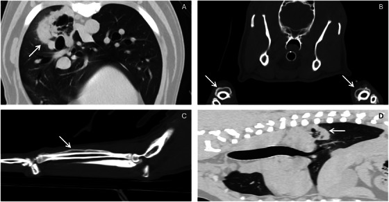
Computed tomography (CT) findings in a dog with pulmonary histiocytic sarcoma and hypertrophic osteopathy. (A) Transverse CT image of the thorax (lung window). A large pulmonary mass is present dorsally within the right caudal lung lobe (arrow). The lesion is well marginated, of soft‐tissue attenuation, and cavitary. (B) Transverse CT image at the level of the temporomandibular region including both antebrachia (bone window). Arrows indicate bilateral, symmetric periosteal new bone formation along the diaphyses of the radius and ulna. The periosteal reaction is thick and smooth, with mild circumferential soft‐tissue swelling. (C) Parasagittal CT image of the right forelimb (bone window). The arrow indicates smooth periosteal new bone formation involving the diaphyseal and metaphyseal regions of the ulna, associated with adjacent soft‐tissue swelling. (D) Parasagittal CT image of the thorax (lung window) showing the large pulmonary mass (arrow) in the right caudal lung lobe (same lesion as in panel A).
The dog underwent right caudal lung lobectomy and ipsilateral tracheobronchial lymphadenectomy, and the excised mass and lymph nodes were submitted for histopathological evaluation. After surgery, the owner reported clinical improvement and resolution of pain. Microscopic examination suggested an undifferentiated neoplasm. Immunohistochemical analysis revealed diffuse positivity for IBA‐1, MHC‐II, and vimentin, while neoplastic cells were negative for pan‐cytokeratin and HBME. Based on these findings, a diagnosis of PHS with metastatic lymph node was made. A few days after surgery, the dog was referred to the VTH. At clinical examination, the patient had a body condition score of 5/9 and a muscle condition score of A (Freeman and Becvarova2011). Contrary to what had been reported by the referring veterinarian and the owner prior to surgery, the dog showed no evidence of pain at the post‐surgical oncology consultation according to the Glasgow Composite Measure Pain Scale Short Form (ICMPS‐SF; Della Rocca et al.2018). No other clinically relevant abnormalities were detected. Adjuvant oral lomustine (80 mg/m2PO, q3W) was initiated. However, due to a significant elevation in ALT levels, lomustine‐induced hepatotoxicity was suspected (grade 3 VCOG‐CTCAE v2; LeBlanc et al.2021), leading to its discontinuation. As an alternative, vinorelbine (15 mg/m2, IV, q1W) was administered, and silymarin was prescribed as a supplement for liver support.
After four vinorelbine treatments, a staging work‐up, including haematobiochemical profile and a total‐body CT, was performed. Treatment response was assessed according to RECIST criteria (Nguyen et al.2015). Biochemical results indicated that serum ALT activity was within the reference interval and other laboratory findings were unremarkable. The CT revealed a newly developed 0.4 cm soft‐tissue nodule in the accessory lung lobe, suggestive of metastases, and therefore compatible with progressive disease (PD). Due to the small lesion size, and the risks associated with diagnostic sampling, fine needle aspiration was not performed. HO persisted on CT; however, the dog remained pain‐free (Della Rocca et al.2018). Consequently, vinorelbine was discontinued, and lomustine was reintroduced as a rescue treatment. Radiographic evidence of HO remained unchanged, compared with the previous CT examination, and despite the development of metastatic disease, the dog continued to show no evidence of pain.
A follow‐up CT performed 4 months later revealed further PD, characterised by an increase in size (0.61 cm) of the previously documented pulmonary nodule and the emergence of multiple additional pulmonary nodules, with soft tissue density. HO persisted unchanged with no signs of pain detected (Della Rocca et al.2018). In response, a metronomic chemotherapy protocol was initiated, consisting of thalidomide (2 mg/kg PO, q24h) and cyclophosphamide (10 mg/m2PO, q48h). After 3 months of treatment, a three‐view thoracic radiographic examination showed that the largest pulmonary lesion remained of similar size, compared to the previous CT, while HO persisted unchanged. The dog developed bilateral ocular mucopurulent discharge and episodes of coughing triggered by physical activity. Pain remained unreported, and no additional analgesic treatments were required. The overall survival time was 450 days.
Discussion
To the best of our knowledge, this is the first reported case of HO associated with PHS.
HO is characterised by painful periosteal proliferation and soft tissue swelling and has been described in association with infectious, inflammatory, congenital and neoplastic conditions (Hesselink and van den Tweel1990; Foster and Armstrong2006; Dunn et al.2007; Caywood et al.1985). Nevertheless, it is most frequently recognised as a paraneoplastic syndrome linked to primary or metastatic pulmonary tumours.1Extrapulmonary neoplasms, including urinary bladder and other urinary tract tumours, have also been reported (Grillo et al.2007; Halliwell and Ackerman1974). The pathogenesis of HO remains incompletely understood. One widely accepted hypothesis suggests that stimulation of the vagus nerve induces peripheral vasodilation, leading to increased periosteal perfusion and subsequent bone proliferation. This theory is supported by reports of regression of periosteal lesions following vagotomy (Flavell1956). An additional proposed mechanism involves abnormal passage of platelet aggregates and megakaryocytes into the systemic circulation. Under normal physiological conditions, these elements are fragmented within the pulmonary capillary bed; however, in HO, this filtering function may be bypassed, allowing them to reach the peripheral circulation (Dickinson and Martin1987). In the distal limbs, these cells may release platelet‐derived growth factor, promoting fibroblast proliferation, neovascularisation and increased vascular permeability, ultimately leading to periosteal new bone formation (Dickinson and Martin1987; Martinez‐Lavin2007; Martinez‐Lavin et al.2008; Thomas1996). Vascular endothelial growth factor (VEGF) has also been implicated in the pathogenesis of HO. Released by platelets, VEGF is a potent mediator of angiogenesis and osteoblastic differentiation, and its expression is often upregulated in hypoxic or neoplastic environments (Martinez‐Lavin2007). Increased circulating VEGF levels have been documented in both primary and secondary forms of HO, particularly in cases associated with pulmonary malignancies (Martinez‐Lavin et al.2008). An alternative hypothesis implicates chronic peripheral hypoxaemia, secondary to congenital or acquired arteriovenous shunting, as a contributing factor to periosteal new bone formation. This process may be mediated through upregulation of VEGF, which stimulates angiogenesis and osteoblastic activity (Midy and Plouët1994). VEGF expression may be enhanced by local hypoxia or impaired pulmonary inactivation, resulting in increased peripheral concentrations of circulating vasodilators and growth factors (Thomas1996). Furthermore, VEGF is constitutively produced and released by megakaryocytes and platelets (Möhle et al.1997). Histiocytic sarcoma is a malignant neoplasm arising from dendritic cells or macrophages (Skorupski et al.2007; Dervisis et al.2017) and may present as either a localised or disseminated form (Dervisis et al.2017; Klahn et al.2011; Murray et al.2022). Dose‐intense chemotherapy is consistently recommended, both as neoadjuvant therapy in localised disease and as the gold standard for disseminated forms. Lomustine remains the most commonly used chemotherapeutic agent (Skorupski et al.2007); however, recent studies have also demonstrated the efficacy of vinorelbine, which shows activity against this specific histotype and favourable pulmonary tissue penetration (Vigevani et al.2025; Rinaldi et al.2023).
Consistent with previous reports, this case further supports the hypothesis that tumour histotype may be less relevant than tumour location in the development of HO (Withers et al.2015; Halliwell and Ackerman1974; Roux et al.2022). In the literature, HO is regarded as a paraneoplastic or parainflammatory syndrome in which pulmonary disease acts as the primary trigger through neurogenic, vascular and humoral mechanisms. This concept explains why removal or effective treatment of the pulmonary lesion frequently results in regression of the clinical signs associated with HO (Pourmorteza et al.2015; Thapa2025). In this case report, the authors aim to place particular emphasis on pain management in this patient. Resolution of pain occurred after surgical removal of the primary pulmonary mass. Following surgery, the dog did not exhibit pain at any subsequent follow‐up examinations, despite persistent radiographic evidence of HO on both CT and radiographic imaging. Notably, pain remained absent even as the disease progressed with the development of new metastatic lesions. The absence of pain was consistently observed both during dose‐intense chemotherapy and during metronomic treatment. These findings suggest that pain resolution was not primarily related to the specific chemotherapeutic agents administered but rather to removal of the initial large pulmonary tumour mass. Emerging evidence indicates that cancer pain is not solely a consequence of structural lesions such as bone metastases but is also strongly mediated by the tumour microenvironment. Mediators released by cancer, stromal and immune cells within the tumour microenvironment, including cytokines and chemokines, contribute to neuroinflammation and nociceptor sensitisation independent of metastatic anatomical damage. This supports the notion that the absence of pain in the present case may be more closely related to changes in the tumour microenvironment following removal of the primary tumour than to the mere presence of radiographically detectable skeletal lesions (Santoni et al.2022; Varrassi et al.2025). In conclusion, HO appears to be a paraneoplastic syndrome primarily related to the location of the primary tumour rather than to tumour histotype. Pain management in affected patients may be influenced by tumour burden; therefore, surgical debulking of the primary tumour may be a reasonable therapeutic approach in selected cases.
Author Contributions
Valentina Rinaldi: conceptualisation, writing – original draft, methodology, investigation.Paolo Emidio Crisi: validation, supervision, resources.Alessia Luciani: supervision.Martina Rosto: investigation.Francesca Ridolfi: investigation.Riccardo Finotello: writing – review and editing.Andrea Boari: supervision.
Funding
The authors have nothing to report.
Conflicts of Interest
The authors declare no conflicts of interest.
References
- Brodey, R. S. 1971. “Hypertrophic Osteoarthropathy in the Dog: A Clinicopathologic Survey of 60 Cases. ”Journal of the American Veterinary Medical Association159, no. 10: 1242–1256.
- Caywood, D. D. , B. A. Kramek, D. A. Feeney, andG. RJohnston. 1985. “Hypertrophic Osteopathy Associated With a Bronchial Foreign Body and Lobar Pneumonia in a Dog. ”Journal of the American Veterinary Medical Association186, no. 7: 698–700.
- Della Rocca, G. , R. Colpo, J. Reid, A. Di Salvo, andM. Scott. 2018. “Creation and Validation of the Italian Version of the Glasgow Composite Measure Pain Scale‐Short Form (ICMPS‐SF). ”Veterinaria Italiana54, no. 3: 251–260. . doi.org/10.12834/VetIt.699.3421.3
- Dervisis, N. G. , M. Kiupel, Q. Qin, andL. Cesario. 2017. “Clinical Prognostic Factors in Canine Histiocytic Sarcoma. ”Veterinary and Comparative Oncology15, no. 4: 1171–1180. . doi.org/10.1111/vco.12252
- Dickinson, C. J. , andJ. F. Martin. 1987. “Megakaryocytes and Platelet Clumps as the Cause of Finger Clubbing. ”Lancet2, no. 8573: 1434–1435. . doi.org/10.1016/s0140-6736(87)91132-9
- Dunn, M. E. , L. Blond, D. Letard, andR. DiFruscia. 2007. “Hypertrophic Osteopathy Associated With Infective Endocarditis in an Adult Boxer Dog. ”Journal of Small Animal Practice48, no. 2: 99–103. . doi.org/10.1111/j.1748-5827.2006.00159.x
- Flavell, G. 1956. “Reversal of Pulmonary Hypertrophic Osteoarthropathy by Vagotomy. ”Lancet270, no. 6911: 260–262. . doi.org/10.1016/s0140-6736(56)91186-2
- Foster, W. K. , andJ. A. Armstrong. 2006. “Hypertrophic Osteopathy Associated With PulmonaryEikenella corrodensInfection in a Dog. ”Journal of the American Veterinary Medical Association228, no. 9: 1366–1369. . doi.org/10.2460/javma.228.9.1366
- WSAVA Nutritional Assessment Guidelines Task Force Members. Freeman, L. , I. Becvarova, et al. 2011. “WSAVA Nutritional Assessment Guidelines. ”Journal of Small Animal Practice52, no. 7: 385–396. . doi.org/10.1111/j.1748-5827.2011.01079.x
- Grillo, T. P. , C. V. S. Brandão, M. J. Mamprim, C. M. N. de Jesus, T. C. Santos, andB. W. Minto. 2007. “Hypertrophic Osteopathy Associated With Renal Pelvis Transitional Cell Carcinoma in a Dog. ”Canadian Veterinary Journal48, no. 7: 745–747.
- Halliwell, W. H. , andN. Ackerman. 1974. “Botryoid Rhabdomyosarcoma of the Urinary Bladder and Hypertrophic Osteoarthropathy in a Young Dog. ”Journal of the American Veterinary Medical Association165, no. 10: 911–913.
- Hesselink, J. W. , andJ. G. van den Tweel. 1990. “Hypertrophic Osteopathy in a Dog With a Chronic Lung Abscess. ”Journal of the American Veterinary Medical Association196, no. 5: 760–762.
- Kim, Y. R. , andJ. H. Kim. 2024. “Hypertrophic Osteopathy Concurrent With an Aberrant Right Subclavian Artery in a Dog. ”Veterinary Sciences11, no. 6: 263. . doi.org/10.3390/vetsci11060263
- Klahn, S. L. , B. E. Kitchell, andN. G. Dervisis. 2011. “Evaluation and Comparison of Outcomes in Dogs With Periarticular and Nonperiarticular Histiocytic Sarcoma. ”Journal of the American Veterinary Medical Association239, no. 1: 90–96. . doi.org/10.2460/javma.239.1.90
- LeBlanc, A. K. , M. Atherton, R. T. Bentley, et al. 2021. “Veterinary Cooperative Oncology Group‐Common Terminology Criteria for Adverse Events (VCOG‐CTCAE v2) Following Investigational Therapy in Dogs and Cats. ”Veterinary and Comparative Oncology19, no. 2: 311–352. . doi.org/10.1111/vco.12677
- Martinez‐Lavin, M. 2007. “Exploring the Cause of the Most Ancient Clinical Sign of Medicine: Finger Clubbing. ”Seminars in Arthritis and Rheumatism36, no. 6: 380–385. . doi.org/10.1016/j.semarthrit.2006.11.004
- Martinez‐Lavin, M. , A. Vargas, andM. Rivera‐Viñas. 2008. “Hypertrophic Osteoarthropathy: A Palindrome With a Pathogenic Connotation. ”Current Opinion in Rheumatology20, no. 1: 88–91. . doi.org/10.1097/BOR.0b013e3282f14a5a
- Midy, V. , andJ. Plouët. 1994. “Vasculotropin/Vascular Endothelial Growth Factor Induces Differentiation in Cultured Osteoblasts. ”Biochemical and Biophysical Research Communications199, no. 1: 380–386. . doi.org/10.1006/bbrc.1994.1240
- Möhle, R. , D. Green, M. A. Moore, R. L. Nachman, andS. Rafii. 1997. “Constitutive Production and Thrombin‐Induced Release of Vascular Endothelial Growth Factor by Human Megakaryocytes and Platelets. ”PNAS94, no. 2: 663–668. . doi.org/10.1073/pnas.94.2.663
- Murray, C. A. , J. L. Willcox, M. De, C. H. Souza, et al. 2022. “Outcome in Dogs With Curative‐Intent Treatment of Localized Primary Pulmonary Histiocytic Sarcoma. ”Veterinary and Comparative Oncology20, no. 2: 458–464. . doi.org/10.1111/vco.12791
- Nguyen, S. M. , D. H. Thamm, D. M. Vail, andC. A. London. 2015. “Response Evaluation Criteria for Solid Tumours in Dogs (v1. 0): A Veterinary Cooperative Oncology Group (VCOG) Consensus Document. ”Veterinary and Comparative Oncology13, no. 3: 176–183. . doi.org/10.1111/vco.12032
- Pourmorteza, M. , S. J. Baumrucker, A. Al‐Sheyyab, andM. Da Silva. 2015. “Hypertrophic Pulmonary Osteoarthropathy: A Rare but Treatable Condition in Palliative Medicine. ”Journal of Pain and Symptom Management50, no. 2: 263–267. . doi.org/10.1016/j.jpainsymman.2015.02.005
- Rinaldi, V. , R. Finotello, A. Boari, C. Emanuele, andC. Paolo Emidio. 2023. “Vinorelbine as First‐Line Treatment in Stage IV Canine Primary Pulmonary Carcinoma. ”Veterinary Sciences10, no. 12: 664. . doi.org/10.3390/vetsci10120664
- Roux, F. A. , E. Deseille, M. Fusellier, M. Rohel, J. Branchereau, andJ. Y. Deschamps. 2022. “Hypertrophic Osteopathy Associated With a Prostatic Adenocarcinoma in a Castrated Dog. ”Veterinary Sciences9, no. 9: 466. . doi.org/10.3390/vetsci9090466
- Santoni, A. , M. Santoni, andE. Arcuri. 2022. “Chronic Cancer Pain: Opioids Within Tumor Microenvironment Affect Neuroinflammation, Tumor and Pain Evolution. ”Cancers14, no. 9: 2253. . doi.org/10.3390/cancers14092253
- Skorupski, K. A. , C. A. Clifford, M. C. Paoloni, et al. 2007. “CCNU for the Treatment of Dogs With Histiocytic Sarcoma. ”Journal of Veterinary Internal Medicine21, no. 1: 121–126. 21[121: cfttod]2. 0. co; 2. doi.org/10.1892/0891-6640(2007)
- Thapa, B, N. Mahendraker, andK. Ramphul. 2025. Paraneoplastic Syndromes. StatPearls Publishing. .
- Thomas, K. A. 1996. “Vascular Endothelial Growth Factor, a Potent and Selective Angiogenic Agent. ”Journal of Biological Chemistry271, no. 2: 603–606. . doi.org/10.1074/jbc.271.2.603
- Varrassi, G. , M. L. G. Leoni, G. Farì, et al. 2025. “Neurobiology of Cancer Pain: A Narrative Review. ”OBM Neurobiology9, no. 4: 315. . doi.org/10.21926/obm.neurobiol.2504315
- Vigevani, N. , C. Chalfon, A. Guillén, et al. 2025. “Vinorelbine as a Rescue Treatment for Canine Histiocytic Sarcoma: A Multicentre Retrospective Study of 18 Dogs. ”Veterinary and Comparative Oncology23, no. 3: 358–365. . doi.org/10.1111/vco.13060
- Withers, S. S. , E. G. Johnson, W. T. N. Culp, C. O. Rodriguez, K. A. Skorupski, andR. B. Rebhun. 2015. “Paraneoplastic Hypertrophic Osteopathy in 30 Dogs. ”Veterinary and Comparative Oncology13, no. 3: 157–165. . doi.org/10.1111/vco.12026
- Wylie, K. B. , D. D. Lewis, R. D. Pechman, D. Y. Cho, andA. Roy. 1993. “Hypertrophic Osteopathy Associated WithMycobacterium fortuitumPneumonia in a Dog. ”Journal of the American Veterinary Medical Association202, no. 12: 1986–1988.
Republished from the open web under CC-BY. Authors: Rinaldi V, Ridolfi F, Finotello R, Rosto M, Luciani A, Boari A, Crisi PE. Read the original.