Interaction Between Rec8 and Mis4 Is Required for Axis-Loop Chromatin Formation and Homologous Chromosome Recombination During Meiosis.
The Rec8 cohesin complex is required for the pairing and recombination of homologous chromosomes during meiosis, as well as for the cohesion of sister chromatids. In the fission yeast Schizosaccharomyces pombe, we previously identified a rec8-F204S mutant that lost the ability to assemble the axis-loop chromatin structure without losing sister chromatid cohesion. This mutant showed reduced meiotic recombination, indicating that pairing and recombination of homologous chromosomes require the formation of the axis-loop chromatin structure mediated by the Rec8 cohesin complex. Loading of the Rec8 cohesin complex onto chromatin is mediated by Mis4 (NIPBL in humans; Scc2 in yeast). In this study, to elucidate the functions of Mis4, we identified a mis4-LR mutant (L1150S and R1159G) that reproduced the phenotypes of the rec8-F204S mutant, which is defective in chromatin axis formation and homologous recombination while retaining sister chromatid cohesion. These mutation sites (Mis4-L1150, Mis4-R1159, and Rec8-F204) are all localized at the interaction surface between Mis4 and Rec8. Biochemical analysis revealed that the Mis4-LR mutant protein exhibited reduced Rec8-binding activity. Considering that the mis4-LR mutant phenocopied the rec8-F204S mutant, our results demonstrate that the Mis4-Rec8 interaction is required for proper formation of Rec8-dependent meiotic chromosome axis.
Introduction
Meiosis is a process of fundamental importance for sexually reproducing eukaryotes, in which one round of DNA replication is followed by two consecutive rounds of chromosome segregation to produce haploid gametes that are inherited by offspring. During this process, homologous chromosomes pair and subsequently undergo recombination. Homologous chromosomes are segregated in the first meiotic division and sister chromatids are segregated in the second meiotic division. Crossovers formed by reciprocal recombination constitute a physical link between homologous chromosomes, ensuring their correct segregation (Zickler and Kleckner1999; Hunter2015). Failure of this process is a major cause of miscarriage or developmental disorders caused by chromosome trisomy, such as Down syndrome, in humans (Nagaoka et al.2012; Webster and Schuh2017).
The mitotic Rad21 cohesin complex establishes cohesion between a pair of replicated sister chromatids. The cohesin complex consists of four protein components: two Structural Maintenance of Chromosomes (SMC) components, one kleisin component, and one stromal antigen (SA) component (Nasmyth2001; Mehta et al.2013; Ishiguro2019; Sakuno and Hiraoka2022); these components are called SMC1α, SMC3, RAD21, and SA1 or SA2, respectively, in human cells. Cohesin complexes are also involved in the formation of higher‐order chromatin structures by bringing together distant regions of DNA, for which a DNA loop extrusion model has been proposed (Mizuguchi et al.2014; Sanborn et al.2015; Fudenberg et al.2016; Rao et al.2017; Davidson et al.2019; Kim et al.2019; Davidson and Peters2021). Cohesin complexes are recruited to chromatin by NIPBL (Mis4 inS. pombe) (Higashi et al.2020; Kurokawa and Murayama2020; Alonso‐Gil and Losada2023), and dissociated from chromatin by PDS5 and WAPL (Pds5 and Wpl1 inS. pombe) (Ding et al.2006; Peters and Nishiyama2012; Tedeschi et al.2013; Challa et al.2016; Crawley et al.2016; Haarhuis et al.2017; Hong et al.2019; Silva et al.2020; Viera et al.2020).
While the regulated segregation of sister chromatids in the mitotic cell cycle is mediated by the mitotic cohesin complex, meiosis‐specific cohesin variants appear upon entry into meiosis to orchestrate meiosis‐specific chromosome events (Nasmyth2001; Revenkova and Jessberger2006). Replacement of the kleisin subunit during meiosis occurs in most organisms, in which the mitotic kleisin RAD21 is replaced by REC8 (Mehta et al.2013; Nasmyth2001; Ishiguro2019; Sakuno and Hiraoka2022). In addition to sister chromatid cohesion, the meiotic Rec8 cohesin complex is required for pairing and recombination of homologous chromosomes (Klein et al.1999; Pasierbek et al.2001; Cai et al.2003; Ishiguro et al.2014; Fukuda et al.2014; Hopkins et al.2014). Rec8‐dependent chromatin structure is a key determinant of pairing and recombination of homologous chromosomes (Ding et al.2006,2016; Sakuno et al.2022; Sakuno and Hiraoka2022).
Recent studies using high‐throughput chromosome conformation capture (Hi‐C) clearly revealed that the Rec8 cohesin complex is essential for constructing meiosis‐specific chromatin axis‐loop structures (Schalbetter et al.2019; Sakuno et al.2022). Using Hi‐C analysis, we delineated meiotic chromosome structures in the fission yeastSchizosaccharomyces pombe, and demonstrated the significance of Rec8‐dependent axis‐loop structures by identifying arec8mutant (rec8‐F204S) that is specifically defective in axis‐loop formation and homologous recombination without affecting sister chromatid cohesion (Sakuno et al.2022).
S. pombeMis4 (NIPBL in human; Scc2 in yeast), a cohesin loader, modulates cohesin functions through its DNA binding, cohesin binding, and ATPase activities (Higashi et al.2020; Kurokawa and Murayama2020; Wei et al.2025). However, it remained unknown how Mis4 is involved in cohesin functions during meiosis. In this study, we isolated amis4mutant that exhibited defects in chromatin axis formation and homologous recombination while retaining sister chromatid cohesion, similarly to therec8‐F204Smutant. Biochemical analysis of the mutant demonstrated the role of Mis4 in meiosis through its interaction with Rec8.
Results
Isolation ofmis4Mutants
In a previous study, we demonstrated that a Rec8 mutant shows defects specifically in chromatin axis formation and homologous chromosome recombination without affecting sister chromatid cohesion. As the responsible mutation, F204S, resides within the Mis4‐binding domain of Rec8, the phenotype may arise from defective interaction between Rec8 and Mis4. Based on this idea, we attempted to identifymis4mutants that are specifically defective in axis formation without impairing cohesion function, similar to therec8‐F204Smutant. To this end, we focused on the Rec8‐binding domain of Mis4 (Figure1a). We used anh90mating‐type strain in amei4∆background to arrest each transformant at meiotic prophase. We also introducedwpl1∆into the host strain to produce a prominent Rec8‐GFP axis (FigureS1a), allowing defects in chromatin axis formation to be readily evaluated by microscopic observation, as described previously (Sakuno et al.2022). Using this strain as a screening host, randomly mutagenizedmis4cDNA fragments encoding the C‐terminal region of Mis4 (amino acid residues 685–1587), generated by error‐prone PCR, were transformed. We screened for mutants that showed defects in axis formation while retaining normal cohesion function by fluorescence microscopy, based on Rec8‐GFP axis morphology.
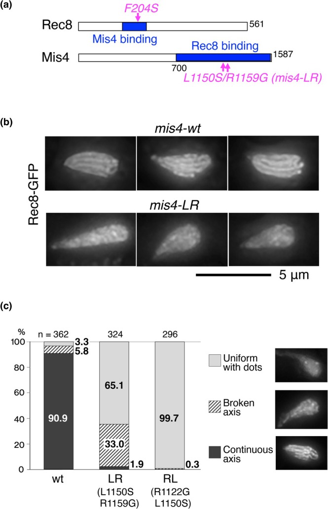
Axis morphology inmis4mutants. (a) Schematic diagram of Mis4 and Rec8. Mutation sites are indicated within the Mis4‐binding domain of Rec8 and Rec8‐binding domain of Mis4. (b) Fluorescence images of the chromatin axis visualized by Rec8‐GFP in wild‐type Mis4 (upper panels) and the Mis4‐LR mutant (lower panels) in amei4∆ wpl1∆background. Larger fields of view containing additional cells are shown in Figure S2a‐c (wild type) and Figure S2d‐f (Mis4‐LR). (c) Axis morphology was categorized into three groups: Continuous axis, Broken axis, and Uniform with dots. Cells in each category were counted in at least three independent cultures. The numbers on the graph indicate the percentage of each category. The numbers of cells examined are shown at the top of the graph.
Chromatin Axis Formation
Among approximately 3000 viable transformants in themis4mutant library, we first identified two mutants, m8 and m24, that displayed defects in chromatin axis formation (FigureS1b) compared withmis4wild‐type cells (Figure1bupper panels). The m8 mutant contained amino acid substitutions G1018R, L1150S, E1361G, and C1457R, whereas the m24 mutant contained R1122G and R1159G. We next examined the effect of each individual amino acid substitution. Among the four mutations in m8, onlymis4‐L1150Sshowed defects in axis formation (FigureS1b). Among the two mutations in m24, bothmis4‐R1122Gandmis4‐R1159Gshowed little effect on the axis (FigureS1b). We then generated double mutants combining these substitutions. Among them, the double mutantsmis4‐L1150S/R1159G(designatedmis4‐LR) andmis4‐R1122G/L1150S(designatedmis4‐RL) exhibited severe defects in axis formation (Figure1b, FigureS2d‐fformis4‐LR; FigureS1b formis4‐RL). To further assess degrees of defects in chromatin axis morphology in these mutants, axis morphology was classified into three categories (continuous axis, broken axis, and uniform with dots), and their populations were counted in each mutant (Figure1c). Wild‐type cells predominantly showed the continuous axis. In contrast,mis4‐LRcells showed increased populations of broken‐axis and uniform‐with‐dots categories, andmis4‐RLcells predominantly showed the uniform‐with‐dots category (Figure1c). Thus, the LR mutation (L1150S/R1159G) confers severe defects in chromatin axis formation whereas the RL mutation (R1122G/L1150S) largely abolishes chromatin axis formation.
Homologous Recombination and Sister Chromatid Cohesion
We next characterized the effects of thesemis4mutations on recombination and sister chromatid cohesion. Homologous recombination and sister chromatid cohesion are both impaired inrec8Δcells. Recombination frequency between theura1andlys3loci was measured in these mutants. The recombination frequency was significantly reduced to a level comparable to that ofrec8Δin the double mutantsmis4‐LR(Figure2a) andmis4‐RL(FigureS3a), but was only moderately reduced inmis4‐L1150S(FigureS3a). We next examined sister chromatid cohesion using alacOinsertion at thecut3locus visualized by LacI‐GFP (cut3‐GFP), in a heterozygous strain. In this assay, cells with a single GFP focus represent intact sister chromatid cohesion, whereas those with two foci indicate loss of cohesion (Figure2b).mis4‐LRcells showed a high proportion of single foci, similar to wild type, in contrast torec8Δcells (Figure2b), indicating that sister chromatid cohesion is retained inmis4‐LR. In contrast,mis4‐RLcells showed an increased proportion of cells with two foci, indicating loss of sister chromatid cohesion (FigureS3b). Taken together, these results identifymis4‐LR(L1150S/R1159G) as a mutant specifically defective in chromatin axis formation while retaining normal cohesion function. Therefore, we focused on this mutant for further experiments.
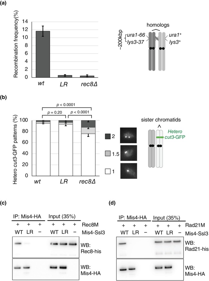
Characterization ofmis4mutants. (a) Recombination frequency. Inset: Schematic of the assay. Recombination frequency between theura1andlys3loci was measured. Recombination frequency (%) in wild‐type and Mis4‐LR cells. Data represent three independent experiments, with > 700 cells analyzed in each experiment. Error bars indicate standard deviation. (b) Sister chromatid cohesion. Inset: Schematic diagram of the assay, cohesion at the cut3‐GFP locus was evaluated by counting fluorescent foci (1, 1.5, or 2 per nucleus). Percentage of cells with each focus number in wild‐type and Mis4‐LR cells. Data represent three independent experiments, with > 130 cells analyzed in each experiment. Error bars indicate standard deviation.pvalues were obtained from chi‐square test for statistical significance. (c) Biochemical analysis of the Mis4 mutant protein. Co‐immunoprecipitation of wild‐type and mutant Mis4 with Rec8M (residues 104–450). Purified Mis4‐Ssl3 was incubated with His‐tagged Rec8M and immunoprecipitated using an anti‐HA antibody via a C‐terminal HA tag on Mis4. Rec8M and Mis4 in input and immunoprecipitated fractions were detected by western blotting with anti‐His or anti‐HA antibody, respectively. (d) Same as (c), except that His‐tagged Rad21M (residues 88–550) was used instead of Rec8M.
Biochemical Analysis of the Purified Mis4‐LR Mutant Protein
mis4‐LRandrec8‐F204Smutants showed similar defects in chromatin axis formation during meiotic prophase, suggesting that themis4‐LRmutations affect physical interaction with Rec8. To test this, we purified the Mis4‐Ssl3 complex and a truncated Rec8 lacking the N‐ and C‐termini (Rec8M) and confirmed their physical interaction using an in vitro pull‐down assay. Purified Mis4‐Ssl3 and Rec8M were incubated together, and Mis4‐Ssl3 was immunoprecipitated using a HA tag fused to Mis4. This resulted in co‐precipitation of Rec8M, detected via a histidine tag fused to its C‐terminus (Figure2c). In contrast to wild‐type Mis4‐Ssl3, the Rec8M signal was markedly reduced when the LR mutant Mis4‐Ssl3 complex was used. We also found that interaction between Mis4 and Rad21 was weakened by the LR mutation (Figure2d). These results indicate that Mis4 directly interacts with Rec8 and Rad21, and the LR mutations weaken interactions both with the meiotic cohesin Rec8 and mitotic cohesin Rad21.
Prediction of the Mis4‐Rec8 Interaction
The crystal structure of Scc2 (a homolog of Mis4) has been determined in the thermophilic fungusChaetomium thermophilum(Kikuchi et al.2016); this protein (CtScc2) adopts a U‐shaped structure composed of α‐helix domains. Amino acid sequences spanning the mutation sites are shown for CtScc2 andS. pombeMis4 (SpMis4) (Figure3a). The crystal structure of CtScc2 was generated from the atomic coordinates of PDB 5T8V (Kikuchi et al.2016), and Figure3bshows its C‐terminal region, which contains the predicted Rec8‐binding region.
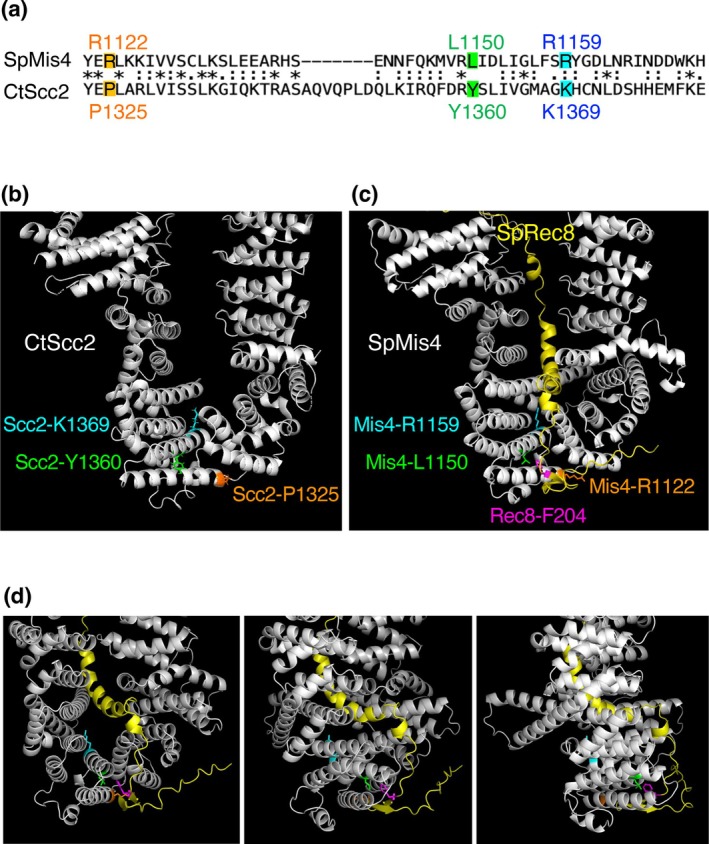
Interaction between Mis4 and Rec8. (a) Amino acid sequences ofS. pombeMis4 (SpMis4) andC. thermophilusScc2 (CtScc2) spanning the mutation sites. (b) Structure of CtScc2 based on PDB entry 5T8V (Kikuchi et al.2016). The C‐terminal region involved in Rec8 binding is shown. Residues of P1325, Y1360, and R1369 of CtScc2, corresponding to R1122, L1150, and R1159 of SpMis4, are indicated by orange, green, and blue, respectively. (c) Structure of SpMis4 and SpRec8 predicted by AlphaFold 3. The Rec8‐binding domain of SpMis4 (residues 700–1587) and the Mis4‐binding domain of SpRec8 (residues 141–280) are shown in white and yellow, respectively. Residues Mis4‐R1122, Mis4‐L1150, Mis4‐R1159, and Rec8‐F204 are shown in orange, green, blue, and magenta, respectively. (d) Views of (c) from different rotation angles: 30°, 60°, and 90° around the y‐axis, from left to right. The same color scheme as in (c) is used. A rotating model is shown in Supporting Information MovieS1.
We predicted the structure of SpMis4 using AlphaFold3 and obtained a U‐shaped structure similar to that of CtScc2, in which the Mis4‐binding domain of Rec8 is positioned between the U‐shaped arms of Mis4 (Figure3c). The mutation sites Mis4‐R1122, Mis4‐L1150, Mis4‐R1159, and Rec8‐F204 are all located within the cleft formed by the U‐shaped domains (Figure3c,d). Correspondingly, P1325, Y1360, and K1369 in CtScc2 (equivalent to R1122, L1150, and R1159 in SpMis4) are also located in this cleft (Figure3b). This predicted model of Mis4 and Rec8 also supports the potential significance of the Mis4‐Rec8 interaction.
Discussion
Meiotic cohesin Rec8 functions in both sister chromatid cohesion and homologous chromosome recombination. Our studies have demonstrated thatrec8‐F204Sandmis4‐LRmutants separate these closely associated functions (Sakuno et al.2022; this study). A recent study reported a human cohesin mutant with the opposite phenotype, that is, defective in sister chromatid cohesion but normal in DNA loop formation (Nagasaka et al.2023). These observations suggest that sister chromatid cohesion and DNA loop formation are mediated by distinct mechanisms.
Possible Mechanisms for Functional Specificity of the Mutations
Mis4 promotes cohesin loading onto DNA via its DNA‐binding activity and interactions with cohesin, concomitant with activation of cohesin's ATPase (Higashi et al.2020; Kurokawa and Murayama2020). Mis4 binds to DNA via two distinct α‐helix domains, both of which are located distantly from the mutations found in this study (Kurokawa and Murayama2020). Therefore, these mutations of Mis4 (R1122G, L1150S, and R1159G) are not expected to affect its DNA binding.
Themis4‐LRmutations (L1150S/R1159G) weaken interaction with Rec8 but retain sister chromatid cohesion. DNA loop formation, which underlies chromatin axis‐loop formation, likely requires a stable interaction between Rec8 and Mis4. Weakened Mis4‐Rec8 interaction may permit DNA entrapment by cohesin sufficient to mediate a basal level of sister chromatid cohesion in both mitosis and meiosis, but is insufficient to support DNA loop formation in meiosis. When combined with L1150S, the R1122G mutation causes defects in sister chromatid cohesion, and axis formation defects more severe than R1159G (comparemis4‐RLwithmis4‐LR; Figure1c, FigureS3b). These results suggest that the R1122 residue of Mis4 plays a more significant role in cohesin function than R1159. Broken axis observed inmis4‐LR(Figure1c) may be sufficient for sister chromatid cohesion, but insufficient for efficient homologous recombination. This is also consistent with the fact thatmis4‐LRcauses defects in meiosis involving Rec8, but not in mitosis, which involves Rad21. However, we cannot exclude the possibility that meiotic phenotypes may be influenced by chromatin structure produced with Rad21 in vegetative cells. The functional specificity of these mutations could be elucidated through further investigation of mutants affecting DNA binding, ATPase activities, and cohesin binding.
Biological Significance of the Mis4‐Rec8 Interaction
We have identified Mis4 and Rec8 mutations that affect cohesin functions and mapped these mutation sites to the interaction surface between Mis4 and Rec8. This Mis4‐Rec8 interaction surface is also conserved in the interaction between human NIPBL and Rad21 (Shi et al.2020). Furthermore, NIPBL mutations associated with Cornelia de Lange syndrome occur within this interaction surface and impair cohesin‐mediated DNA loop extrusion (Panarotto et al.2022). Therefore, these residues of NIPBL/Mis4 play an important role in modulating cohesin function through interactions with Rec8 and Rad21.
Experimental Procedures
Schizosaccharomyces pombeStrains
All media and growth conditions were as described previously (Moreno et al.1991). Complete medium supplemented with uracil, lysine, adenine, histidine, and leucine (YE5S), minimal medium (EMM2), and sporulation‐inducing medium (SPA and SSA) were used unless otherwise stated. The strains used in this study are listed in TableS1.
Deletion of endogenous genes and tagging with 3 × flag tag were performed using a PCR‐based gene targeting method forS. pombe, using thehphMX6(hygR),natMX6(natR), andbsdRgenes as selection markers. To generate strains ofmis4point mutations with a 3 × flag tag, the coding region corresponding to the C‐terminal 685 amino acids ofmis4+(OLI3462), together with a sequence extending 528 bp downstream of the stop codon (OLI3202), was amplified by colony PCR usingmis4‐3 × flag‐tagged strain (YS119) as a template. The amplified fragment was cloned into pUC119 using the NEBuilder system (New England Biolabs) to generate pUC119‐mis4C‐wt‐3flag‐nat‐3UTR. Subsequently, additional plasmids were constructed in which the selection marker of pUC119‐mis4C‐wt‐3flag‐nat‐3UTR was replaced with either hyg or bsd (pUC119‐mis4C‐wt‐3flag‐hyg‐3UTR or pUC119‐mis4C‐wt‐3flag‐bsd‐3UTR). Target sequences ofmis4+were then mutated using the KOD One Master Mix Blue (TOYOBO) to introduce amino acid substitutions. The C‐terminal portion of themis4+open reading frame, including the mutation site and marker gene with themis4+terminator, was amplified by PCR and transformed into the wild‐typemis4+locus. Chromosomal integration of the mutated fragment was selected by the appropriate marker, and correct replacement was confirmed by PCR and direct sequencing.
Mis4 Mutant Screening
Mutations were introduced into the Rec8‐binding domain (amino acid residues 685–1587) of Mis4 by error‐prone PCR (FigureS1a). Error‐prone PCR ofmis4+cDNA in pUC119‐mis4C‐wt‐3flag‐nat‐3UTR by Ex Taq DNA Polymerase (TaKaRa Bio) was performed using multiple conditions: varying MnCl2concentrations (final 0.05, 0.15, 0.25, and 0.3 mM) and the addition of 1 mM dGTP or dATP to 0.2 mM of dNTP mix (eight conditions in total), using a reverse primer containing the 3 × flag sequence (OLI3198). In parallel, a PCR fragment containing the 3 × flag sequence, the nat resistance markernatMX6(natR), and themis4+terminator was amplified from pUC119‐mis4C‐wt‐3flag‐nat‐3UTR. These fragments were assembled by PCR using KOD One Master Mix Blue (TOYOBO), with the flag sequence serving as an overlapping region. The resulting PCR products (mis4mutant library) were used to transform host strains for screening. Theh90mating‐type strain in themei4∆ wpl1∆background was used as the host strain:mei4∆to arrest each transformant at meiotic prophase on sporulation medium plates (Horie et al.1998), andwpl1∆to produce a prominent Rec8‐GFP axis (Sakuno et al.2022). More than 95% of blasticidin‐resistant colonies obtained after transformation showed correct integration, as confirmed by PCR. Rec8‐GFP signals during meiotic prophase were examined in colonies that exhibited relatively normal growth, thereby generating amis4mutant library suitable for screening.
Preparation of Meiotic Cells
For microscopic observation of Rec8‐GFP or Rec8‐GFP,h90cells cultured on YE plates at 26°C overnight were streaked on SSA plates or suspended in 20 mg/mL of leucine and spotted on SPA or SSA plates. Cells were incubated at 26°C for 6–10 h to observe the horsetail stage.
ForlacO/lacI‐GFP‐based cohesion assays,h+andh−cells were mixed in the presence of 20 mg/mL leucine and spotted onto SSA or SPA plates. Diploid cells were grown in EMM2 liquid medium containing 5 mg/mL NH4Cl at a density of 5 × 10⁶cells/mL at 25°C, then resuspended in EMM2 (1% glucose) medium lacking NH4Cl at a density of 1 × 10⁷cells/mL and cultured at 25°C for 15 h.
Fluorescence Microscopy
Fluorescence images were obtained using an Axioplan2 microscope (Carl Zeiss) equipped with a Quantix cooled CCD camera (Photometrics) and a DeltaVision microscope system (GE Healthcare Inc.) equipped with a CoolSNAP HQ2cooled CCD camera (Photometrics) and a 60× Plan‐ApoN SC oil immersion objective lens (NA = 1.40; Olympus). The brightness of the images was adjusted using Fiji software without changing the gamma settings.
Recombination Assay
For intergenic recombination analysis, haploid strains carrying appropriate markers were mated and sporulated at 26°C on SSA plates for 30 h. Spores were isolated using β‐glucuronidase solution (Wako) and plated on YE medium. Colonies were then replica‐plated onto selective media [EMM2‐uracil, EMM2‐lysine, EMM2‐lysine, YE5S containing hygromycin B (FUJIFILM) or YE5S containing clonNAT (WERNER BioAgents)] and YE5S containing phloxine B (Wako) to select haploid cells. Recombination frequency in theura1‐lys3interval was determined by assessing lysine auxotrophy among uracil prototrophic colonies (n> 700, replicated three times).
Immunoprecipitation
Wild‐type Mis4 and the Mis4‐LR mutant (L1150S/R1159G) proteins were expressed inS. pombecells and purified as heterodimer complexes with Ssl3 (Mis4‐Ssl3), as described previously (Murayama and Uhlmann2014).S. pombeRad21M (residues 88–550, fused to a C‐terminal 6 × His tag) was expressed inE. coliand purified as described previously (Kurokawa and Murayama2020). To purify Rec8M (residues 104–450), the corresponding cDNA was fused to a GST tag at the N‐terminus and a 7 × His tag at the C‐terminus and cloned into the pGEX6P plasmid. The plasmid was introduced intoE. colistrain Rosetta 2 (DE3) and cultured in 1 L LB‐Miller broth to OD600= 0.5 at 30°C. Isopropyl β‐D‐1‐thiogactopyranoside (IPTG) was added to a final concentration of 0.5 mM, and cells were further cultured for 2 h at 30°C. Cells were resuspended in R buffer (25 mM Tris–HCl, pH 7.5, 10% (v/v) glycerol) containing 0.3 M NaCl, 0.01% (w/v) NP‐40, and EDTA‐free cOmplete protease inhibitor cocktail (Merck) and disrupted by sonication. After clarification by centrifugation at 280,000 ×gfor 30 min, the lysate (50 mL) was incubated with 1 mL bed volume of Glutathione Sepharose 4 Fast Flow resin (Cytiva) for 1 h. The resin was washed with 80 mL of R buffer containing 0.3 M NaCl and 0.01% (w/v) NP‐40 and bound proteins were eluted with 25 mM glutathione in the same buffer, followed by purification using 0.5 mL bed volume of NiNTA metal affinity resin (Qiagen). After 1‐h incubation, the resin was washed with 10 mL of R buffer containing 0.3 M NaCl and 0.01% (w/v) NP‐40. The resin was resuspended in 5 mL of the same buffer containing 5 mM ATP and 5 mM MgCl2and incubated for 30 min. The resin was further washed with 10 mL of R buffer containing 0.3 M NaCl. Bound proteins were eluted with 200 mM imidazole in the same buffer. After concentration by ultrafiltration (Amicon Ultra‐4, Y‐10, Merck), the eluate was dialyzed against R buffer containing 250 mM NaCl and 0.5 mM tris(2‐carboxyethyl) phosphine (TCEP), and the aliquots were flash‐frozen by liquid nitrogen and stored at −80°C.
Purified Mis4‐Ssl3 (150 nM) was incubated with Rec8M (150 nM) or Rad21M (180 nM) in IP buffer (35 mM Tris–HCl, pH 7.5, 100 mM NaCl, 1 mM MgCl2, 0.01% (w/v) Tween 20 and 5% (v/v) glycerol) at 30°C for 10 min. Anti‐HA bound protein A‐conjugated magnetic beads (Thermo Fisher) were added and incubated at 4°C for 1 h to immunoprecipitate Mis4‐Ssl3 via the HA tag. Beads were washed twice with IP buffer, and bound proteins were analyzed by SDS‐PAGE, followed by western blotting.
Prediction of Structure and Interaction of Proteins
The structure and interaction between Mis4 and Rec8 were predicted using AlphaFold 3 (Abramson et al.2024). Coordinates were visualized using PyMOL (version 3.0.3). The Rec8‐binding domain of Mis4 (amino acid residues 700–1587) and the Mis4‐binding domain of Rec8 (amino acid residues 141–280) are shown in Figure1c.
Author Contributions
Yasuto Murayama:methodology, investigation, validation, formal analysis, writing – review and editing, writing – original draft, visualization, funding acquisition.Takeshi Sakuno:conceptualization, methodology, data curation, investigation, validation, formal analysis, resources, writing – review and editing, writing – original draft.Yasushi Hiraoka:conceptualization, methodology, data curation, validation, formal analysis, supervision, investigation, funding acquisition, writing – review and editing, writing – original draft, resources, visualization.Tokuko Haraguchi:conceptualization, formal analysis, supervision, funding acquisition, resources, writing – original draft, writing – review and editing, validation.
Funding
This work was supported by Japan Society for the Promotion of Science (JP23K05636, JP18H05528, JP24H02282, JP25H00989).
Conflicts of Interest
The authors declare no conflicts of interest.
Acknowledgments
This study was supported by JSPS KAKENHI Grant numbers: JP24H02282, JP25H00989 (to Y.M.), JP18H05528 (to T.H.), and JP23K05636 (to Y.H.).
Sakuno, T. , Murayama Y., Haraguchi T., and Hiraoka Y.. 2026. “Interaction Between Rec8 and Mis4 Is Required for Axis‐Loop Chromatin Formation and Homologous Chromosome Recombination During Meiosis.” Genes to Cells 31, no. 4: e70128. 10.1111/gtc.70128.
Data Availability Statement
The data that support the findings of this study are available from the corresponding author upon reasonable request.
Associated Data
Data Availability Statement
The data that support the findings of this study are available from the corresponding author upon reasonable request.
References
- Abramson, J. , Adler J., Dunger J., et al. 2024. “Accurate Structure Prediction of Biomolecular Interactions With AlphaFold 3.” Nature 630, no. 8016: 493–500. 10.1038/s41586-024-07487-w. doi.org/10.1038/s41586-024-07487-w
- Alonso‐Gil, D. , and Losada A.. 2023. “NIPBL and Cohesin: New Take on a Classic Tale.” Trends in Cell Biology 33, no. 10: 860–871. 10.1016/j.tcb.2023.03.006. doi.org/10.1016/j.tcb.2023.03.006
- Cai, X. , Dong F., Edelmann R. E., and Makaroff C. A.. 2003. “The Arabidopsis SYN1 Cohesin Protein Is Required for Sister Chromatid Arm Cohesion and Homologous Chromosome Pairing.” Journal of Cell Science 116: 2999–3007. 10.1242/jcs.00601. doi.org/10.1242/jcs.00601
- Challa, K. , Lee M. S., Shinohara M., Kim K. P., and Shinohara A.. 2016. “Rad61/Wpl1 (Wapl), a Cohesin Regulator, Controls Chromosome Compaction During Meiosis.” Nucleic Acids Research 44, no. 7: 3190–3203. 10.1093/nar/gkw034. doi.org/10.1093/nar/gkw034
- Crawley, O. , Barroso C., Testori S., et al. 2016. “Cohesin‐Interacting Protein WAPL‐1 Regulates Meiotic Chromosome Structure and Cohesion by Antagonizing Specific Cohesin Complexes.” eLife 5: e10851. 10.7554/eLife.10851. doi.org/10.7554/eLife.10851
- Davidson, I. F. , Bauer B., Goetz D., Tang W., Wutz G., and Peters J. M.. 2019. “DNA Loop Extrusion by Human Cohesin.” Science 366, no. 6471: 1338–1345. 10.1126/science.aaz3418. doi.org/10.1126/science.aaz3418
- Davidson, I. F. , and Peters J. M.. 2021. “Genome Folding Through Loop Extrusion by SMC Complexes.” Nature Reviews. Molecular Cell Biology 22, no. 7: 445–464. 10.1038/s41580-021-00349-7. doi.org/10.1038/s41580-021-00349-7
- Ding, D. Q. , Haraguchi T., and Hiraoka Y.. 2016. “A Cohesin‐Based Structural Platform Supporting Homologous Chromosome Pairing in Meiosis.” Current Genetics 62, no. 3: 499–502. 10.1007/s00294-016-0570-x. doi.org/10.1007/s00294-016-0570-x
- Ding, D. Q. , Sakurai N., Katou Y., et al. 2006. “Meiotic Cohesins Modulate Chromosome Compaction During Meiotic Prophase in Fission Yeast.” Journal of Cell Biology 174, no. 4: 499–508. 10.1083/jcb.200605074. doi.org/10.1083/jcb.200605074
- Fudenberg, G. , Imakaev M., Lu C., Goloborodko A., Abdennur N., and Mirny L. A.. 2016. “Formation of Chromosomal Domains by Loop Extrusion.” Cell Reports 15, no. 9: 2038–2049. 10.1016/j.celrep.2016.04.085. doi.org/10.1016/j.celrep.2016.04.085
- Fukuda, T. , Fukuda N., Agostinho A., Hernandez‐Hernandez A., Kouznetsova A., and Hoog C.. 2014. “STAG3‐Mediated Stabilization of REC8 Cohesin Complexes Promotes Chromosome Synapsis During Meiosis.” EMBO Journal 33, no. 11: 1243–1255. 10.1002/embj.201387329. doi.org/10.1002/embj.201387329
- Haarhuis, J. H. I. , van der Weide R. H., Blomen V. A., et al. 2017. “The Cohesin Release Factor WAPL Restricts Chromatin Loop Extension.” Cell 169, no. 4: 693–707.e14. 10.1016/j.cell.2017.04.013. doi.org/10.1016/j.cell.2017.04.013
- Higashi, T. L. , Eickhoff P., Sousa J. S., et al. 2020. “A Structure‐Based Mechanism for DNA Entry Into the Cohesin Ring.” Molecular Cell 79, no. 6: 917–933.e9. 10.1016/j.molcel.2020.07.013. doi.org/10.1016/j.molcel.2020.07.013
- Hong, S. , Joo J. H., Yun H., Kleckner N., and Kim K. P.. 2019. “Recruitment of Rec8, Pds5 and Rad61/Wapl to Meiotic Homolog Pairing, Recombination, Axis Formation and S‐Phase.” Nucleic Acids Research 47, no. 22: 11691–11708. 10.1093/nar/gkz903. doi.org/10.1093/nar/gkz903
- Hopkins, J. , Hwang G., Jacob J., et al. 2014. “Meiosis‐Specific Cohesin Component, Stag3 Is Essential for Maintaining Centromere Chromatid Cohesion, and Required for DNA Repair and Synapsis Between Homologous Chromosomes.” PLoS Genetics 10, no. 7: e1004413. 10.1371/journal.pgen.1004413. doi.org/10.1371/journal.pgen.1004413
- Horie, S. , Watanabe Y., Tanaka K., et al. 1998. “TheSchizosaccharomyces pombemei4+Gene Encodes a Meiosis‐Specific Transcription Factor Containing a Forkhead DNA‐Binding Domain.” Molecular and Cellular Biology 18, no. 4: 2118–2129. 10.1128/MCB.18.4.2118. doi.org/10.1128/MCB.18.4.2118
- Hunter, N. 2015. “Meiotic Recombination: The Essence of Heredity.” Cold Spring Harbor Perspectives in Biology 7, no. 12: a016618. 10.1101/cshperspect.a016618. doi.org/10.1101/cshperspect.a016618
- Ishiguro, K. 2019. “The Cohesin Complex in Mammalian Meiosis.” Genes to Cells 24, no. 1: 6–30. 10.1111/gtc.12652. doi.org/10.1111/gtc.12652
- Ishiguro, K. , Kim J., Shibuya H., et al. 2014. “Meiosis‐Specific Cohesin Mediates Homolog Recognition in Mouse Spermatocytes.” Genes & Development 28, no. 6: 594–607. 10.1101/gad.237313.113. doi.org/10.1101/gad.237313.113
- Kikuchi, S. , Borek D. M., Otwinowski Z., Tomchick D. R., and Yu H.. 2016. “Crystal Structure of the Cohesin Loader Scc2 and Insight Into Cohesinopathy.” Proceedings of the National Academy of Sciences of the United States of America 113, no. 44: 12444–12449. 10.1073/pnas.1611333113. doi.org/10.1073/pnas.1611333113
- Kim, Y. , Shi Z., Zhang H., Finkelstein I. J., and Yu H.. 2019. “Human Cohesin Compacts DNA by Loop Extrusion.” Science 366, no. 6471: 1345–1349. 10.1126/science.aaz4475. doi.org/10.1126/science.aaz4475
- Klein, F. , Mahr P., Galova M., et al. 1999. “A Central Role for Cohesins in Sister Chromatid Cohesion, Formation of Axial Elements, and Recombination During Yeast Meiosis.” Cell 98, no. 1: 91–103. 10.1016/S0092-8674(00)80609-1. doi.org/10.1016/S0092-8674(00)80609-1
- Kurokawa, Y. , and Murayama Y.. 2020. “DNA Binding by the Mis4Scc2Loader Promotes Topological DNA Entrapment by the Cohesin Ring.” Cell Reports 33, no. 6: 108357. 10.1016/j.celrep.2020.108357. doi.org/10.1016/j.celrep.2020.108357
- Mehta, G. D. , Kumar R., Srivastava S., and Ghosh S. K.. 2013. “Cohesin: Functions Beyond Sister Chromatid Cohesion.” FEBS Letters 587, no. 15: 2299–2312. 10.1016/j.febslet.2013.06.035. doi.org/10.1016/j.febslet.2013.06.035
- Mizuguchi, T. , Fudenberg G., Mehta S., et al. 2014. “Cohesin‐Dependent Globules and Heterochromatin Shape 3D Genome Architecture inS. pombe.” Nature 516, no. 7531: 432–435. 10.1038/nature13833. doi.org/10.1038/nature13833
- Moreno, S. , Klar A., and Nurse P.. 1991. “Molecular Genetic Analysis of Fission YeastSchizosaccharomyces pombe.” Methods in Enzymology 194: 795–823. 10.1016/0076-6879(91)94059-l. doi.org/10.1016/0076-6879(91)94059-l
- Murayama, Y. , and Uhlmann F.. 2014. “Biochemical Reconstitution of Topological DNA Binding by the Cohesin Ring.” Nature 505, no. 7483: 367–371. 10.1038/nature12867. doi.org/10.1038/nature12867
- Nagaoka, S. I. , Hassold T. J., and Hunt P. A.. 2012. “Human Aneuploidy: Mechanisms and New Insights Into an Age‐Old Problem.” Nature Reviews. Genetics 13, no. 7: 493–504. 10.1038/nrg3245. doi.org/10.1038/nrg3245
- Nagasaka, K. , Davidson I. F., Stocsits R. R., et al. 2023. “Cohesin Mediates DNA Loop Extrusion and Sister Chromatid Cohesion by Distinct Mechanisms.” Molecular Cell 83, no. 17: 3049–3063.e6. 10.1016/j.molcel.2023.07.024. doi.org/10.1016/j.molcel.2023.07.024
- Nasmyth, K. 2001. “Disseminating the Genome: Joining, Resolving, and Separating Sister Chromatids During Mitosis and Meiosis.” Annual Review of Genetics 35: 673–745. 10.1146/annurev.genet.35.102401.091334. doi.org/10.1146/annurev.genet.35.102401.091334
- Panarotto, M. , Davidson I. F., Litos G., Schleiffer A., and Peters J. M.. 2022. “Cornelia de Lange Syndrome Mutations in NIPBL Can Impair Cohesin‐Mediated DNA Loop Extrusion.” Proceedings of the National Academy of Sciences of the United States of America 119, no. 18: e2201029119. 10.1073/pnas.2201029119. doi.org/10.1073/pnas.2201029119
- Pasierbek, P. , Jantsch M., Melcher M., Schleiffer A., Schweizer D., and Loidl J.. 2001. “ACaenorhabditis elegansCohesion Protein With Functions in Meiotic Chromosome Pairing and Disjunction.” Genes & Development 15, no. 11: 1349–1360. 10.1101/gad.192701. doi.org/10.1101/gad.192701
- Peters, J. M. , and Nishiyama T.. 2012. “Sister Chromatid Cohesion.” Cold Spring Harbor Perspectives in Biology 4, no. 11: a011130. 10.1101/cshperspect.a011130. doi.org/10.1101/cshperspect.a011130
- Rao, S. S. P. , Huang S. C., Glenn St Hilaire B., et al. 2017. “Cohesin Loss Eliminates All Loop Domains.” Cell 171, no. 2: 305–320.e24. 10.1016/j.cell.2017.09.026. doi.org/10.1016/j.cell.2017.09.026
- Revenkova, E. , and Jessberger R.. 2006. “Shaping Meiotic Prophase Chromosomes: Cohesins and Synaptonemal Complex Proteins.” Chromosoma 115, no. 3: 235–240. 10.1007/s00412-006-0060-x. doi.org/10.1007/s00412-006-0060-x
- Sakuno, T. , and Hiraoka Y.. 2022. “Rec8 Cohesin: A Structural Platform for Shaping the Meiotic Chromosomes.” Genes (Basel) 13, no. 2: 200. 10.3390/genes13020200. doi.org/10.3390/genes13020200
- Sakuno, T. , Tashiro S., Tanizawa H., et al. 2022. “Rec8 Cohesin‐Mediated Axis‐Loop Chromatin Architecture Is Required for Meiotic Recombination.” Nucleic Acids Research 50, no. 7: 3799–3816. 10.1093/nar/gkac183. doi.org/10.1093/nar/gkac183
- Sanborn, A. L. , Rao S. S., Huang S. C., et al. 2015. “Chromatin Extrusion Explains Key Features of Loop and Domain Formation in Wild‐Type and Engineered Genomes.” Proceedings of the National Academy of Sciences of the United States of America 112, no. 47: E6456–E6465. 10.1073/pnas.1518552112. doi.org/10.1073/pnas.1518552112
- Schalbetter, S. A. , Fudenberg G., Baxter J., Pollard K. S., and Neale M. J.. 2019. “Principles of Meiotic Chromosome Assembly Revealed inS. cerevisiae.” Nature Communications 10, no. 1: 4795. 10.1038/s41467-019-12629-0. doi.org/10.1038/s41467-019-12629-0
- Shi, Z. , Gao H., Bai X. C., and Yu H.. 2020. “Cryo‐EM Structure of the Human Cohesin‐NIPBL‐DNA Complex.” Science 368, no. 6498: 1454–1459. 10.1126/science.abb0981. doi.org/10.1126/science.abb0981
- Silva, M. C. C. , Powell S., Ladstätter S., et al. 2020. “Wapl Releases Scc1‐Cohesin and Regulates Chromosome Structure and Segregation in Mouse Oocytes.” Journal of Cell Biology 219, no. 4: e201906100. 10.1083/jcb.201906100. doi.org/10.1083/jcb.201906100
- Tedeschi, A. , Wutz G., Huet S., et al. 2013. “Wapl Is an Essential Regulator of Chromatin Structure and Chromosome Segregation.” Nature 501, no. 7468: 564–568. 10.1038/nature12471. doi.org/10.1038/nature12471
- Viera, A. , Berenguer I., Ruiz‐Torres M., et al. 2020. “PDS5 Proteins Regulate the Length of Axial Elements and Telomere Integrity During Male Mouse Meiosis.” EMBO Reports 21, no. 6: e49273. 10.15252/embr.201949273. doi.org/10.15252/embr.201949273
- Webster, A. , and Schuh M.. 2017. “Mechanisms of Aneuploidy in Human Eggs.” Trends in Cell Biology 27, no. 1: 55–68. 10.1016/j.tcb.2016.09.002. doi.org/10.1016/j.tcb.2016.09.002
- Wei, Q. , Wang L., Zhang Y., Abulimiti S., Wang J., and Xu X.. 2025. “Site‐To‐Site Mutational Dissection of Fission Yeast Cohesin Reveals Its Dynamics.” G3: Genes, Genomes, Genetics 15, no. 7: jkaf111. 10.1093/g3journal/jkaf111. doi.org/10.1093/g3journal/jkaf111
- Zickler, D. , and Kleckner N.. 1999. “Meiotic Chromosomes: Integrating Structure and Function.” Annual Review of Genetics 33: 603–754. 10.1146/annurev.genet.33.1.603. doi.org/10.1146/annurev.genet.33.1.603
Republished from the open web under CC-BY. Authors: Sakuno T, Murayama Y, Haraguchi T, Hiraoka Y. Read the original.