Lymphatic Embolization for Lymphatic Leakage After Pelvic Lymph Node Dissection: Case Report.
Introduction Radical cystectomy with pelvic lymph node dissection for muscle-invasive bladder cancer may be complicated by postoperative lymphatic leakage when high-output drainage persists despite conservative management. We describe treatment of diffuse lymphoascites after pelvic lymph node dissection using upstream lymphatic embolization. Case presentation A man in his 70s developed lymphatic leakage with drainage > 1000 mL/day after radical cystectomy and pelvic lymph node dissection. Lymphoscintigraphy showed bilateral iliac leakage with diffuse intraperitoneal spread, making local sclerotherapy unsuitable. Intranodal lymphangiography via bilateral inguinal nodes identified iliac lymphatic duct leaks. Upstream embolization with a N-butyl cyanoacrylate-ethiodized oil mixture through multiple inguinal nodes rapidly reduced drainage, allowing drain removal by day 4 and discharge by day 10 without persistent lymphedema. Conclusion Upstream lymphatic embolization is a minimally invasive option for refractory high-output lymphatic leakage after pelvic lymph node dissection.
Introduction
Radical cystectomy with pelvic lymph node dissection (PLND) is a standard curative treatment for muscle‐invasive bladder cancer (MIBC), but postoperative lymphatic leakage can prolong hospitalization and delay recovery. Reported incidences vary with definitions and diagnostic criteria, but rates of 1%–26% have been described [1,2]. High‐output lymphatic leakage was defined as persistent drainage exceeding 1000 mL/day [3]. Conservative management, including drainage and dietary modification, is usually first‐line but is often insufficient in cases with diffuse peritoneal leakage. Lymphatic embolization using N‐butyl cyanoacrylate (NBCA) via intranodal lymphangiography, including upstream injection techniques, has recently emerged as a minimally invasive treatment option [4,5]. We report a case of refractory postoperative lymphatic leakage after radical cystectomy and PLND successfully treated with upstream lymphatic embolization using NBCA.
The patient was a man in his 70s with MIBC, clinically staged as cT4N0M0, who underwent robot‐assisted radical cystectomy, open ileocolic conduit urinary diversion, and bilateral PLND including the external iliac, obturator, and internal iliac lymph nodes. Two intra‐abdominal drains were placed in the pelvic cavity.
On postoperative day (POD) 6, the total drainage output reached 1389 mL/day. Conservative management, including a low‐fat diet, was initiated. However, drainage remained persistently above 1000 mL/day. On POD 10, serum albumin was 2.7 g/dL. Drain fluid analysis revealed non‐chylous, lymphocyte‐predominant serous fluid, consistent with postoperative non‐chylous lymphatic leakage. Mild bilateral lower extremity edema was also noted. On POD 11, lymphoscintigraphy demonstrated bilateral pelvic lymphatic leakage with extensive intraperitoneal dissemination (Figure1). This helped confirm the bilateral and diffuse nature of the leakage before embolization.
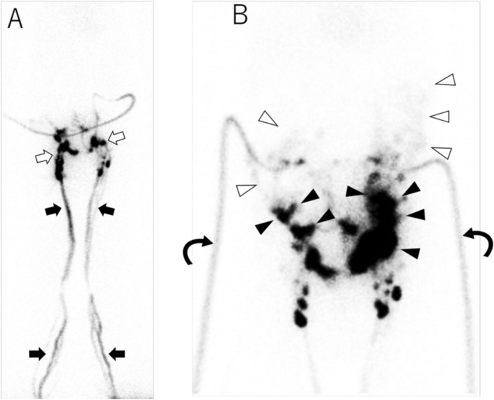
Lymphoscintigraphy after interdigital injection of 99mTc‐labeled human serum albumin–diethylenetriaminepentaacetic acid. (A) At 30 min after tracer injection, bilateral lower limb lymphatic channels (black arrows) and inguinal lymph nodes (white arrows) are visualized, whereas the lumbar lymphatic trunks and thoracic duct are not seen. (B) At 90 min, tracer leakage into the pelvic cavity (black arrowheads) with diffuse spread within the peritoneal cavity (white arrowheads) and accumulation along the drainage tubes (curved arrows) is observed.
On POD 12, intranodal lymphangiography and lymphatic embolization were performed. Bilateral inferior superficial inguinal lymph nodes were punctured under ultrasound guidance with 23‐gauge needles, and 7 mL of ethiodized oil was injected on the right and 6 mL on the left (Figure2A). Consistent with the lymphoscintigraphy findings, contrast leakage from the pelvic level with accumulation in the peritoneal cavity was observed (Figure2B), and lymphatic ducts cranial to the iliac level were not visualized, suggesting bilateral leakage at the pelvic lymphatic ducts.
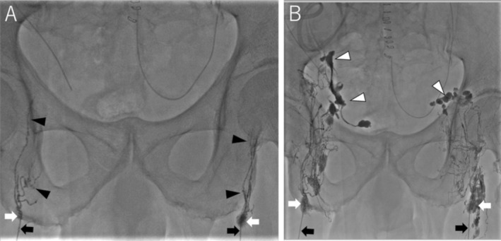
Intranodal lymphangiography performed via the bilateral inguinal lymph nodes. Both inguinal lymph nodes (white arrows) were punctured with 23‐gauge Cathelin needles (black arrows), and ethiodized oil was injected. (A) Fluoroscopic image obtained immediately after injection, showing opacification of the efferent lymphatic channels (black arrowheads). (B) After injection of 7 mL of ethiodized oil on the right side and 6 mL on the left side, contrast extravasation (white arrowheads) into the pelvic cavity is observed bilaterally, indicating pelvic lymphatic leakage.
Under image guidance, three additional lymph nodes located cranial to the initially opacified nodes (one left superior and two right‐sided inguinal nodes) were selectively punctured for upstream injection. Upstream injection of the embolic material was performed from all five accessed lymph nodes, including the two initially punctured nodes and the three additional nodes. A mixture of NBCA and ethiodized oil at a 1:8 ratio was used as the embolic agent. To reduce embolization‐related pain, 1% lidocaine hydrochloride was injected intranodally via the same upstream route before NBCA administration, and a total of 10 mL of NBCA–Lipiodol was injected from the five nodes (Figure3) (VideoS1). Post‐embolization fluoroscopy confirmed that the embolic material had reached the leakage sites in the bilateral pelvic cavity (Figure4). No acute complications were observed in relation to the embolization procedure.
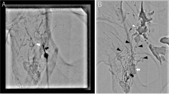
Lymphatic embolization using an upstream injection technique via a lymph node located cranial to the leakage site. (A) Under fluoroscopic guidance, an opacified inguinal lymph node (white arrow), located upstream from the leakage site, is punctured with a 23‐gauge Cathelin needle (black arrow). (B) A mixture of N‐butyl cyanoacrylate and ethiodized oil at a 1:8 ratio is injected by upstream injection through the punctured node. The embolic material fills the targeted lymph node (white arrow) and efferent lymphatic channels (black arrowheads) and reaches the pelvic leakage site (white arrowhead).
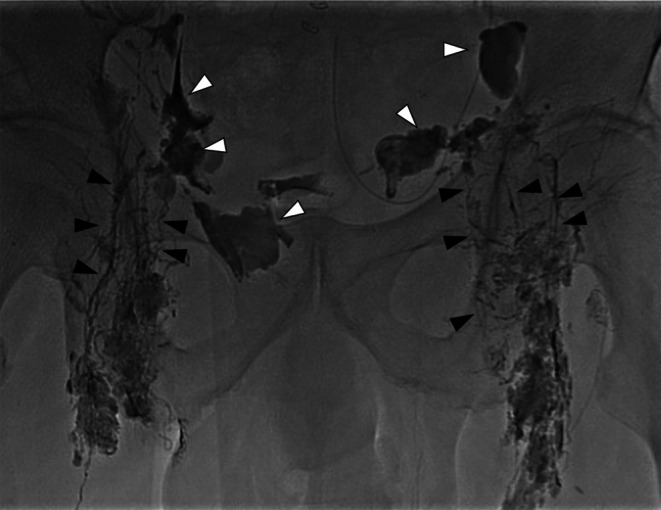
Post‐embolization fluoroscopy after lymphatic embolization using an upstream injection technique. A mixture of N‐butyl cyanoacrylate and ethiodized oil at a 1:8 ratio was injected upstream from the leakage sites. The lymphatic channels located cranial to the leakage points are filled with embolic material (black arrowheads). Previously extravasated Lipiodol and embolic material are seen within the pelvis along the leakage sites (white arrowheads).
Following lymphatic embolization, a marked reduction in drain output was observed from the next day. All pelvic drains were removed by post‐embolization day 4, and no reaccumulation of ascitic fluid was noted. Although bilateral lower extremity edema transiently worsened, the patient remained ambulatory, and activities of daily living were not impaired. He was discharged on post‐embolization day 10. At the 3‐month follow‐up, edema had improved and lymphoscintigraphy showed no tracer leakage or signs of secondary lymphedema. At 6 months, there was no recurrence of ascites.
Discussion
Lymphatic leakage is a known complication following radical cystectomy and PLND for MIBC. Although reported incidences vary with diagnostic criteria, several series have described postoperative lymphatic leakage or lymphocele in 1%–26% of patients [1,2]. While many cases are low‐output and self‐limiting, high‐output or clinically significant leakage can cause malnutrition and immunodeficiency. Conservative management remains the first‐line approach, but is often insufficient in patients with diffuse intraperitoneal leakage, as in the present case.
Recently, lymphatic embolization using NBCA via intranodal lymphangiography, including upstream injection techniques, has emerged as a minimally invasive option for refractory lymphatic leakage after pelvic surgery [4,5,6,7,8,9,10]. However, awareness of this technique among urologists appears limited, and reports focusing specifically on lymphatic complications after PLND are scarce. Our case demonstrates that upstream lymphatic embolization can effectively control massive lymphoascites without the need for reoperation.
Lymphatic leakage can be broadly categorized into localized, encapsulated collections such as lymphoceles and non‐encapsulated free leakage into the peritoneal cavity [11]. Sclerotherapy is well suited to localized lymphoceles but is unsuitable for free peritoneal leakage, in which sclerosants disperse widely. NBCA‐based lymphatic embolization has been reported to be effective in both lymphoceles and diffuse lymphorrhea [4,5,6,7,8,9]. Recent series have reported high clinical success rates for intranodal NBCA embolization in high‐output postoperative leakage, with fewer treatment sessions and shorter treatment duration compared with sclerotherapy [6,7,8]. In our patient, high‐output drainage rapidly decreased after embolization, allowing drain removal within 4 days. Precise identification of bilateral iliac leakage and selective puncture of multiple inguinal lymph nodes for upstream injection likely contributed to the early therapeutic success.
Lower limb lymphedema is one of the most frequently reported complications after lymphatic embolization, although its incidence is relatively low and most cases respond to conservative measures [7,8,10]. Soga et al. demonstrated collateral lymphatic pathways in many patients with lower limb lymphedema [12], and Koehler et al. and Kariya et al. suggested the presence of lymphatico‐venous anastomoses [13,14]. These findings may help explain why procedure‐related lymphedema is often mild and reversible. In the present case, lower extremity edema worsened transiently after embolization but gradually improved.
This report describes a single case; therefore, the findings cannot be generalized to all patients with postoperative lymphatic leakage. Nevertheless, our experience suggests that upstream lymphatic embolization is a valuable minimally invasive option for lymphatic leakage after PLND.
Consent
Informed consent was obtained from the patient.
Conflicts of Interest
The authors declare no conflicts of interest.
Acknowledgments
The authors have nothing to report.
Data Availability Statement
The data that support the findings of this study are available on request from the corresponding author. The data are not publicly available due to privacy or ethical restrictions.
Associated Data
Data Availability Statement
The data that support the findings of this study are available on request from the corresponding author. The data are not publicly available due to privacy or ethical restrictions.
References
- Lawrentschuk N., Colombo R., Hakenberg O. W., et al., “Prevention and Management of Complications Following Radical Cystectomy for Bladder Cancer,” European Urology 57, no. 6 (2010): 983–1001, 10.1016/j.eururo.2010.02.024. doi.org/10.1016/j.eururo.2010.02.024
- Novotny V., Hakenberg O. W., Wiessner D., et al., “Perioperative Complications of Radical Cystectomy in a Contemporary Series,” European Urology 51, no. 2 (2007): 397–401, 10.1016/j.eururo.2006.06.014. doi.org/10.1016/j.eururo.2006.06.014
- Shin Y. S., Han K., Lee J., et al., “Lymphatic Embolization for Early Post‐Operative Lymphatic Leakage After Radical Cystectomy for Bladder Cancer,” PLoS One 19, no. 9 (2024): e0305240. doi.org/10.1371/journal.pone.0305240
- Chu H. H., Shin J. H., Kim J. W., Noh S. Y., Yang W. J., and Park S., “Lymphangiography and Lymphatic Embolization for the Management of Pelvic Lymphocele After Radical Prostatectomy in Prostatic Cancer,” Cardiovascular and Interventional Radiology 42, no. 6 (2019): 873–879, 10.1007/s00270-019-02209-6. doi.org/10.1007/s00270-019-02209-6
- Kariya S., Nakatani M., Ono Y., et al., “Nodal Lymphangiography and Embolization for Postoperative Lymphatic Leakage,” Interventional Radiology 10 (2025): e20240012, 10.22575/interventionalradiology.2024-0012. doi.org/10.22575/interventionalradiology.2024-0012
- Lee K., Chang S. J., Won J. H., et al., “Intranodal Lymphangiography and Embolization for the Treatment of Early Postoperative Lymphatic Leaks After Pelvic Surgery,” Journal of Vascular and Interventional Radiology 34, no. 4 (2023): 591–599.e1, 10.1016/j.jvir.2022.12.020. doi.org/10.1016/j.jvir.2022.12.020
- Seyferth E. R., Itkin M., and Nadolski G. J., “Intranodal Glue Embolization for Postoperative Lymphatic Leaks in the Groin and Pelvis: Comparison With Sclerotherapy,” Journal of Vascular and Interventional Radiology 34, no. 4 (2023): 600–606, 10.1016/j.jvir.2022.12.031. doi.org/10.1016/j.jvir.2022.12.031
- Kim S. W., Hur S., Kim S. Y., et al., “The Efficacy of Lymph Node Embolization Using N‐Butyl Cyanoacrylate Compared to Ethanol Sclerotherapy in the Management of Symptomatic Lymphorrhea After Pelvic Surgery,” Journal of Vascular and Interventional Radiology 30, no. 2 (2019): 195–202.e1, 10.1016/j.jvir.2018.09.038. doi.org/10.1016/j.jvir.2018.09.038
- Ten Hove A. S., Tjiong M. Y., and Zijlstra I. A. J., “Treatment of Symptomatic Postoperative Pelvic Lymphoceles: A Systematic Review,” European Journal of Radiology 134 (2021): 109459, 10.1016/j.ejrad.2020.109459. doi.org/10.1016/j.ejrad.2020.109459
- Moussa A. M., Camacho J. C., Maybody M., et al., “Percutaneous Lymphatic Embolization as Primary Management of Pelvic and Retroperitoneal Iatrogenic Lymphoceles,” Journal of Vascular and Interventional Radiology 32, no. 11 (2021): 1529–1535, 10.1016/j.jvir.2021.07.007. doi.org/10.1016/j.jvir.2021.07.007
- Santos E. and Moussa A. M., “Chylous Ascites and Lymphoceles: Evaluation and Interventions,” Semin Intervent Radiol 37, no. 3 (2020): 274–284, 10.1055/s-0040-1715168. doi.org/10.1055/s-0040-1715168
- Soga S., Onishi F., Jinzaki M., Mikoshi A., Minabe T., and Shinmoto H., “Analysis of Collateral Lymphatic Circulation in Patients With Lower Limb Lymphedema Using Magnetic Resonance Lymphangiography,” Journal of Vascular Surgery. Venous and Lymphatic Disorders 9, no. 2 (2021): 471–481, 10.1016/j.jvsv.2020.07.024. doi.org/10.1016/j.jvsv.2020.07.024
- Koehler P. R. and Schaffer B., “Peripheral Lymphatico‐Venous Anastomoses. Report of Two Cases,” Circulation 35, no. 2 (1967): 401–404. doi.org/10.1161/01.cir.35.2.401
- Kariya S., Komemushi A., Nakatani M., Yoshida R., Kono Y., and Tanigawa N., “Intranodal Lymphangiogram: Technical Aspects and Findings,” Cardiovascular and Interventional Radiology 37, no. 6 (2014): 1606–1610, 10.1007/s00270-014-0901-0. doi.org/10.1007/s00270-014-0901-0
Republished from the open web under CC-BY. Authors: Oshima R, Kariya S, Nakatani M, Ono Y, Maruyama T, Tanaka Y, Sugiura K, Tanigawa N. Read the original.