Prevalence, Associated Factors, and Temporal Trends of Brucella Detection Across Human and Animal Hosts in Bangladesh: A 25-Year Meta-Analysis.
Background Brucellosis remains an important zoonotic disease in Bangladesh, yet evidence on its prevalence across human and animal hosts is fragmented and heterogeneous. Objectives This study aims to synthesize evidence on Brucella detection across human and animal hosts in Bangladesh and to evaluate associated study-level factors and temporal patterns. Methods The data were extracted from eligible studies published between 2000 and 2024 following a systematic review framework. Pooled prevalence was estimated using a random-effects meta-analysis. Heterogeneity was assessed using Cochran's Q and I 2 statistics. Subgroup meta-analyses and univariable meta-regression were conducted to examine variation by host species, diagnostic method, and study-level factors. Forecasting was performed using ARIMA models on annual study-reported Brucella detection, with model performance evaluated using AIC, MAE, and RMSE. Results The pooled Brucella detection across hosts was 3.75% (95% CI: 3.07-4.56), with substantial heterogeneity (I 2 = 89.5%). Detection was the highest in dogs (7.49%) and the lowest in horses (1.78%)). Among evaluated variables, abortion history was the only study-level factorsignificantly associated with higher Brucella positivity, with aborted animals showing approximately 10 times higher odds of detection compared to non-aborted animals (OR = 10.09, 95% CI: 4.64-21.93, p Conclusion Brucella exposure and detection persist across multiple host species in Bangladesh, with variation largely driven by methodological and host-related differences. These findings underscore the need for standardized diagnostic and reporting approaches and support integrated One Health surveillance to improve interpretation and control strategies in Bangladesh.
Introduction
Brucellosis is one of the major global zoonotic diseases, caused by Gram‐negative bacteria of the genusBrucella, and is responsible for substantial economic losses in the livestock industry worldwide (Rossetti et al.2017; SciELO—Public Health2025).Brucellainfects almost half a million humans worldwide each year (Seleem et al.2010; Pappas et al.2006). However, it is recognized as a neglected zoonosis by the World Health Organization (Franc et al.2018) and the World Organization for Animal Health (Zhang et al.2019) because the health system does not prioritize this disease. It mainly invades the reproductive tract of domestic animals and can be transmitted vertically and horizontally in them (Moreno2014; Tadesse2016). Contact with infected animals and drinking milk without pasteurization can cause the transmission of this pathogen to humans (Moreno2014). Pet animals, such as dogs, can be infected by ingesting vaginal discharge from diseased animals (M. A. Islam et al.2013).
Brucellosis is endemic in Bangladesh, as in some other developing countries, including Africa (Maurice et al.2013; Ducrotoy et al.2017; Vhoko et al.2018), Central America (Moreno2002), Latin America (Lucero et al.2008), and the Middle East (Refai2002; Musallam et al.2016). Clinical pictures of brucellosis vary widely among humans and animals. In animals, clinical signs are abortion, stillbirth, low survival rate of calves, delayed calving, reduced milk production, and infertility in male animals (Khan and Zahoor2018). In humans, symptoms are weakness, pain in joints and muscles, enlargement of the liver and spleen, headache, and undulant fever, accompanied by a negligible death rate, with the infection lasting for several years (Madkour2001). Humans with brucellosis most often have a fever that is mistaken for malaria, which is another endemic disease in Bangladesh (M. J. Corbel2006).
Bangladesh is a South Asian country with a fast‐growing livestock industry where almost every family has domestic or pet animals in their houses. They hold a close relationship with them, particularly with sheep and goats, which is the principal source of human brucellosis (Moreno2014), although humans can acquire this pathogen from a wide range of animal species. Existing studies on the prevalence of human brucellosis showed remarkable differences in the approximate range from 2.51% in Kathmandu, Nepal (Pokhrel et al.2021), to 44.7% in the Sylhet district of Bangladesh (Akhtar et al.2020). A meta‐analysis between 2008 and 2017 with 22 studies on brucellosis found 17%, 2%, 33%, and 3% prevalence of brucellosis in bovine, caprine, porcine, and sheep‐goat, respectively, in the northern region of India (Barman et al.2020). These findings may be relevant to Bangladesh due to its geographical proximity and nearly similar socio‐economic settings, but still not enough for policymakers to make control strategies.
For accurate estimation, a systematic evaluation of available studies is necessary, as individual studies cannot reliably represent the overall magnitude of a disease across a broader geographic area. Despite the endemicity of brucellosis in Bangladesh, comprehensive, nationally synthesized evidence onBrucelladetection across human and animal hosts remains limited. In particular, variation in reported prevalence by host species, demographic characteristics, and diagnostic methods has not been systematically evaluated. Therefore, the objective of this study was to synthesize available evidence onBrucelladetection across human and animal hosts in Bangladesh and to examine associated study‐level factors and temporal patterns.
Methods
Systematic Review and Meta‐Analysis
Study Design and Search Strategy
This study employed a systematic review and meta‐analysis to synthesize existing articles on the evidence ofBrucelladetection across human and animal hosts in Bangladesh and to examine associated study‐level factors and temporal patterns. The review was conducted in accordance with the Preferred Reporting Items for Systematic Reviews and Meta‐Analyses (PRISMA) guidelines to ensure transparency and reproducibility (Moher et al.2015) (TableS1). A comprehensive literature search was conducted using bibliographic databases, including PubMed, and Scopus supplemented by targeted searches in Google Scholar, which served as a search engine to identify additional relevant evidence. The search covered predefined combinations of keywords such as ‘Brucella’, ‘Brucella detection’, ‘prevalence’, ‘seroprevalence’, ‘risk factors’, and ‘Bangladesh’ (Table1), adapted from previously published systematic reviews (Musallam et al.2016; Mamun et al.2025; H. Talukder et al.2023; Raquib et al. 2022,2025). The study protocol was registered in the PROSPERO database (ID: CRD420251082670).
Table: Algorithm for study search to identify published articles on prevalence of brucellosis in humans and animals.
Eligibility Criteria
Eligible studies were included if they reported the prevalence ofBrucelladetection in human or animal species in Bangladesh and provided sufficient extractable data using recognized diagnostic methods, including the Rose Bengal Test (RBT), enzyme‐linked immunosorbent assay (ELISA), and polymerase chain reaction (PCR). These methods capture different aspects ofBrucelladetection: RBT and ELISA are widely used serological screening tools for population‐level surveillance, whereas PCR enables direct detection ofBrucellaDNA and is considered a confirmatory method for identifying active infection (Dadar et al.2026).
Included studies were cross‐sectional or case‐control observational studies published in English between January 2000 and December 2024 and available in full text. Grey literature (e.g., government reports and academic theses) was reviewed to assess the completeness of the evidence but was excluded from the quantitative meta‐analysis due to the lack of peer review. Studies with insufficient or non‐extractable data, as well as those from non‐peer‐reviewed sources, were excluded. Review articles, conference proceedings, case reports, editorials, theses, and opinion papers were also excluded. Duplicate publications and studies conducted outside Bangladesh were removed. Study selection was performed independently by two authors, with disagreements resolved through discussion with a third author. The reference lists of the included studies were manually screened to identify additional relevant articles.
Data Extraction
Data extraction was conducted systematically using a predefined data extraction form to ensure consistency and accuracy. Information was extracted independently from each eligible study and recorded in a Microsoft Excel spreadsheet. Extracted variables included the first author's name, year of publication, study location (district), study design, host species (e.g., human, cattle, goat), sample size, and reported prevalence or seroprevalence ofBrucelladetection. The details of diagnostic methods used, including PCR, ELISA, RBT, standard agglutination test (SAT), and milk ring test (MRT), were recorded. When multiple diagnostic tests were reported within a single study, the test with the highest reported diagnostic performance was selected for prevalence extraction based on the following predefined hierarchy: RT‐PCR > conventional PCR > ELISA > RBT > SAT > MRT, consistent with previous studies (Asaad and Alqahtani2012; Getachew et al.2016; Sadhu et al.2015; Sharma et al.2016). Additional extracted variables included sex, breed, abortion history, and other study‐reported factors examined in relation toBrucelladetection. These variables were treated as study‐level associated factors. Information on sampling and data collection methods was also recorded to support assessment of study quality and comparability.
Quality Assessment of Selected Studies
Assessment and assurance of the quality of the selected studies were done through a checklist devised by the Joanna Briggs Institute (JBI) for studies reporting prevalence data (JBI2020). A scoring approach was employed where one point was allotted for the presence of each of the following items in the selected article: study objective, sampling area, study period, sample close representation of the general population, random sampling, sample size calculation, diagnostic techniques used, correct analysis, categorized by sex, and consistent methodology.
Case definition:Brucellapositivity was defined based on detection ofBrucellaexposure or presence using serological (e.g., ELISA, RBT) or molecular (PCR) diagnostic methods as reported in the original studies. Serological methods were interpreted as evidence of prior exposure, whereas PCR‐based methods were considered indicative of current infection. Clinical diagnosis of brucellosis was not required for inclusion, and therefore pooled estimates reflect laboratory/kit‐based evidence of exposure or detection rather than clinically confirmed disease.
Data Analysis
The meta, metafor, and dmetar packages in R (version 4.4.2) were used to estimate pooledBrucelladetection. All prevalence estimates were logit‐transformed before meta‐analysis to stabilize variances.After analysis, results are back‐transformed. A random‐effects meta‐analysis was used as the primary analytical approach to account for between‐study variability. Statistical heterogeneity among studies was assessed using Cochran'sQtest and quantified using theI2statistic, which represents the proportion of total variability attributable to between‐study heterogeneity rather than sampling error (Higgins2003). Values ofI2greater than 50% were interpreted as indicating substantial heterogeneity. Given the expected methodological diversity across studies, random‐effects models were applied throughout.
Forest plots were used to visually present pooled prevalence estimates and confidence intervals (Barendregt et al.2013; Li et al.2020). Potential small‐study effects were assessed using funnel plots (Figure3) and Egger's regression test (Table2) (Egger et al.1997). Sensitivity analyses, including leave‐one‐out analysis and trim‐and‐fill procedures, were conducted to evaluate the robustness of the pooled estimates. To explore potential sources of heterogeneity, subgroup meta‐analyses were performed by host species (cattle, buffalo, sheep, goat, horse, pig, dog, and human), diagnostic method (PCR, ELISA, RBT, SAT, and others), geographic location, and sex. Additional subgroup analyses for animal studies considered breed (local, exotic, crossbred), abortion history (yes/no), and pregnancy status (yes/no), where data were available. To further examine study‐level associations with reported prevalence, univariable meta‐regression analyses were conducted. Univariable meta‐regression was performed using a generalized linear mixed‐effects model with a logit‐transformed prevalence as the effect size. Between‐study heterogeneity was estimated using maximum likelihood, and the results were expressed as odds ratios (ORs) with 95% confidence intervals.
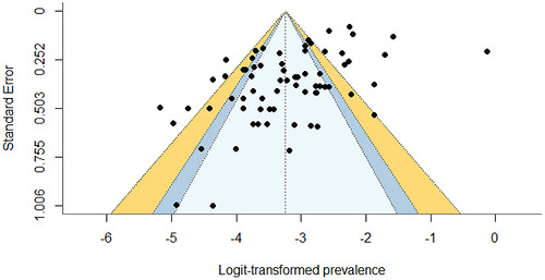
Funnel plot assessing publication bias in the meta‐analysis ofBrucellaprevalence in Bangladesh.
Table: Meta‐regression ofBrucellaassociation in different categories.
Forecast Analysis
Exploratory time‐series analyses were conducted using autoregressive integrated moving average (ARIMA) models applied to annual study‐reported prevalence estimates. These analyses were performed solely to illustrate patterns in published data over time and do not represent population‐level surveillance or disease forecasts. Given heterogeneity in study design, diagnostics, and sampling frames, the results were interpreted cautiously as descriptive trend projections. The Box‐Jenkins approach, using autoregressive moving average ARMA or ARIMA models in time‐series analysis, was used to determine whether the time series fits its historical values in order to provide forecasts. Two evaluation measures have been used in this study, which are mean absolute error (MAE) and root mean square error (RMSE), to assess the efficacy of the model. The forecasting technique used averages to analyze several functions to distinguish and predict values. The remaining terms in equations (1) and (2) were used to describe these discrepancies between prognostic values and practical values ([Evaluating forecast accuracy (MAE, RMSE, MAPE) 2025](#ref-Evaluating forecast accuracy (MAE, RMSE, MAPE) 2025)).
The final models ARIMA (1,0,1) for goat and cattle, respectively, were selected based on the lowest Akaike Information Criterion (AIC) value and the highestR2value, as shown in Table3. Exploratory ARIMA models suggested divergent patterns in reportedBrucelladetection for goats and cattle; however, substantial volatility in predicted values indicates sensitivity to heterogeneous input data and limits epidemiological interpretation.
Table: Model selection for goat and cattle.
Results
Characteristics of the Included Studies
The systematic literature search identified a total of 320 articles. After removal of duplicates and initial screening, 112 studies were assessed based on titles and abstracts, followed by full‐text evaluation of potentially eligible studies. Ultimately, 54 studies met the inclusion criteria and were included in the meta‐analysis, as summarized in the PRISMA flow diagram (Figure1). In 54 identified studies, a total of 24,966 samples were identified. The highest number of articles (N= 32) had prevalence data about cattle (M. S. Ahasan et al.2010; Maruf et al.2019; M.K. M.A Rahman et al.2019; Md. S. Ahasan et al.2017; M.K.M.A. Rahman et al.2017; Md. S. Rahman2012; S. Sikder et al.2012; M. A. S. Sarker et al.2018; Sarker, Begum, et al.2017; Faruque et al.2021; M. S. Islam et al.2018; Md. S. Rahman et al.2013; K. M. R. Amin et al.2005;M.S Rahman et al.2006; MAS et al.2014; M. Rahnan et al.2019; M. Rahman et al.2009; M. Rahman et al.2011; M. Rahman et al.2010; M. Islam et al.2007; M. M. Rahman et al.2020; Nahar and Ahmed1970; Deb Nath et al.2023; Dey et al.2014; K. Amin et al.2004; Hassan et al.2014; Sarker, Sarker, et al.2017; S. Islam et al.2021; Belal and Ansari2013; M. A. Islam et al.2014; Md. S. Rahman et al.2012; M. S. Rahman et al.2014) with the highest number of samples (n= 13,343). Twelve, 10, and 6 studies were found to have prevalence estimation of brucellosis in goats (Md. S. Ahasan et al.2017; Md. S. Rahman2012; M. Rahman et al.2011; M. K. M. A. Rahman et al.2013; Md. S. Rahman et al.2011; Uddin et al.2007; M.S. Rahman et al.2013; M. Islam et al.2012; Shafy et al.2017; Mollah2019; Akhter et al.2014; Munsi et al.2021), sheep (Md. S. Rahman2012; M. Rahman et al.2011; M. K. M. A. Rahman et al.2013; Md. S. Rahman et al.2011; Shafy et al.2017; Akhter et al.2014; M. Rahman et al.2024; M. Ahsan et al.2014; M. Rahman et al.2013; Gani et al.2016), and buffalo (Md. S. Rahman2012; M. Rahman et al.2011; M. A. S. Sarker, et al.2017; M. A. Islam et al.2014; M. S. Rahman et al.2014; M. Rahman et al.2014), respectively, brucellosis of horses (Millat et al.2018) and pig (M. S. Rahman et al.2013) wasidentifiedin one study each, and two studies had prevalence data about dog (B. Talukder et al.2012; M.S. Rahman et al.2015). Only eight studies reported human brucellosis, comprising a total sample size of 2982 individuals (A. K. M. A. Rahman et al.2017; M. Rahnan et al.2019; M. M. Rahman et al.2020; Nahar and Ahmed1970; Akhtar et al.2020; Rasheduzzaman et al.2020; A. K. M. A. Rahman et al.2012; A. A. Rahman et al.2016). The majority of included studies (n= 48) were published between 2010 and 2019, whereas only six studies were published between 2000 and 2009.
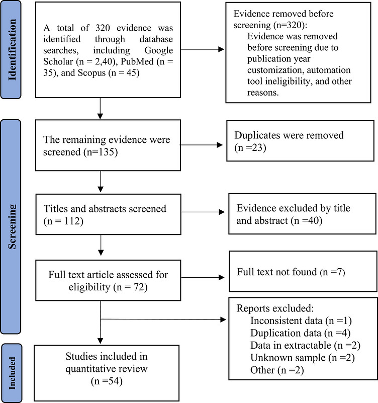
PRISMA flow diagram of literature search and study selection (Moher et al.2015).
BrucellaDetection in Humans and Animals
Subgroup analyses were conducted by host species, sex, breed, diagnostic method, abortion history, and pregnancy status. Between‐study variance (τ2) varied across subgroups and was the highest among studies involving humans, male host, crossbred animals, RT‐PCR‐based diagnostics, animals without a reported abortion history, and non‐pregnant subjects, indicating substantial residual heterogeneity within these categories.
Overall, the pooled prevalence ofBrucelladetection across human and animal hosts was 3.75% (95% confidence interval [CI]: 3.07–4.56), with substantial heterogeneity (I2= 89.5%,p< 0.001). Species‐specific subgroup analysis showed the highest pooledBrucelladetection in dogs (7.49%, 95% CI: 3.23–16.43). Lower pooled estimates were observed for humans (2.941%, 95% CI: 1.00–8.31 (Figure2), and horses (1.78%, 95% CI: 0.44–6.85) (Table4). Breed‐specific analysis indicated higher detection in crossbred animals (5.19%, 95% CI: 3.54–7.55).Brucelladetection estimates varied across diagnostic methods, with higher pooled estimates observed in studies using theBrucellaantigen kit (5.13%, 95% CI: 3.15–8.27), followed by RBT (4.07%, 95% CI: 3.30–4.99), and ELISA (3.35%, 95% CI: 2.31–4.82). These differences likely reflect variation in diagnostic sensitivity, study populations, and sampling strategies rather than test performance alone. Sex and reproductive‐status‐specific analyses showed higher prevalence among female animals (4.15%, 95% CI: 3.46–5.46), pregnant animals (8.80%, 95% CI: 6.64–11.91), and animals with a history of abortion (30.13%, 95% CI: 20.60–41.74) (Table4).
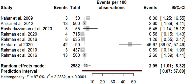
Study‐level and pooled prevalence estimates of humanBrucellain Bangladesh.
Table: Pooled prevalence ofBrucelladetection in humans and animals.
Factors Associated WithBrucellaDetection
Univariable meta‐regression analyses were conducted to explore study‐level factors associated withBrucelladetection (Table5). Among all evaluated variables, abortion history showed a strong association withBrucelladetection, with animals having a history of abortion exhibiting markedly higher odds ofBrucelladetection compared with those without abortion history (OR = 10.09, 95% CI: 4.64–21.93). Physiological status also demonstrated a statistically significant association at both the between‐group and within‐group levels. Pregnant animals had higher odds ofBrucelladetection compared with non‐pregnant animals (OR = 1.71, 95% CI: 1.03–2.84). In contrast, species, sex, breed, diagnostic test, and sample type did not show statistically significant between‐group differences inBrucellaprevalence.Although some species (e.g., dogs, pigs, and buffalo) showed higher estimated odds of Brucella detection compare to horses. No significant differences were observed across diagnostic test types or sample type.
Table: Publication bias assessment with Egger's test.
Publication Bias Assessment
The funnel plot of logit‐transformed prevalence against standard error showed an overall triangular distribution, with most studies falling within the expected pseudo 95% confidence limits; however, some asymmetry was visually apparent, particularly among smaller studies (Figure3). Egger's test provided statistical evidence of small‐study effects, with a significant intercept (bias coefficient = −0.56, 95% CI: −0.80 to −0.31;p< 0.001). The significant slope coefficient (2.25, 95% CI: 2.00 to 2.49;p< 0.001) further indicates a strong relationship between study precision and reported effect sizes. Together, these findings suggest the presence of publication bias or other small‐study effects in the included literature.
Spatial Distribution ofBrucellaDetection
District‐level choropleth mapping revealed marked spatial heterogeneity inBrucelladetection across Bangladesh (Figure4). HigherBrucelladetection clusters were predominantly observed in the northeastern and northern regions of the country. Sylhet district exhibited the highest reportedBrucelladetection (31.88%), followed by Sherpur (11.83%) and Netrokona (11.11%), placing these districts within the very high prevalence category. In contrast, several districts demonstrated comparatively low prevalence levels, including Sunamganj (1.90%) and Lalmonirhat (2.14%), which fell within the very low prevalence category. Many districts, particularly in central and southeastern Bangladesh, showed either low‐to‐medium detection or lacked sufficient data for estimation. Overall, the spatial patterns indicate substantial geographic variation inBrucelladetection across Bangladesh.
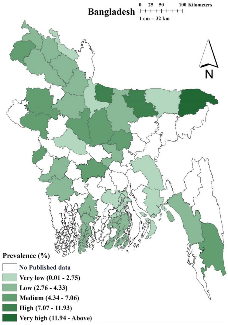
Map showing pooled prevalence ofBrucellain different districts of Bangladesh.
Trend Analysis and Forecasting
The temporal analysis of goatBrucelladetection from 2005 to 2024 showed substantial interannual variability, with no evidence of a stable monotonic trend.The period was selected because no comparable published data were available prior to 2005.Brucelladetection fluctuated markedly over time, characterized by intermittent peaks and troughs, indicating an unstable disease pattern rather than a consistent increase or decrease (Figure5). Forecasting of goatBrucelladetection for the period 2025–2035 likewise suggested continued variability, with alternating years of low and relatively high predicted prevalence (Figure5). The lowest forecasted prevalence was observed in 2026 (point estimate: 0.95%), whereas pronounced peaks were projected for 2027 (9.70%), 2029 (8.59%), and 2030 (8.04%). From 2031 onward, the model indicated a gradual decline in detection, reaching lower predicted levels by 2034 (1.82%), followed by a modest increase in 2035 (3.27%) (TableS1).
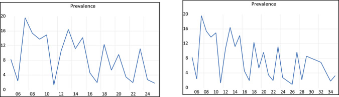
Temporal trend (2005–2024) and forecast (2025–2035) ofBrucelladetection in goats of Bangladesh.
In contrast to goats, time‐series analysis of cattleBrucelladetection from 2003 to 2024 revealed an overall increasing tendency, although substantial interannual variability was evident throughout the study period (Figure5). Reported prevalence fluctuated markedly between years, with the lowest observedBrucelladetection occurring in 2006 (approximately 4%) and a pronounced peak in 2022 (around 25%), indicating an unstable but upward‐leaning disease pattern. Forecasts for the period 2025–2035 suggested that cattleBrucelladetection is likely to remain relatively high and variable (Figure6; TableS1). Point estimates indicated prevalence values largely ranging between ∼16% and ∼28%, with the highest projected prevalence occurring around 2032, exceeding 25%. Although short‐term declines were projected in some years, the overall forecast did not indicate a sustained reduction, instead suggesting persistent transmission with notable fluctuations. The model demonstrated a good fit to the observed data, as indicated by low MAE and RMSE values. For cattle, the MAE and RMSE were 0.019 and 0.074, respectively, while for goats, the corresponding values were 0.145 and 0.102 (Table6). These relatively small error estimates suggest that the model adequately captured the underlying temporal patterns and is suitable for short‐ to medium‐term forecasting. Nevertheless, given the observed temporal variability, forecasted estimates should be interpreted with appropriate caution.
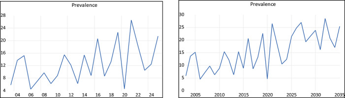
Temporal trend (2003–2024) and forecast (2025–2035) ofBrucelladetection in cattle.
Table: Summarization of the MAE and RMSE for the goat and cattle models.
Discussion
This systematic review and meta‐analysis synthesized savailable articles onBrucelladetection across human and animal hosts in Bangladesh, providing a quantitative yet context‐dependent estimate of detection at 3.75% (95% CI: 3.07–4.56). Importantly, this estimate should not be interpreted as a direct measure of disease burden, but rather as a structured summary of heterogeneous evidence derived from diverse diagnostic and study designs. A key finding is thatBrucelladetection operates within a multi‐host system, with relatively comparable estimates across major livestock species. This pattern suggests widespread exposure across host communities rather than species‐specific susceptibility, consistent with ecological dynamics of endemic zoonoses. This study found a pooled Brucella detection of 3.75% across all species in Bangladesh, which is lower than that reported in some other systematic reviews and meta‐analyses conducted in Iran (Khoshnood et al.2022) and Africa (Simpson et al.2021). This difference may be attributed to several factors, including variations in host species, temperature, humidity, test accuracy, and underreporting. Although higher estimates were observed in dogs and lower estimates in horses, these results are constrained by limited sample sizes and wide CIs, reducing inferential strength and precluding strong conclusions about species‐specific differences. Since the current study included very few studies that reportedBrucelladetection in dogs and horses, these findings cannot represent the exact burden of brucellosis in those species. Besides, various livestock species showed persistentBrucelladetection estimates in Bangladesh, suggesting thatBrucellaspp. exists in the region as a multi‐host infection system rather than being driven by a single dominant species. The relatively comparable prevalence estimates across cattle, goats, sheep, buffalo, and pigs indicate widespread exposure rather than species‐specific susceptibility. Additionally, the reported species‐specific seroprevalence of brucellosis in Bangladesh is lower than that reported in other studies conducted in India (Khoshnood et al.2022), Pakistan (Simpson et al.2021), and Nepal (Acharya et al.2016). Although these countries share a similar ecological and socio‐economic setting, this discrepancy may be attributable to underreporting.
Study heterogeneity emerged as a dominant driver of variation, particularly with respect to diagnostic approach. Serological assays (e.g., ELISA, RBT) capture historical exposure, whereas PCR‐based methods detect current presence of bacterial DNA, leading to fundamentally different epidemiological interpretations (Dadar et al.2025). Consequently, variation observed across studies is more plausibly attributable to differences in management practices, herd structure, and surveillance intensity than to intrinsic host biological susceptibility. Accordingly, differences in reported prevalence across studies likely reflect diagnostic context rather than true variation in transmission intensity, reinforcing that pooled estimates are inherently method‐dependent. This distinction is critical in endemic systems, where chronic exposure and repeated circulation can inflate serological signals relative to active infection.
Breed and sex specific differences were observed but were not statistically significant in meta‐regression. Similarly, higher prevalence in females and crossbred animals was observed, consistent with previous studies (Tadesse2016; Hassan et al.2014; Mcdermott et al.2013; Dadar et al.2025). Diagnostic method was a major source of heterogeneity. Higher estimates from serological assays compared to PCR reflect the detection of past exposure rather than active infection. This discrepancy is well documented in endemic settings (Freire et al.2024; Yagupsky et al.2019) and highlights that prevalence estimates are strongly influenced by diagnostic approach. Among evaluated variables, abortion history and physiological status emerged as the consistent correlate ofBrucellapositivity, supporting well‐established biological mechanisms linkingBrucellaspp. to reproductive tissues (M.J. Corbel et al.2006; Alsaif et al.2018). However, this association should be interpreted cautiously as an epidemiological linkage rather than causal evidence, as abortion both results from infection and increases the likelihood of detection through targeted testing. In contrast, variables such as sex and breed did not retain significance after accounting for study‐level heterogeneity, suggesting that these factors likely act as proxies for exposure intensity, management practices, or sampling bias rather than independent determinants of infection (Tadesse2016; Alsaif et al.2018).
Spatial and temporal patterns further illustrate the limitations of inference from heterogeneous literature. Higher reported prevalence in northern and northeastern regions likely reflects greater livestock density and research activity, rather than true geographic hotspots of transmission (Musallam et al.2016). Similarly, the observed increase in cattle and decrease in goats over time should be interpreted with caution, as these trends are derived from study‐reported data rather than standardized surveillance systems. Such apparent trends may therefore arise from shifting diagnostic practices, study focus, and sampling strategies, rather than underlying epidemiological change (Munsi et al.2021; Stanley and Jarrell1989).
Overall, these findings demonstrate thatBrucellaprevalence estimates are highly context‐dependent, shaped by diagnostic methods, study design, and sampling structure. Univariable associations should therefore be interpreted as indicators of exposure structure rather than definitive risk factors, and pooled estimates should be viewed as summaries of heterogeneous evidence rather than precise population‐level metrics. This study provides one of the first integrated syntheses across human and multiple animal hosts in Bangladesh, offering a baseline for future research and policy. Moving forward, progress in understandingBrucellaepidemiology will depend on standardized diagnostics, improved geographic coverage, and longitudinal surveillance capable of distinguishing true transmission dynamics from methodological artifacts.
Conclusion
This study shows thatBrucelladetection remains present across human and animal hosts in Bangladesh, with a pooled prevalence estimate of 3.75%. Subgroup analyses suggest higher detection in dogs, females, and crossbred animals; however, these differences were not statistically significant. Among study‐level variables, abortion history was the only correlate ofBrucellapositivity. Temporal trends showed an increase in cattle and a decrease in goats. The spatial patterns varied across studies, likely reflecting differences in study design, diagnostics, and coverage rather than true epidemiological trends. Overall, these findings highlight the need for cautious interpretation of pooled estimates and emphasize the importance of standardized methods in futureBrucellaresearch.
Recommendations
FutureBrucellastudies in Bangladesh should prioritize standardized diagnostic and reporting frameworks to improve comparability and reduce methodological heterogeneity in pooled analyses. Transparent reporting of reproductive history, particularly abortion status, should be emphasized, given its consistent association withBrucellapositivity across studies. Improved geographic coverage and more balanced host representation are needed to minimize spatial and species‐level biases and strengthen inference on prevalence patterns. The interpretation of prevalence estimates should explicitly account for diagnostic context, avoiding direct comparisons across fundamentally different detection methods. Where data permit, future studies should consider integrated analyses across animal and human hosts to better characterize shared exposure pathways and risk structures. This gap reflects surveillance, diagnostic, and study design limitations rather than biological differences. Finally, time‐series and forecasting analyses should be treated as descriptive summaries of published studies rather than proxies for structured surveillance, given their sensitivity to heterogeneous study inputs. These recommendations are intended to enhance the quality, consistency, and interpretability of futureBrucellaresearch in Bangladesh, without extending beyond the evidence generated in this study.
Author Contributions
Radwan Raquib: conceptualization, methodology, formal analysis, writing – review & editing.Farhaj Ahammed Arnob: investigation, validation, data curation.Riyadh Hossain: conceptualization, methodology, formal analysis, writing – review & editing.Ahsan Raquib: formal analysis, visualization, investigation, writing – original draft.Tajul Islam Mamun: methodology, supervision, writing – original draft, writing – review & editing. All authors read and approved of the final manuscript.
Funding
The authors have nothing to report.
Ethics Statement
Ethical approval was not required for this study, as it involved secondary data analysis and did not include live animals and human subjects.
Data Availability Statement
None.
Associated Data
Data Availability Statement
None.
References
- Acharya, K. P. , Niroula N., and Kaphle K.. 2016. “Overview of Brucellosis in Nepal.” Epidemiol Health 38: e2016042. 10.4178/epih.e2016042. doi.org/10.4178/epih.e2016042
- Ahasan, M. S. , Rahman M., Song H.‐J.. Ahasan M. S., Rahman M. S., and Song H. J.. 2010. “A Sero‐Surveillance ofBrucellaspp. Antibodies and Individual Risk Factors of Infection in Cattle of Bangladesh.” Korean Journal of Veterinary Service 33, no. 2: 121–128.
- Ahasan, M. S. , Rahman M. S., Rahman A., and Berkvens D.. 2017. “Bovine and Caprine Brucellosis in Bangladesh: Bayesian Evaluation of Four Serological Tests, True Prevalence, and Associated Risk Factors in Household Animals.” Tropical Animal Health and Production 49: 1–11. 10.1007/s11250-016-1151-1. doi.org/10.1007/s11250-016-1151-1
- Ahsan, M. S. , Rahman M., Das G. C., Rahman M. S., and Ali M. L.. 2014. “Seroprevalence of brucellosis in sheep in Mymensingh and Netrokona district of Bangladesh.” International Journal of Natural and Social Sciences 1: 33–40.
- Akhtar, J. , Chowdhury O. A., Das P., and Sinha S. P.. 2020. “Sero‐Prevalence of human Brucellosis in High Risk Individuals and Normal Individuals of Sylhet District in Bangladesh.” Bangladesh Medical Research Council Bulletin 46: 35–40. 10.3329/bmrcb.v46i1.47467. doi.org/10.3329/bmrcb.v46i1.47467
- Akhter, L. , Islam M. A., Das S., Khatun M. M., and Islam M. A.. 2014. “Seroprevalence of Brucellosis and Its Associated Risk Factors in Sheep and Goat in the Farms and Slaughter House in Mymensingh.” Bangladesh Microbes Health 3: 25–28. 10.3329/mh.v3i1.19778. doi.org/10.3329/mh.v3i1.19778
- Alsaif, M. , Dabelah K., Featherstone R., and Robinson J. L.. 2018. “Consequences of Brucellosis Infection During Pregnancy: A Systematic Review of the Literature.” International Journal of Infectious Diseases 73: 18–26. 10.1016/j.ijid.2018.05.023. doi.org/10.1016/j.ijid.2018.05.023
- Amin, K. , Rahman M., Sarkar S., Kabir S. M. L., and Akand M.. 2004. “Serological Epidemiology of Brucellosis in Cattle of Mymensingh district of Bangladesh.” Journal of Animal and Veterinary Advances 3: 898–900.
- Amin, K. M. R. , Rahman M. B., Rahman M. S., Han J. C., Park J. H., and Chae J. S.. 2005. “Prevalence ofBrucellaAntibodies in Sera of Cows in Bangladesh.” Journal of Veterinary Science 6: 223–226.
- Asaad, A. M. , and Alqahtani J. M.. 2012. “Serological and Molecular Diagnosis of Human Brucellosis in Najran, Southwestern Saudi Arabia.” Journal of Infection and Public Health 5: 189–194. doi.org/10.1016/j.jiph.2012.02.001
- Barendregt, J. J. , Doi S. A., Lee Y. Y., Norman R. E., and Vos T.. 2013. “Meta‐Analysis of Prevalence.” Journal of Epidemiology and Community Health 67: 974–978. 10.1136/jech-2013-203104. doi.org/10.1136/jech-2013-203104
- Barman, N. N. , Patil S. S., Kurli R., et al. 2020. “Meta‐Analysis of the Prevalence of Livestock Diseases in North Eastern Region of India.” Veterinary World 13: 80–91. 10.14202/vetworld.2020.80-91. doi.org/10.14202/vetworld.2020.80-91
- Belal, S. , and Ansari A.. 2013. “Seroprevalence ofBrucella abortusAntibodies in the Cattle Population in the Selected Upazilas of Sirajgonj District.” Bangladesh Journal of Veterinary Medicine 11: 127–130.
- Corbel, M. J. , Food and Agriculture Organization of the United Nations . World Health Organization & World Organisation for Animal Health (2006). Brucellosis in humans and animals. World Health Organization.https://iris.who.int/handle/10665/43597.
- Dadar, M. , Fakhri Y., Shahali Y., Tittarelli M., Sacchini F., and De Massis F.. 2025. “Global Epidemiology and Diagnostic Insights Into Canine Brucellosis: A Comprehensive Meta‐Analysis and Meta‐Regression.” One Health 21: 101225. 10.1016/j.onehlt.2025.101225. doi.org/10.1016/j.onehlt.2025.101225
- Dadar, M. , Rebollada‐Merino A., and Shahali Y.. 2026. “Diagnostic Tools and Techniques forBrucellaDetection.” In Brucellosis, 195–212. Elsevier. 10.1016/B978-0-443-30067-7.00028-0. doi.org/10.1016/B978-0-443-30067-7.00028-0
- Deb Nath, N. , Ahmed S. S. U., Malakar V., Hussain T., Chandra Deb L., and Paul S.. 2023. “Sero‐Prevalence and Risk Factors Associated With Brucellosis in Dairy Cattle of Sylhet District, Bangladesh: A Cross‐Sectional Study.” Veterinary Medicine and Science 9: 1349–1358. 10.1002/vms3.1100. doi.org/10.1002/vms3.1100
- Dey, S. , Rahman M., Rima U., et al. 2013. “Serological and Pathological Investigation of Brucellosis in Dairy Cows of Mymensingh district, Bangladesh.” Bangladesh Journal of Veterinary Medicine 11, no. 2: 107–112. 10.3329/bjvm.v11i2.19124. doi.org/10.3329/bjvm.v11i2.19124
- Ducrotoy, M. , Bertu W. J., Matope G., et al. 2017. “Brucellosis in Sub‐Saharan Africa: Current Challenges for Management, Diagnosis and Control.” Acta Tropica 165: 179–193. doi.org/10.1016/j.actatropica.2015.10.023
- Egger, M. , Smith G. D., Schneider M., and Minder C.. 1997. “Bias in Meta‐Analysis Detected by a Simple, Graphical Test.” BMJ 315: 629–634. doi.org/10.1136/bmj.315.7109.629
- Evaluating forecast accuracy (MAE, RMSE, MAPE) . 2025. Intro to Time Series Class Notes.https://fiveable.me/intro‐time‐series/unit‐8/evaluating‐forecast‐accuracy‐mae‐rmse‐mape/study‐guide/ijqkb0CAqRaHLBFi.
- Faruque, M. , Ahad A., Hossain M., Sayeed M. A., and Hoque M.. 2021. “Molecular Detection and Epidemiology ofBrucellain Dairy Cattle of.” Bangladesh 8: 49–54. 10.60015/bjvas.vi.94. doi.org/10.60015/bjvas.vi.94
- Franc, K. A. , Krecek R. C., Häsler B. N., and Arenas‐Gamboa A. M.. 2018. “Brucellosis Remains a Neglected Disease in the Developing World: A Call for Interdisciplinary Action.” BMC Public Health 18: 1–9. doi.org/10.1186/s12889-017-5016-y
- Freire, M. L. , Machado De Assis T. S., Silva S. N., and Cota G.. 2024. “Diagnosis of Human Brucellosis: Systematic Review and Meta‐Analysis.” Public Library of Science Neglected Tropical Diseases 18: e0012030. 10.1371/journal.pntd.0012030. doi.org/10.1371/journal.pntd.0012030
- Gani, M. O. , Munsi M. N., Ershaduzzaman M., Rahman A. A., Sultana S., and Alam M. S.. 2016. “Seroprevalence of Ovine Brucellosis in Bangladesh.” Asian Journal of Medical and Biological Research 2: 13–18. 10.3329/ajmbr.v2i1.27563. doi.org/10.3329/ajmbr.v2i1.27563
- Getachew, T. , Getachew G., Sintayehu G., Getenet M., and Fasil A.. 2016. “Bayesian Estimation of Sensitivity and Specificity of Rose Bengal, Complement Fixation, and Indirect ELISA Tests for the Diagnosis of Bovine Brucellosis in Ethiopia.” Veterinary Medicine International 2016, no. 1: 8032753. 10.1155/2016/8032753. doi.org/10.1155/2016/8032753
- Hassan, A. A. , Uddin M. B., Islam M. R., Cho H.‐S., and Hossain M. M.. 2014. “Serological Prevalence of Brucellosis of Cattle in Selected Dairy Farms in Bangladesh.” Korean Journal of Veterinary Research 54: 239–243. 10.14405/kjvr.2014.54.4.239. doi.org/10.14405/kjvr.2014.54.4.239
- Higgins, J. P. T. 2003. “Measuring Inconsistency in Meta‐Analyses.” BMJ 327: 557–560. 10.1136/bmj.327.7414.557. doi.org/10.1136/bmj.327.7414.557
- Islam, M. , Samad M., and Rahman A.. 2012. “Risk Factors Associated with Prevalence of Brucellosis in Black Bengal Goats in Bangladesh.” Bangladesh Journal of Veterinary Medicine 8: 141–147. 10.3329/bjvm.v8i2.11198. doi.org/10.3329/bjvm.v8i2.11198
- Islam, M. , Siddiqui M., Baki M., Majumder S., Parrish J., and Shamsuddin M.. 2007. “Screening Some Major Communicable Diseases of AI Bulls in Bangladesh.” Livestock Research for Rural Development 19: 1–9.
- Islam, M. A. , Akter L., Khatun M. M., and Islam M. A.. 2014. “Seroprevalence of Brucellosis and Its Associated Risk Factors in Bovine at Greater Mymensingh District of Bangladesh.” Microbes Health 2: 12–14. 10.3329/mh.v2i1.17256. doi.org/10.3329/mh.v2i1.17256
- Islam, M. A. , Khatun M. M., Werre S. R., Sriranganathan N., and Boyle S. M.. 2013. “A Review ofBrucellaSeroprevalence Among Humans and Animals in Bangladesh With Special Emphasis on Epidemiology, Risk Factors and Control Opportunities.” Veterinary Microbiology 166: 317–326. doi.org/10.1016/j.vetmic.2013.06.014
- Islam, M. S. , Islam M. A., Khatun M. M., Saha S., Basir M. S., and Hasan M.‐M.. 2018. “Molecular Detection ofBrucellaspp. From Milk of Seronegative Cows From some Selected Area in Bangladesh.” Journal of Pathogens 2018, no. 1: 9378976. 10.1155/2018/9378976. doi.org/10.1155/2018/9378976
- Islam, S. , Barua S. R., Moni S. P., Islam A., Rahman A., and Chowdhury S.. 2021. “Seroprevalence and Risk Factors for Bovine Brucellosis in the Chittagong Metropolitan Area of Bangladesh.” Veterinary Medicine and Science 7: 86–98. 10.1002/vms3.348. doi.org/10.1002/vms3.348
- JBI . 2020. “Systematic Reviews of Prevalence and Incidence.” InJBI Reviewer's Manual. 10.46658/JBIMES-20-06. doi.org/10.46658/JBIMES-20-06
- Khan, M. Z. , and Zahoor M.. 2018. “An Overview of Brucellosis in Cattle and Humans, and Its Serological and Molecular Diagnosis in Control Strategies.” Tropical Medicine and Infectious Disease 3: 65. 10.3390/tropicalmed3020065. doi.org/10.3390/tropicalmed3020065
- Khoshnood, S. , Pakzad R., Koupaei M., et al. 2022. “Prevalence, Diagnosis, and Manifestations of Brucellosis: A Systematic Review and Meta‐Analysis.” Frontiers in Veterinary Science 9: 976215. 10.3389/fvets.2022.976215. doi.org/10.3389/fvets.2022.976215
- Li, X. , Ni H.‐B., Ren W.‐X., Jiang J., Gong Q.‐L., and Zhang X.‐X.. 2020. “Seroprevalence of Toxoplasma Gondii in Horses: A Global Systematic Review and Meta‐Analysis.” Acta Tropica 201: 105222. 10.1016/j.actatropica.2019.105222. doi.org/10.1016/j.actatropica.2019.105222
- Lucero, N. , Ayala S., Escobar G., and Jacob N.. 2008. “BrucellaIsolated in Humans and Animals in Latin America From 1968 to 2006.” Epidemiology and Infection 136: 496–503. doi.org/10.1017/S0950268807008795
- Madkour, M. M. 2001. Madkour's Brucellosis. Springer Berlin Heidelberg. 10.1007/978-3-642-59533-2. doi.org/10.1007/978-3-642-59533-2
- Mamun, T. I. , Rahman J., Hossain M. J., Hasan R., Neoaj M. T., and Mia M. M.. 2025. “Global Prevalence of Chlamydia Infection Among Wild Birds as Public Health Concerns: A Systematic Review and Meta‐Analysis.” Bulgarian Journal of Veterinary Medicine 28: 1–17. 10.15547/bjvm.2023-0060. doi.org/10.15547/bjvm.2023-0060
- Maruf, A. , Yasmin F., Yeasmin F., et al. 2019. “Assessment of Haemato‐Biochemical and Therapeutic Responses of Chronic Brucellosis in Crossbred Dairy Cows in Bangladesh.” Journal of Veterinary Medical and One Health Research 1: 211–229. 10.36111/jvmohr.2019.1(2).0013. doi.org/10.36111/jvmohr.2019.1(2).0013
- MAS, S. , Rahman M., Islam M. T., Rahman A., MB R., and Rahman M.. 2014. “Prevalence of Brucellosis in Dairy Cattle in Organized and Smallholder Farms in some Selected Areas of Bangladesh.” Bangladesh Journal of Veterinary Medicine 12: 167–171.
- Maurice, N. A. , Wungak S. Y., Gana B. A., et al. 2013. “Seroprevalence of Bovine Brucellosis in Northern Plateau state, North Central Nigeria.” Asian Pacific Journal of Tropical Medicine 3: 337–340.
- Mcdermott, J. J. , Grace D., and Zinsstag J.. 2013. “Economics of Brucellosis Impact and Control in Low‐Income Countries.” Revue Scientifique et Technique ‐ Office International des Epizooties 32: 249–261. 10.20506/rst.32.1.2197. doi.org/10.20506/rst.32.1.2197
- Millat, M. , Shafy N. M., Sharmy S. T., et al. 2018. “Seroprevalence of Equine Brucellosis: First Report in Bangladesh.” Bangladesh Journal of Veterinary Medicine 16: 103–106. 10.3329/bjvm.v16i1.37383. doi.org/10.3329/bjvm.v16i1.37383
- Moher, D. , Shamseer L., Clarke M., et al. 2015. “Preferred Reporting Items for Systematic Review and Meta‐Analysis Protocols (PRISMA‐P) 2015 Statement.” Cochrane Reviews 4: 1. 10.1186/2046-4053-4-1. doi.org/10.1186/2046-4053-4-1
- Mollah, M. L. 2019. “Seroprevalence and Risk Factors of Brucellosis in Black Bengal Goats at Coastal Region of Bangladesh.” International Journal of Veterinary Science .6: 56–61.https://www.academia.edu/77541459/Seroprevalence_and_Risk_Factors_of_Brucellosis_in_Black_Bengal_Goats_at_Coastal_Region_of_Bangladesh.
- Moreno, E. 2002. “Brucellosis in Central America.” Veterinary Microbiology 90: 31–38. doi.org/10.1016/s0378-1135(02)00242-0
- Moreno, E. 2014. “Retrospective and Prospective Perspectives on Zoonotic Brucellosis.” Frontiers in Microbiology 5: 213. doi.org/10.3389/fmicb.2014.00213
- Munsi, M. , Akther S., Rahman M., Hassan M., Ali M., and Ershaduzzaman M.. 2021. “Seroprevalence of Brucellosis in Goats in some Selected Areas of Bangladesh.” Journal of Advanced Veterinary and Animal Research 8: 123. 10.5455/javar.2021.h494. doi.org/10.5455/javar.2021.h494
- Musallam, I. I. , Abo‐Shehada M. N., Hegazy Y. M., Holt H. R., and Guitian F. J.. 2016. “Systematic Review of Brucellosis in the Middle East: Disease Frequency in Ruminants and Humans and Risk Factors for Human Infection.” Epidemiology and Infection 144: 671–685. doi.org/10.1017/S0950268815002575
- Nahar, A. , and Ahmed M. U.. 1970. “Sero‐Prevalence Study of Brucellosis in Cattle and Contact Human in Mymensingh District.” Bangladesh Journal of Veterinary Medicine 7: 269–274. 10.3329/bjvm.v7i1.5071. doi.org/10.3329/bjvm.v7i1.5071
- Pappas, G. , Papadimitriou P., Akritidis N., Christou L., and Tsianos E. V.. 2006. “The New Global Map of Human Brucellosis.” The Lancet Infectious Diseases 6: 91–99. 10.1016/S1473-3099(06)70382-6. doi.org/10.1016/S1473-3099(06)70382-6
- Pokhrel, A. , Rayamajhee B., Khadka S., et al. 2021. “Seroprevalence and Clinical Features of Scrub Typhus Among Febrile Patients Attending a Referral Hospital in Kathmandu, Nepal.” Tropical Medicine and Infectious Disease 6: 78. 10.3390/tropicalmed6020078. doi.org/10.3390/tropicalmed6020078
- Rahman, M. S. , Sarker M. A. S., Rahman A., et al. 2014. “The Prevalence ofBrucella abortusDNA in Seropositive Bovine Sera in Bangladesh.” African Journal of Microbiology Research 8: 3856–3860. 10.5897/AJMR2014.6031. doi.org/10.5897/AJMR2014.6031
- Rahman, A. , Dirk B., Fretin D., et al. 2012. “Seroprevalence and Risk Factors for Brucellosis in a High‐Risk Group of Individuals in Bangladesh.” Foodborne Pathogens and Disease 9: 190–197. 10.1089/fpd.2011.1029. doi.org/10.1089/fpd.2011.1029
- Rahman, A. , Saegerman C., Berkvens D., et al. 2017. “Brucella abortusIs Prevalent in Both Humans and Animals in Bangladesh.” Zoonoses Public Health 64: 394–399. 10.1111/zph.12344. doi.org/10.1111/zph.12344
- Rahman, A. , Saegerman C., Berkvens D., et al. 2013. “Bayesian Estimation of True Prevalence, Sensitivity and Specificity of Indirect ELISA, Rose Bengal Test and Slow Agglutination Test for the Diagnosis of Brucellosis in Sheep and Goats in Bangladesh.” Preventive Veterinary Medicine 110: 242–252. 10.1016/j.prevetmed.2012.11.029. doi.org/10.1016/j.prevetmed.2012.11.029
- Rahman, A. , Smit S., Devleesschauwer B., et al. 2019. “Bayesian Evaluation of Three Serological Tests for the Diagnosis of Bovine Brucellosis in Bangladesh.” Epidemiology and Infection 147: e73. 10.1017/S0950268818003503. doi.org/10.1017/S0950268818003503
- Rahman, A. A. , Berkvens D., Saegerman C., et al. 2016. “Seroprevalence of Brucellosis in Patients With Prolonged Fever in Bangladesh.” Journal of Infection in Developing Countries 10: 939–946. 10.3855/jidc.6844. doi.org/10.3855/jidc.6844
- Rahman, M. , Ahsan M. D., Das G. C., and Rahman M. S.. 2014. “Seroprevalence of Brucellosis in Buffaloes in Bagerhat and Mymensingh District, Bangladesh.” International Journal of Natural and Social Sciences 1: 75–80.
- Rahman, M. , Akther S., Haque M., Ali M., Zihadi M., and Rahman M.. 2024. “Ovine Brucellosis in Bangladesh: Seroprevalence and Its Associated Risk Factors.” Journal of Research in Veterinary Sciences 3: 28. 10.5455/JRVS.20240519053238. doi.org/10.5455/JRVS.20240519053238
- Rahman, M. , Alam N., Rahman A., et al. 2009. “Seroprevalence of SpecificBrucellaDetection of Cattle in Bangladesh Agricultural University Veterinary Clinics and Its Surrounding Areas.” Korean Journal of Veterinary Service 32, no. 3: 219–225.
- Rahman, M. , Faruk M. O., Her M., et al. 2011. “Prevalence of Brucellosis in Ruminants in Bangladesh.” Veterinarni Medicina 56, no. 8: 379–385.
- Rahman, M. , Huque F., Ahasan M. S., et al. 2010. “Indirect Enzyme Linked Immunosorbent Assay for the Diagnosis of Brucellosis in Cattle.” Korean Journal of Veterinary Service 33, no. 2: 113–119.
- Rahman, M. , Rahaman M., Islam M., et al. 2013. “Seroprevalence of Brucellosis in Sheep in the Gaibandha District of Bangladesh.” Progressive Agriculture 23: 25–32. 10.3329/pa.v23i1-2.16562. doi.org/10.3329/pa.v23i1-2.16562
- Rahman, M. M. , Rahman M. S., Rahman A., et al. 2020. “Sero‐Molecular Epidemiology and Rick Factors Analysis of Brucellosis in Human and Lactating Cows of Military Dairy Farms in Bangladesh.” Journal of Veterinary Medical and One Health Research 2, no. 18. 10.36111/jvmohr.2020.2(1).0018. doi.org/10.36111/jvmohr.2020.2(1).0018
- Rahman, M. S. 2012. “Brucellosis Among Ruminants in Some Districts of Bangladesh Using Four Conventional Serological Assays.” African Journal of Microbiology Research 6, no. 22: 4775–4781. 10.5897/AJMR12.475. doi.org/10.5897/AJMR12.475
- Rahman, M. S. , Azad M. K., Ahasan M. S., et al. 2013. “Prevalence and Diagnostic Test Comparison of Brucellosis in Cattle in Pabna and Mymensingh Districts of Bangladesh.” Pakistan Journal of Scientific and Industrial Research 56: 147–153. 10.52763/PJSIR.BIOL.SCI.56.3.2013.147.153. doi.org/10.52763/PJSIR.BIOL.SCI.56.3.2013.147.153
- Rahman, M. S. , Chakrabartty A., Islam M. T., et al. 2012. “Seroprevalence of Brucellosis in Cattle in Selected Area of Bangladesh and Comparison Between Rose Bengal Test and i‐ELISA Used for the Screening of Brucellosis.” Korean Journal of Veterinary Service 35: 133–137. 10.7853/kjvs.2012.35.2.133. doi.org/10.7853/kjvs.2012.35.2.133
- Rahman, M. S. , Hahsin M. F. A., Ahasan M. S., et al. 2011. “Brucellosis in Sheep and Goat of Bogra and Mymensingh Districts of Bangladesh.” Korean Journal of Veterinary Research 51: 277–280. 10.14405/kjvr.2011.51.4.277. doi.org/10.14405/kjvr.2011.51.4.277
- Rahman, M. S. , Han J., Park J., Lee J., Eo S., and Chae J.. 2006. “Prevalence of Brucellosis and Its Association With Reproductive Problems in Cows in Bangladesh.” The Veterinary Record 159: 180–182. 10.1136/vr.159.6.180. doi.org/10.1136/vr.159.6.180
- Rahman, M. S. , Kabir S. L., and Rahman M. S.. 2015. “Seroprevalence of Canine Brucellosis in Dhaka City Corporation Area, Bangladesh.” Asian Journal of Medical and Biological Research 1: 17–21.
- Rahman, M. S. , Mithu S., Islam M. T., et al. 2013. “Prevalence of Brucellosis in Black Bengal Goats in Bangladesh.” Bangladesh Journal of Veterinary Medicine 10: 51–56. 10.3329/bjvm.v10i1-2.15646. doi.org/10.3329/bjvm.v10i1-2.15646
- Rahman, M. S. , Nuruzzaman M., Ahasan M. S., et al. 2013. “Prevalence of Brucellosis in Pigs: The First Report in Bangladesh.” Bangladesh Journal of Veterinary Medicine 10: 75–80. 10.3329/bjvm.v10i1-2.15649. doi.org/10.3329/bjvm.v10i1-2.15649
- Rahnan, M. , Rahman A., Hussaini S., et al. 2019. “First Report of Brucellosis in Dairy Cattle and Humans in Military Farms in Bangladesh.” EC Veterinary Science 4: 01–05.
- Raquib, A. , Raquib R., Jamil S., Hossain A., al‐Mamun F., and Mamun M. A.. 2022. “Knowledge, Attitudes, and Practices toward the Prevention of COVID‐19 in Bangladesh: A Systematic Review and Meta‐Analysis.” Frontiers in Medicine 9: 856156. 10.3389/fmed.2022.856156. doi.org/10.3389/fmed.2022.856156
- Raquib, A. , Salman A., Ziaulhaq A., et al. 2025. “Geospatial Distribution of HIV Seroprevalence among Blood Donors in South Asian Countries: A Systematic Review and Meta‐Analysis.” Aids and Behavior 29: 689–701. 10.1007/s10461-024-04551-4. doi.org/10.1007/s10461-024-04551-4
- Rasheduzzaman, M. , Hossain M. A., Paul S. K., et al. 2020. “Serological and Molecular Epidemiology of Human Brucellosis in Mymensingh Region Bangladesh.” Bangladesh Journal of Medical Microbiology 14: 19–24. 10.3329/bjmm.v14i2.57794. doi.org/10.3329/bjmm.v14i2.57794
- Refai, M. 2002. “Incidence and Control of Brucellosis in the Near East Region.” Veterinary Microbiology 90: 81–110. doi.org/10.1016/s0378-1135(02)00248-1
- Rossetti, C. A. , Arenas‐Gamboa A. M., and Maurizio E.. 2017. “Caprine Brucellosis: A Historically Neglected Disease With Significant Impact on Public Health.” Public Library of Science Neglected Tropical Diseases 11: e0005692. 10.1371/journal.pntd.0005692. doi.org/10.1371/journal.pntd.0005692
- Sadhu, D. B. , Panchasara H. H., Chauhan H. C., Sutariya D. R., Parmar V. L., and Prajapati H. B.. 2015. “Seroprevalence and Comparison of Different Serological Tests for Brucellosis Detection in Small Ruminants.” Veterinary World 8: 561–566. 10.14202/vetworld.2015.561-566. doi.org/10.14202/vetworld.2015.561-566
- Sarker, M. A. S. , Begum M. M., Rahman M. F., et al. 2018. “Conventional PCR Based Detection ofBrucella abortusInfected Cattle in some Selected Areas of Bangladesh.” Bangladesh Journal of Veterinary Medicine 16: 39–44. 10.3329/bjvm.v16i1.37372. doi.org/10.3329/bjvm.v16i1.37372
- Sarker, M. A. S. , Rahman M. S., Begum M. M., et al. 2017. “Milk Ring, Rose Bengal Tests and Conventional PCR Based Detection ofBrucella abortusInfected Dairy Cattle in Bangladesh.” African Journal of Microbiology Research 11: 1505–1509. 10.5897/AJMR2017.8672. doi.org/10.5897/AJMR2017.8672
- Sarker, M. A. S. , Sarker R. R., Begum M. M., et al.2017. “Seroprevalence and Molecular Diagnosis ofBrucella abortusandBrucella melitensisin Bangladesh.” Bangladesh Journal of Veterinary Medicine 14: 221–226. 10.3329/bjvm.v14i2.31400. doi.org/10.3329/bjvm.v14i2.31400
- SciELO—Public Health . 2025. “Human Health Benefits From Livestock Vaccination for Brucellosis: Case Study Human Health Benefits From Livestock Vaccination for Brucellosis: Case Study.”.https://www.scielosp.org/article/bwho/2003.v81n12/867‐876/en/.
- Seleem, M. N. , Boyle S. M., and Sriranganathan N.. 2010. “Brucellosis: A Re‐Emerging Zoonosis.” Veterinary Microbiology 140: 392–398. 10.1016/j.vetmic.2009.06.021. doi.org/10.1016/j.vetmic.2009.06.021
- Shafy, N. M. , Ahmed B. S., Sarker R. R., et al. 2017. “Serological Prevalence of Ovine and Caprine Brucellosis in Bangladesh.” Bangladesh Journal of Veterinary Medicine 14: 209–213. 10.3329/bjvm.v14i2.31398. doi.org/10.3329/bjvm.v14i2.31398
- Sharma, H. K. , Kotwal S. K., Singh D. K., et al. 2016. “Seroprevalence of Human Brucellosis in and Around Jammu, India, Using Different Serological Tests.” Veterinary World 9: 742–746. 10.14202/vetworld.2016.742-746. doi.org/10.14202/vetworld.2016.742-746
- Sikder, S. , Rahman A., Faruque R., et al. 2012. “Brucellosis: An Epidemiological Study at Chittagong, Bangladesh.” Pakistan Veterinary Journal 32: 499–502.
- Simpson, G. , Thompson P. N., Saegerman C., et al. 2021. “Brucellosis in Wildlife in Africa: A Systematic Review and Meta‐Analysis.” Scientific Reports 11: 5960. 10.1038/s41598-021-85441-w. doi.org/10.1038/s41598-021-85441-w
- Stanley, T. D. , and Jarrell S. B.. 1989. “Meta‐Regression Analysis: A Quantitative Method of Literature Surveys.” Journal of Economic Surveys 3: 161–170. 10.1111/j.1467-6419.1989.tb00064.x. doi.org/10.1111/j.1467-6419.1989.tb00064.x
- Tadesse, G. 2016. “Brucellosis Seropositivity in Animals and Humans in Ethiopia: A Meta‐Analysis.” Public Library of Science Neglected Tropical Diseases 10, no. 10: e0005236. 10.1371/journal.pntd.0005006. doi.org/10.1371/journal.pntd.0005006
- Talukder, B. , Samad M., and Rahman A.. 2012. “Comparative Evaluation of Commercial Serodiagnostic Tests for the Seroprevalence Study of Brucellosis in Stray Dogs in Bangladesh.” Bangladesh Journal of Veterinary Medicine 9: 79–83. 10.3329/bjvm.v9i1.11217. doi.org/10.3329/bjvm.v9i1.11217
- Talukder, H. , Roky S. A., Debnath K., Sharma B., Ahmed J., and Roy S.. 2023. “Prevalence and Antimicrobial Resistance Profile of Salmonella Isolated From Human, Animal and Environment Samples in South Asia: A 10‐Year Meta‐Analysis.” Journal of Epidemiology and Global Health 13: 637–652. 10.1007/s44197-023-00160-x. doi.org/10.1007/s44197-023-00160-x
- Uddin, M. J. , Rahman M., Hossain M., et al. 2007. “Relation Between Brucellosis and Husbandry Practices in Goats in Bangladesh.” Korean Journal of Veterinary Service 30, no. 2: 259–267.
- Vhoko, K. , Iannetti S., Burumu J., Ippoliti C., Bhebhe B., and DeMassis F.. 2018. “Estimating the Prevalence of Brucellosis in Cattle in Zimbabwe From Samples Submitted to the Central Veterinary Laboratory Between 2010 and 2014.” Veterinaria Italiana 54: 21–27. doi.org/10.12834/VetIt.1111.6191.2
- Yagupsky, P. , Morata P., and Colmenero J. D.. 2019. “Laboratory Diagnosis of Human Brucellosis.” Clinical Microbiology Reviews 33: e00073–19. 10.1128/CMR.00073-19. doi.org/10.1128/CMR.00073-19
- Zhang, N. , Zhou H., Huang D.‐S., and Guan P.. 2019. “Brucellosis Awareness and Knowledge in Communities Worldwide: A Systematic Review and Meta‐Analysis of 79 Observational Studies.” Public Library of Science Neglected Tropical Diseases 13: e0007366. 10.1371/journal.pntd.0007366. doi.org/10.1371/journal.pntd.0007366
Republished from the open web under CC-BY. Authors: Raquib R, Arnob FA, Hossain R, Raquib A, Mamun TI. Read the original.