Synthesis, Characterization, Antioxidant Evaluation, Cytotoxicity Studies, and Molecular Docking of Novel Sulfonamide Derivatives.
A series of novel sulfonamide derivatives (4a-i) was synthesized via a three-step sequence involving Schiff base formation, sodium borohydride reduction to the corresponding secondary amines (2a-i), and subsequent treatment with p-toluenesulfonyl chloride in the presence of sodium carbonate. The structures of all intermediates and final products were unambiguously confirmed by FT-IR, 1 H NMR, 13 C NMR, and HRMS analyses. The antioxidant activity of the target compounds was assessed using the DPPH radical scavenging assay with BHT as a reference standard. Among the synthesized derivatives, compound 4d exhibited the most pronounced antioxidant activity, surpassing BHT at concentrations of 37.5 and 62.5 µg/mL. The cytotoxicity of the compounds was evaluated against human ovarian carcinoma (A2780) and prostate cancer (LNCaP) cell lines using the MTT assay, with docetaxel as a positive control. Compounds 4g, 4h, and 4i demonstrated the highest cytotoxic potency against both cell lines, with compound 4h displaying a log IC 50 value of 1.343 µg/mL against LNCaP cells. Structure-activity relationship analysis suggests that the methoxy substituent enhances cytotoxic activity, likely through increased lipophilicity and electron-donating effects facilitating interactions with biological targets. Molecular docking studies were performed to further rationalize the observed biological activities.
Introduction
Sulfonamides constitute one of the most versatile and pharmacologically significant classes of organic compounds, with a rich history dating back to the discovery of Prontosil in the 1930s, which heralded the modern era of antibacterial chemotherapy [1]. The sulfonamide moiety has since been recognized as a privileged pharmacophore, appearing in numerous therapeutic agents spanning antibacterial, anticancer, antiviral, anti‐inflammatory, CA enzyme inhibitor and diuretic drug classes [2,3,4,5,6]. In recent years, considerable attention has been directed toward the design and synthesis of novel sulfonamide derivatives endowed with multifunctional biological properties, particularly antioxidant and anticancer activities, which remain pressing therapeutic needs in contemporary medicinal chemistry [7].
Oxidative stress, arising from an imbalance between reactive oxygen species (ROS) production and the cellular antioxidant defense mechanisms, has been implicated in the pathogenesis of a wide spectrum of chronic diseases including cancer, cardiovascular disorders, neurodegenerative diseases, and diabetes [8]. The development of potent synthetic antioxidants capable of scavenging free radicals represents a viable strategy for mitigating oxidative damage. In parallel, the global burden of cancer continues to escalate, underscoring the urgent need for the discovery of novel cytotoxic agents with improved selectivity and efficacy [4]. We report here in the preparation of a series of novel sulfonamide derivatives (4a–i) through a sequential Schiff base formation–reduction–sulfonylation strategy. The synthesized compounds were fully characterized by spectroscopic methods and evaluated for their antioxidant activity using the DPPH radical scavenging assay and for their in vitro cytotoxicity against human ovarian carcinoma (A2780) and prostate cancer (LNCaP) cell lines using the MTT assay. Molecular docking studies were additionally performed to provide mechanistic insights into the observed biological activities [9,10].
Experimental Section
Chemical Synthesis
General Information
All of the analytical‐grade chemicals and solvents used were acquired from Sigma Aldrich. Using tetramethylsilane as an internal standard,1H NMR and13C NMR spectra were captured in dimethyl sulfoxide (DMSO‐d6)‐d6 using Advance 400 and 101 MHz spectrometers. Thermo Scientific Orbitrap mass spectrometer used to obtain the mass of the compounds. Recorded FTIR spectra were on KBr, (cm−1). A Gallenkamp brand melting point determination device was used to obtain the melting point of the compounds. Every experiment was conducted at the Department of Chemistry, Inonu University in Malatya, Turkey.
General Procedure for the Synthesis of Schiff Base
Schiff base intermediates (1a–i) were synthesized as reported literature [11].
General Procedure for the Preparation of Secondary Amine (2a–i)
To a mixture of Schiff base (1a–i) in MeOH at 0°C was added NaBH4by portions and allowed to stirred for 5 h. Upon the completion of the reaction, the MeOH was evaporated and crushed ice was added to precipitate the product. The product was filtered washed with cold water to afford (2a–I) [12].
N‐(4‐Bromobenzyl)−4‐chloroaniline (2a)
Yield: 88%, m.p:. 78.2–78.9°C.1H NMR (400 MHz, CDCl3) δ 7.49 (d,J= 8.2 Hz, 2H, Ar‐H), 7.25 (d,J= 8.1 Hz, 2H, Ar‐H), 7.13 (d,J= 8.6 Hz, 2H, Ar‐H), 6.55 (d,J= 8.6 Hz,2H, Ar‐H), 4.30 (s, 2H, –CH2NH–).13C NMR (101 MHz, CDCl3) δ 146.2, 137.9, 131.8, 129.2, 129.0, 122.5, 121.1, 114.1 (Ar‐C), 47.8 (CH2NH–).
N‐(4‐Bromobenzyl)−4‐methylaniline (2b)
Yield: 88%, m.p:. 64.3–64.8°C.1H NMR (400 MHz, CDCl3) δ 7.48 (d,J= 8.3 Hz, 2H, Ar‐H), 7.27 (d,J= 8.2 Hz, 2H, Ar‐H), 7.02 (d,J= 8.2 Hz, 2H, Ar‐H), 6.57 (d,J= 8.3 Hz, 2H, Ar‐H), 4.30 (br, 2H, –CH2NH–)), 2.27(s, 3H, CH3Ph–).13C NMR (101 MHz, CDCl3) δ 145.4, 138.7, 131.7, 129.8, 129.1, 120.9, 113.1 (Ar‐C), 48.1(–CH2NH), 20.4 (CH3Ph–).
N‐(4‐Chlorobenzyl)−4‐chloroaniline (2c)
Yield: 81%, m.p:. 72.1–72.8°C.1H NMR (400 MHz, CDCl3) δ 7.32 (q,J= 8.5 Hz, 4H, Ar‐H), 7.14 (d,J= 8.7 Hz, 2H, Ar‐H), 6.55 (d,J= 8.7 Hz, 2H, Ar‐H), 4.31 (br, 2H, ‐CH2NH‐).13C NMR (101 MHz, CDCl3) δ 146.3, 137.5, 133.1, 129.1, 128.9, 128.7, 122.5, 114.0 (Ar‐C), 47.69 (–CH2NH).
N‐(4‐Chlorobenzyl)−4‐methylaniline (2d)
Yield: 86%, m.p:. 62.4–63.1°C.1H NMR (400 MHz, CDCl3) δ 7.33 (s, 4H, Ar‐H), 7.02 (d,J= 8.1 Hz, 2H, Ar‐H), 6.57 (d,J= 8.3 Hz, 2H, Ar‐H), 4.32 (br, 2H, –CH2NH–), 2.27 (s, 3H, CH3Ph–).13C NMR (101 MHz, CDCl3) δ 145.5, 138.2, 132.8, 129.8, 128.7, 127.1, 113.1(Ar‐C), 48.0 (–CH2NH), 20.4(CH3Ph–).
4‐Chloro‐N‐(4‐nitrobenzyl)aniline (2e)
Yield: 87%, m.p:. 98.4–99.7°C.1H NMR (400 MHz, CDCl3) δ 8.22 (d,J= 8.6 Hz, 2H, Ar‐H), 7.54 (d,J= 8.6 Hz, 2H, Ar‐H), 7.13 (d,J= 8.8 Hz, 2H, Ar‐H), 6.53 (d,J= 8.8 Hz, 2H, Ar‐H), 4.48 (br, 3H, –CH2NH–).13C NMR (101 MHz, CDCl3) δ 147.3, 146.9, 145.8, 129.3, 127.7, 124.0, 122.9, 114.1 (Ar‐C), 47.7 (–CH2NH).
4‐Methyl‐N‐(4‐nitrobenzyl)aniline (2 f)
Yield: 87%, m.p:. 73.8–74.5°C.1H NMR (400 MHz, CDCl3) δ 8.21 (d,J= 8.5 Hz, 2H, Ar‐H), 7.56 (d,J= 8.5 Hz, 2H, Ar‐H), 7.01 (d,J= 8.1 Hz, 2H, Ar‐H), 6.54 (d,J= 8.2 Hz, 2H, Ar‐H), 4.48 (br, 2H, CH2NH–), 2.27 (s, 3H, CH3Ph–).13C NMR (101 MHz, CDCl3) δ 147.7, 147.2, 145.0, 129.9, 127.7, 123.9, 113.1 (Ar‐C), 48.0 (–CH2NH), 20.4(CH3Ph–).
4‐Chloro‐N‐(4‐methoxybenzyl)aniline (2g)
Yield: 91%, m.p:. 65.9–66.7°C.1H NMR (400 MHz, CDCl3) δ 7.30 (d,J= 8.4 Hz, 2H, Ar‐H), 7.14 (d,J= 8.7 Hz, 2H, Ar‐H), 6.91 (d,J= 8.5 Hz, 2H, Ar‐H), 6.58 (d,J= 8.7 Hz, 2H, Ar‐H), 4.25 (br, 2H, CH2NH–), 3.83 (s, 3H, –OCH3).13C NMR (101 MHz, CDCl3) δ 159.0, 146.6, 130.8, 129.1, 128.8, 122.1, 114.1, 114.0 (Ar‐C), 55.33 (–OCH3), 47.91 (–CH2NH).
N‐(4‐Methoxybenzyl)−4‐methylaniline (2h)
Yield: 95%, m.p:. 70.6–71.1°C.1H NMR (400 MHz, CDCl3) δ 7.32 (d,J= 8.4 Hz, 2H, Ar‐H), 7.02 (d,J= 8.3 Hz, 2H, Ar‐H), 6.91 (d,J= 8.5 Hz, 2H, Ar‐H), 6.61 (d,J= 8.3 Hz, 2H, Ar‐H), 4.27 (br, 2H, CH2NH–), 3.84 (s, 3H, –OCH3), 2.28 (s, 3H, CH3Ph–).13C NMR (101 MHz, CDCl3) δ 158.8, 146.0, 131.6, 129.8, 128.8, 126.8, 114.0, 113.1 (Ar‐C), 55.3 (–OCH3), 48.2 (–CH2NH), 20.4 (CH3Ph–).
N‐(4‐Bromobenzyl)−2‐(1H‐indol‐3‐yl)ethan‐1‐amine (2i)
Yield: 63%, m.p:. 86.2–86.8°C.1H NMR (400 MHz, DMSO‐d6) δ 10.78 (s, 1H, indole NH), 7.48 (dd,J= 6.5, 1.9 Hz, 3H, Ar‐H), 7.31 (dd,J= 14.3, 8.2 Hz, 3H, Ar‐H), 7.13 (s, 1H, Ar‐H), 7.08–7.03 (m, 1H, Ar‐H), 6.99–6.94 (m, 1H, Ar‐H), 3.72 (d,J= 6.3 Hz, 2H, CH2NH–), 2.89–2.81 (m, 2H, CH2‐CH2), 2.79 (t,J= 6.6 Hz, 2H, CH2‐CH2), 2.08 (t,J= 6.5 Hz, 1H, NH).13C NMR (101 MHz, DMSO‐d6) δ 141.1, 136.7, 131.4, 130.6, 127.7, 123.0, 121.3, 119.8, 118.8, 118.6, 113.1, 111.8, (Ar‐C), 52.6 (–CH2NH) 50.0 (–CH2NH), 26.0 (CH2‐CH2).
General Procedure for the Preparation of Sulfonamide (4a–i)
An equimolar mixture of compounds (2a–i) and sodium carbonate in a THF/H2O (1:1) was stirred for 30 min at room temperature. After which, a solution of methylphenylsulfonyl chloride (0.80 mmol) in THF was added dropwise. Upon the completion of the reaction after 24–48 h as monitored by TLC, the solvent was removed under reduced pressure to afford an oily residue, which was acidified with 20% aqueous HCl. The resulting solid was filtered and recrystallized from ethanol to afford4a–i[13].
N‐(4‐Bromobenzyl)‐N‐(4‐chlorophenyl)−4‐methylbenzenesulfonamide (4a)
Yield: 67%, m.p:. 264.1–264.9°C. IR(KBr)(cm−1): 2908 (C‐H Aliphatic); 1378 (SO2‐N); 721 (C‐Cl); 682 (C‐Br).1H NMR (400 MHz, DMSO‐d6) δ 7.52 (dd,J= 10.1, 8.3 Hz, 4H, Ar‐H), 7.31 (d,J= 8.2 Hz, 2H, Ar‐H), 7.14 (d,J= 8.2 Hz, 4H, Ar‐H), 6.69 (d,J= 8.6 Hz, 2H, Ar‐H), 4.29 (s, 2H, –CH2NH–), 2.30 (s, 3H, CH3Ph–).13C NMR (101 MHz, DMSO‐d6) δ 145.7, 138.4, 131.7, 130.3, 129.2, 128.6, 126.0, 120.6, 115.9 (Ar‐C), 47.3 (CH2NH–), 21.3 (CH3Ph–). HRMS (ESI+, FTMS)m/zfor C20H17BrClNO2S [M]+calcd. 448. 9852, found 448.2414 [M]+.
N‐(4‐Bromobenzyl)−4‐methyl‐N‐(p‐tolyl)benzenesulfonamide (4b)
Yield: 76%, m.p:. 238.0–238.7°C. IR(KBr)(cm−1) 2962 (C‐H Aliphatic); 1343 (SO2‐N); 683 (C‐Br).1H NMR (400 MHz, DMSO‐d6) δ 7.53 (d,J= 8.1 Hz, 2H, Ar‐H), 7.44 (dd,J= 11.9, 8.3 Hz, 4H, Ar‐H), 7.22 (d,J= 8.2 Hz, 2H, Ar‐H), 7.06 (d,J= 8.1 Hz, 2H, Ar‐H), 6.92 (d,J= 8.2 Hz, 2H, Ar‐H), 4.73 (s, 2H, CH2NH–), 2.42 (s, 3H, CH3Ph–), 2.22 (s, H, CH3Ph–).13C NMR (101 MHz, DMSO‐d6) δ 144.1, 137.6, 136.4, 136.3, 135.3, 131.7, 130.8, 130.3, 129.9, 128.6, 127.9, 121.0 (Ar‐C), 53.3 (CH2NH–), 21.5 (CH3Ph–), 21.0 (CH3Ph–). HRMS (ESI+, FTMS)m/zfor C21H20BrNO2S [M]+calcd. 429.0398, found 429.2409 [M]+.
N‐(4‐Chlorobenzyl)‐N‐(4‐chlorophenyl)−4‐methylbenzenesulfonamide (4c)
Yield: 82%, m.p:. 243.8‐244.6°C. IR(KBr)(cm−1) 2995 (C‐H Aliphatic); 1380 (SO2‐N); 746, 797 (2C‐Cl).1H NMR (400 MHz, DMSO‐d6) δ 7.50 (d,J= 8.0 Hz, 2H, Ar‐H), 7.43 – 7.30 (m, 4H, Ar‐H), 7.14 (d,J= 8.4 Hz, 4H, Ar‐H), 6.69 (d,J= 8.7 Hz, 2H, Ar‐H), 4.31 (s, 2H, –CH2NH–), 2.30 (s, 3H, CH3Ph–).13C NMR (101 MHz, DMSO‐d6) δ 145.8, 138.4, 132.1, 129.9, 129.2, 128.8, 128.6, 126.0 (Ar‐C), 47.19 (CH2NH–), 21.27 (CH3Ph–). HRMS (ESI+, FTMS) m/z:[M]+calcd for C20H17Cl2NO2S: 405,0357; found: 405.1320 [M]+.
N‐(4‐Chlorobenzyl)−4‐methyl‐N‐(p‐tolyl)benzenesulfonamide (4d)
Yield: 63%, m.p:. 243.3–243.8°C. IR(KBr)(cm−1) 2578 (C‐H Aliphatic); 1371 (SO2‐N); 747 (C‐Cl).1H NMR (400 MHz, DMSO‐d6) δ 7.50 (d,J= 8.0 Hz, 2H, Ar‐H), 7.48–7.40 (m, 5H, Ar‐H), 7.19 (d,J= 8.0 Hz, 2H, Ar‐H), 7.13 (d,J= 7.9 Hz, 2H, Ar‐H), 7.09 (d,J= 7.1 Hz, 1H, Ar‐H), 4.48 (s, 2H, –CH2NH–), 2.30 (s, 3H, CH3Ph–), 2.27 (s, 3H, CH3Ph–).13C NMR (101 MHz, DMSO‐d6) δ 145.9, 138.3, 133.5, 131.8, 130.4, 129.0, 128.6, 126.0 (Ar‐C), 40.6 (CH2NH–), 21.3 (CH3Ph–), 20.9 (CH3Ph–). HRMS (ESI+, FTMS) m/z: [M]+calcd for C21H20ClNO2S: 385,0903; found: 385.2114 [M]+.
N‐(4‐Chlorophenyl)−4‐methyl‐N‐(4‐nitrobenzyl)benzenesulfonamide (4e)
Yield: 68.03%, m.p:. 247.9–248.6°C. IR(KBr)(cm−1) 2720 (C‐H Aliphatic); 1525 (NO2); 1370 (SO2‐N); 748 (C‐Cl).1H NMR (400 MHz, DMSO‐d6) δ 8.21 (d,J= 8.4 Hz, 2H, Ar‐H), 7.61 (d,J= 8.3 Hz, 2H, Ar‐H), 7.50 (d,J= 7.8 Hz, 2H, Ar‐H), 7.14 (d,J= 7.7 Hz, 2H, Ar‐H), 7.09 (d,J= 8.6 Hz, 2H, Ar‐H), 6.58 (d,J= 8.6 Hz, 2H, Ar‐H), 4.44 (s, 2H, CH2NH–), 2.30 (s, 3H, CH3Ph–).13C NMR (101 MHz, DMSO‐d6) δ 148.8, 147.2, 147.0, 145.9, 138.3, 129.1, 128.6, 128.6, 126.0, 124.0, 120.3, 114.5 (Ar‐C), 46.4 (CH2NH–), 21.2 (CH3Ph–). HRMS (ESI+, FTMS) m/z: [M]+calcd for C20H17ClN2O4S: 416,0598; found: 416.2943 [M]+.
4‐Methyl‐N‐(4‐nitrobenzyl)‐N‐(p‐tolyl)benzenesulfonamide (4f)
Yield: 69%, m.p:. 266.3–266.7°C. IR(KBr)(cm−1) 2919 (C‐H Aliphatic); 1518 (NO2); 1343 (SO2‐N).1H NMR (400 MHz, DMSO‐d6) δ 8.23 (d,J= 8.5 Hz, 2H, Ar‐H), 7.67 (d,J= 8.5 Hz, 2H, Ar‐H), 7.50 (d,J= 7.9 Hz, 2H, Ar‐H), 7.12 (dd,J= 15.6, 8.0 Hz, 4H, Ar‐H), 6.93 (d,J= 7.3 Hz, 2H, Ar‐H), 4.57 (s, 2H, CH2NH–), 2.30 (s, 3H, CH3Ph–), 2.23 (s, 3H, CH3Ph–).13C NMR (101 MHz, DMSO‐d6) δ 147.5, 145.8, 138.4, 130.3, 128.6, 126.0, 124.0 (Ar‐C), 40.7 (CH2NH–), 21.3 (CH3Ph–), 20.8 (CH3Ph–). HRMS (ESI+, FTMS) m/z: [M]+calcd for C21H20N2O4S: 396,1144; found: 396.6066 [M]+.
N‐(4‐Chlorophenyl)‐N‐(4‐methoxybenzyl)−4‐methylbenzenesulfonamide (4g)
Yield: 70%. m.p:. 222.0–222.7°C. IR(KBr)(cm−1) 2950 (C‐HAliphatic); 1364 (SO2‐N); 1034 (OCH3).1H NMR (400 MHz, DMSO‐d6) δ 7.50 (d,J= 7.9 Hz, 2H, Ar‐H), 7.28 (dd,J= 16.8, 8.4 Hz, 4H,) Ar‐H, 7.14 (d,J= 7.8 Hz, 2H, Ar‐H), 6.90 (t,J= 7.2 Hz, 4H, Ar‐H), 4.30 (s, 2H, CH2NH–), 3.74 (s, 3H, –OCH3), 2.30 (s, 3H, CH3Ph–).13C NMR (101 MHz, DMSO‐d6) δ 159.3, 145.8, 138.3, 130.2, 129.4, 128.6, 126.0, 114.3 (Ar‐C), 55.6 (–OCH3), 40.6 (CH2NH–), 21.3 (CH3Ph–). HRMS (ESI+, FTMS) m/z: [M]+calcd for C21H20ClNO3S: 401,0852; found: 401.6417 [M]+.
N‐(4‐Methoxybenzyl)−4‐methyl‐N‐(p‐tolyl)benzenesulfonamide (4h)
Yield: 56%, m.p:. 135.8–136.6°C. IR(KBr)(cm−1) 2920 (C‐HAliphatic); 1361 (SO2‐N); 1028 (OCH3).1H NMR (400 MHz, DMSO‐d6) δ 7.53 (d,J= 8.1 Hz, 2H, Ar‐H), 7.42 (d,J= 8.0 Hz, 2H, Ar‐H), 7.14 (d,J= 8.4 Hz, 2H, Ar‐H), 7.04 (d,J= 8.1 Hz, 2H, Ar‐H), 6.88 (d,J= 8.1 Hz, 2H, Ar‐H), 6.80 (d,J= 8.5 Hz, 2H, Ar‐H), 4.67 (s, 2H, CH2NH–), 3.68 (s, 3H, –OCH3), 2.43 (s, 3H, CH3Ph–), 2.22 (s, 3H, CH3Ph–).13C NMR (101 MHz, DMSO‐d6) δ 159.0, 143.9, 137.4, 136.4, 135.6, 130.2, 129.9, 129.8, 128.7, 128.5, 127.8, 114.1 (Ar‐C), 55.4 (–OCH3), 53.4 (CH2NH–), 21.5 (CH3Ph–), 20.9 (CH3Ph–). HRMS (ESI+, FTMS) m/z:[M]+calcd for C22H23NO3S:381.1399; found: 381.1386 [M]+.
N‐(2‐(1H‐indol‐3‐yl)ethyl)‐N‐(4‐bromobenzyl)−4‐methylbenzenesulfonamide (4i)
Yield: 60%, m.p:. 148.2–148.6°C. IR(KBr)(cm−1) 3424 (N‐H indole); 2925 (CH‐Aliphatic);1347 (SO2‐N).1H NMR (400 MHz, DMSO‐d6) δ 10.81 (s, 1H, indole NH), 7.77 (d,J= 8.2 Hz, 2H, Ar‐H), 7.47–7.38 (m, 6H, Ar‐H), 7.31 (d,J= 8.1 Hz, 1H, Ar‐H), 7.21 (d,J= 7.9 Hz, 1H, Ar‐H), 7.09–6.99 (m, 2H, Ar‐H), 6.93 (t,J= 7.4 Hz, 1H, Ar‐H), 4.39 (s, 2H, CH2NH–), 3.27 (dd,J= 9.6, 6.7 Hz, 2H, CH2NH), 2.68 (dd,J= 9.5, 6.7 Hz, 2H, CH2), 2.41 (s, 3H, CH3Ph–).13C NMR (101 MHz, DMSO‐d6) δ 143.8, 136.9, 136.6, 132.7, 130.5, 130.4, 128.9, 127.4, 127.2, 123.37, 121.4, 118.8, 118.3, 111.9, 110.9 (Ar‐C), 51.3 (–CH2NH), 49.4 (–CH2NH), 25.0 (CH2‐CH2), 21.5 (CH3Ph–). HRMS (ESI+, FTMS) m/z: [M]+calcd for C24H23BrN2O2S: 482,0664; found [M + 1]+:483.0735 [M]+.
Antioxidant Activity
The antioxidant activity was evaluated using the DPPH (2,2‐diphenyl‐1‐picrylhydrazyl) free radical scavenging assay, following a slightly modified previously reported procedure [14,15]. The radical scavenging capacity of the synthesized compounds was determined using DPPH. Solutions of the compounds were prepared in DMSO‐d6 at five different concentrations (12.5, 25.0, 37.5, 62.5, and 125 μg/mL). Each solution was incubated for 20 min at room temperature. After incubation, the absorbance of each sample was measured at 517 nm using a UV–Vis spectrophotometer. The percentage of DPPH radical scavenging activity was calculated using the equation shown below.
Cytotoxicity Studies
Human Cancer Cell Lines and Culture Conditions
The human ovarian cancer cell line A2780 (ovarian carcinoma) and prostate cancer cell line LNCaP (Lymph Node Carcinoma of the Prostate) LNCaP used in this study were sourced from ATCC and cultured in RPMI‐1640 medium with 10% FBS and 1% penicillin/streptomycin at 37°C in a 5% CO2humidified incubator. Experiments were conducted during the logarithmic growth phase using MTT reagent and analytical grade chemicals [16,17].
MTT Assay: The anticancer activity of the synthesized compounds (4a–i) was performed against the A2780 and the LNCaP cell lines using the MTT (3‐(4,5‐dimethylthiazol‐2‐yl)‐2,5‐diphenyltetrazolium bromide) reduction assay. The MTT colorimetric assay was conducted on 96‐well cell culture plates at a density of 1 × 10³per well (final volume: 100 μL), The plates were incubated for 24 h, the medium was replaced with fresh medium (100 µL) containing varying concentrations of the studied compounds (0.1, 1, 10, and 100 μg/mL). After a 24‐h a volume of MTT Solution (0.5 mg/mL, 100 µL), was introduced into each well and subjected to an additional 3 h incubation at a temperature of 37°C. Next, the MTT solution was aspirated, 100 μL of dimethyl sulfoxide (DMSO‐d6) solvent was added to dissolve the resulting formazan crystals and absorbance was measured at 570 nm using a microplate reader (BioTek Synergy HT) [18,19,20].
Molecular Docking
Crystal structures of human carbonic anhydrase II (hCA II, PDB: 8UFX), vascular endothelial growth factor receptor‐2 (VEGFR‐2, PDB: 4ASD), human carbonic anhydrase IX (hCA IX, PDB: 8UFW), and human carbonic anhydrase XII (hCA XII, PDB: 6QNG) were retrieved from the Protein Data Bank. Protein structures were prepared by removing crystallographic water molecules and co‐crystallized ligands prior to docking. The chemical structures of the investigated compounds were constructed and geometry‐optimized using Avogadro and the optimized conformations were used for subsequent docking studies. Protein and ligand preparation was carried out in AutoDockTools v1.5.7 [21] by adding polar hydrogens, assigning appropriate atom types, and calculating Gasteiger charges, and the prepared structures were saved in PDBQT format. Docking simulations were performed using AutoDock Vina v1.2.7 with an exhaustiveness value of 40 and 8 output poses [22]. The docking grid box dimensions were set to 20 × 20 × 20 Å for all targets. Grid centers were defined at (15.46, 20.46, 12.92 Å) for hCA II, (−24.62, −0.38, −10.92 Å) for VEGFR‐2, (15.97, 6.96, 13.60 Å) for hCA IX, and (17.91, 24.93, −12.47 Å) for hCA XII, based on the corresponding native ligand‐binding sites. The docking protocol was validated by re‐docking the co‐crystallized ligands into their respective binding pockets, yielding RMSD values of 1.305 Å for hCA II, 0.84 Å for VEGFR‐2, 2.07 Å for hCA IX, and 1.37 Å for hCA XII, indicating acceptable agreement between the predicted and experimental binding modes (Figure1). Docked conformations and protein‐ligand interactions were visualized and analyzed using Discovery Studio Visualizer (Dassault Systèmes BIOVIA) and UCSF Chimera [23].
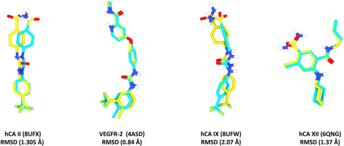
Validation of the docking protocol by re‐docking the co‐crystallized ligands into the active sites of hCA II (PDB: 8UFX), VEGFR‐2 (PDB: 4ASD), hCA IX‐mimic (PDB: 8UFW), and hCA XII (PDB: 6QNG).
Results and Discussion
Chemistry
The synthetic route to the target sulfonamide derivatives4a–iis outlined in Scheme1. The initial step involved the preparation of a series of Schiff bases from the condensation of appropriately substituted aldehydes with primary amines. Subsequent reduction of the Schiff bases with sodium borohydride (NaBH4) in methanol at 0°C for 5 h afforded the corresponding secondary amines2a–iin good yields. The successful reduction was confirmed by1H and13C NMR spectroscopy. In the1H NMR spectra of the reduced intermediates, a diagnostic signal attributable to theN–CH2methylene protons was observed in the aliphatic region (δ 3.72–4.48 ppm), whereas the characteristic imine singlet (δ 8.00–9.00 ppm) was conspicuously absent, thereby confirming the complete reduction of the azomethine bond. The secondary amines2a–iwas subsequently subjected toN‐sulfonylation with methanesulfonyl chloride in the presence of sodium carbonate as a base, employing a THF/H2O biphasic solvent system at ambient temperature for 24–48 h. This mild reaction protocol furnished the desired sulfonamide derivatives4a–iin satisfactory yields.
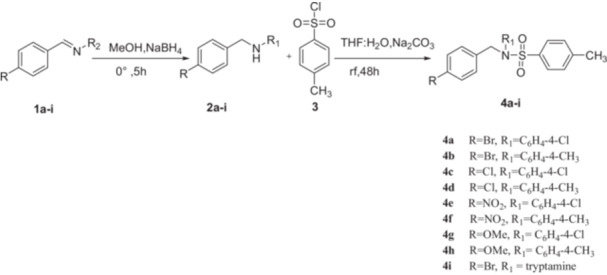
General scheme for the synthesis of sulfonamide (4a–i).
The structures of all target compounds were rigorously established through a combination of FT‐IR,1H NMR,13C NMR, and HRMS analyses. The FT‐IR spectra of the final products exhibited characteristic asymmetric and symmetric S = O stretching vibrations in the 1342–1380 cm−1region, unequivocally confirming the successful incorporation of the sulfonamide functionality. In the representative1H NMR spectrum of compound4b, a diagnostic singlet at δ 2.30 ppm was assigned to the N–SO2CH3methyl protons, while the aromatic protons resonated as doublets and doublets of doublets in the δ 6.69–7.52 ppm region. The13C NMR spectrum revealed aromatic carbon signals spanning δ 115.9–145.9 ppm, with the aliphatic region displaying characteristic resonances at δ 47.3 ppm (N–CH2) and δ 21.3 ppm (Ar–CH3). High‐resolution mass spectrometric analysis provided further structural confirmation; for instance, compound4bdisplayed a molecular ion peak at m/z 448.2414 [M]+, which was in excellent agreement with the calculated molecular formula. All spectroscopic data for the remaining compounds were fully consistent with the proposed structures.
Antioxidant Activity
The antioxidant potential of the synthesized sulfonamide derivatives4a–iwas evaluated by the 2,2‐diphenyl‐1‐picrylhydrazyl (DPPH) free radical scavenging assay, a widely employed method for assessing the hydrogen‐donating ability of potential antioxidant, Butylated hydroxytoluene (BHT) was employed as the reference standard [14,15], and the results are summarized in Table1.
Table: Antioxidant Activities of the sulfonamides derivatives, 4a–i.
Among the tested compounds,4dexhibited remarkably potent antioxidant activity across all concentrations examined. Notably, at 37.5 and 62.5 µg/mL, compound4ddisplayed superior radical scavenging capacity (67.20% and 72.56%, respectively) compared to BHT (65.32% and 69.82%, respectively). At the remaining concentrations, the antioxidant activity of4dremained closely comparable to that of BHT, further underscoring its promising antioxidant potential. Compounds4a,4b,4c,4e,4f,4g, and4hexhibited moderate antioxidant activity, particularly at higher concentrations, whereas compound4idemonstrated the lowest activity among all derivatives evaluated.
The observed structure–activity relationships reveal that the nature and position of substituents on the aromatic ring exert a profound influence on antioxidant efficacy. Compound4d, bearing one chlorine atom and two methyl groups, benefits from a synergistic electronic interplay: the electron‐withdrawing chlorine substituent contributes to the stabilization of the resultant radical species following hydrogen atom transfer, while the electron‐donating methyl groups enhance the electron density of the aromatic system, thereby facilitating radical scavenging. This favorable combination of electronic effects accounts for the superior antioxidant activity of4d. The remaining compounds, functionalized with nitro, methoxy, or bromo substituents, exhibited moderate antioxidant activity, which can be rationalized on the basis of the steric and electronic characteristics of these groups.
Cytotoxicity Studies
The in vitro cytotoxic potential of the sulfonamide derivatives4a–iwas assessed against two human cancer cell lines: A2780 (ovarian carcinoma) and LNCaP (lymph node carcinoma of the prostate) using the MTT colorimetric assay, with docetaxel serving as a positive control. Cells were incubated with the test compounds at concentrations ranging from 0.1 to 100 µg/mL for 24 h, and cell viability was determined. The log IC50values, derived from dose–response curves, are presented in Table2, while detailed cell viability data are provided in Tables3and4.
Table: Log IC50concentrations calculated for LNCaP, A2780 cell lines for the new sulfonamides derviatives (4a–i).
Table: A2780 Cell viability (%) results with compounds 4a–i.
Table: LNCaP Cell viability (%) results with compounds 4a–i.
As shown in Table2, compound4hdemonstrated the most potent cytotoxic activity against LNCaP cells, with a log IC50= 1.343, followed by compound4i(log IC50= 1.686). The remaining compounds displayed moderate cytotoxicity against LNCaP cells, with log IC50values ranging from 1.835 to 2.096. Against the A2780 cell line, compounds4g,4i, and4hexhibited the highest cytotoxic potency, with log IC50= 1.089, 1.164, and 1.585, respectively, while the remaining compounds showed moderate activity (log IC50range: 1.814–2.223).
The structure–activity relationship analysis suggests that the presence of the methoxy substituent in compounds4gand4hplays a critical role in enhancing cytotoxic activity. The methoxy group, acting as an electron‐donating substituent, increases the electron density of the aromatic ring, thereby potentially facilitating interactions with key biological targets. Furthermore, the enhanced lipophilicity conferred by the methoxy group may improve cellular membrane permeability, leading to greater intracellular accumulation of the active compounds [24].
The cell viability data for the A2780 cell line (Table3) revealed that compounds4c,4d, and4gexhibited statistically significant cytotoxic activity (p< 0.05) across all tested concentrations (0.1–100 µg/mL), indicating a broad effective dose range. The remaining derivatives displayed concentration‐dependent cytotoxicity, with significant effects predominantly observed at higher concentrations (10 and 100 µg/mL), with the exception of compound4f, which exhibited significant cytotoxicity only at 100 µg/mL.
For the LNCaP cell line (Table4), the sulfonamide derivatives generally exhibited significant cytotoxicity only at higher concentrations (10 and 100 µg/mL), suggesting a somewhat lower sensitivity of this cell line compared to A2780. Again, compound4fdemonstrated significant activity exclusively at 100 µg/mL. Collectively, these results indicate that the A2780 ovarian carcinoma cell line is generally more susceptible to the cytotoxic effects of the synthesized sulfonamide derivatives than the LNCaP prostate cancer cell line.
Molecular Docking
In order to rationalize the promising anticancer activity of the most potent sulfonamide derivatives (4g, 4h, and4i), molecular docking studies were performed against VEGFR‐2, hCA II, hCA IX‐mimic, and hCA XII. These targets were selected because they represent two complementary pathways that are highly relevant to tumor progression, namely angiogenesis and pH regulation. VEGFR‐2 is a key driver of tumor angiogenesis and remains one of the most validated molecular targets in anticancer therapy, while sulfonamide‐based derivatives have shown considerable promise as VEGFR‐2 inhibitors and, in some cases, as multitarget agents [25,26].
On the other hand, hCA IX and hCA XII are hypoxia‐associated tumor carbonic anhydrase isoforms that facilitate cancer cell adaptation to the acidic tumor microenvironment and are strongly implicated in invasion, metastasis, and treatment resistance [27,28].
hCA II was additionally included as a reference isoform to compare ligand accommodation within the conserved carbonic anhydrase active site and to provide an indication of possible isoform selectivity among the tested compounds. Thus, the selected docking panel was considered suitable for exploring whether the observed biological activity of the synthesized sulfonamides could be associated with interference in VEGFR‐2‐driven angiogenic signaling and/or inhibition of tumor‐associated carbonic anhydrases.
The docking scores of the selected sulfonamide derivatives against VEGFR‐2, hCA II, hCA IX, and hCA XII are presented in Table5. In the present docking set, the co‐crystallized ligands used for protocol validation were BAX for VEGFR‐2/4ASD, WJN for hCA II/8UFX, WJN for hCA IX‐mimic/8UFW, and benzenesulfonamide for hCA XII/6QNG. As shown in Table5, compounds4g–4idisplayed more favorable binding energies than the native ligand in hCA II, with values of −8.83, −8.94, and −9.44 kcal/mol, respectively, compared with −7.95 kcal/mol for the native ligand, suggesting a particularly strong affinity of these derivatives toward this isoform. Against hCA XII, compound4ialso exhibited the most favorable score (−9.22 kcal/mol), surpassing the native ligand (−8.75 kcal/mol), whereas4gand4hshowed slightly weaker but still comparable affinities (−8.15 and −8.17 kcal/mol). In the case of hCA IX, compound4iagain emerged as the best derivative (−8.47 kcal/mol), with a value very close to that of the native ligand (−8.53 kcal/mol), while4gand4hshowed only slightly lower binding affinities (−8.09 and −8.17 kcal/mol). By contrast, all tested compounds were markedly less favored in VEGFR‐2 than the co‐crystallized ligand, giving docking scores between −7.46 and −7.62 kcal/mol versus −12.03 kcal/mol for the native ligand. Overall, these findings indicate that the anticancer‐active sulfonamide derivatives, particularly compound4i, are more likely to exert their activity through preferential interaction with carbonic anhydrase isoforms, especially hCA II and hCA XII, rather than through strong VEGFR‐2 binding under the present docking conditions.
Table: Docking scores of the native ligands and the most active sulfonamide derivatives (4g–4i) against hCA II, VEGFR‐2, hCA IX‐mimic, and hCA XII, expressed as binding energy values (kcal/mol).
To further elucidate the binding modes of the most active sulfonamide derivatives toward the studied enzymes, their interaction profiles were compared with those of the corresponding co‐crystallized ligands. For hCA II (Figure2), the three docked compounds retained several of the key contacts observed for the native ligand, which supports a similar accommodation within the active site. The most consistent shared interactions involved Phe131, Leu198, and Val121, indicating preservation of the same hydrophobic recognition region. In addition, His94 and/or His96 were also maintained by the docked compounds, suggesting that they occupy the catalytic pocket in a manner comparable to the native ligand. Among the tested derivatives,4hshowed the closest overlap with the native ligand, as it preserved the largest number of common contacts, including His94, His96, His119, Thr199, Phe131, Leu198, Val121, and Trp209. These interaction pattern indicates that compounds4g–4ibind hCA II through the same principal anchoring residues involved in native‐ligand recognition.
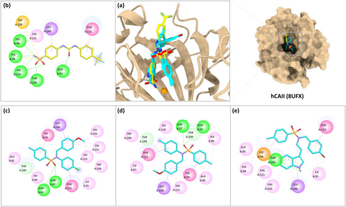
Binding mode analysis of the most active sulfonamide derivatives within the hCA II active site. (a) Superposition of the docked poses of compounds4g–4iwith the co‐crystallized ligand inside the binding pocket of hCA II (PDB: 8UFX). (b) Two‐dimensional interaction diagram of the native ligand. (c–e) Two‐dimensional interaction diagrams of compounds4g, 4h, and4i, respectively.
For VEGFR‐2 (Figure3), the docked compounds showed only limited overlap with the interaction pattern of the native ligand, in agreement with their lower docking scores. The main common contact retained by all three derivatives was Glu885, indicating that this residue represents the most conserved recognition point within the binding site. In addition, compounds4gand4halso shared interaction with Val898, while compound4ireproduced a broader part of the native binding environment through common contacts with Asp1046, Leu1019, and Glu885. Therefore, among the tested derivatives,4idisplayed the closest interaction profile to the co‐crystallized ligand, although the overall similarity remained lower than that observed for the carbonic anhydrase targets.
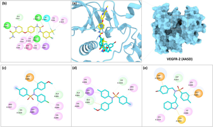
Binding mode analysis of the most active sulfonamide derivatives within the VEGFR‐2 active site. (a) Superposition of the docked poses of compounds4g–4iwith the co‐crystallized ligand inside the binding pocket of VEGFR‐2 (PDB: 4ASD). (b) Two‐dimensional interaction diagram of the native ligand. (c–e) Two‐dimensional interaction diagrams of compounds4g, 4h,and4i, respectively.
For hCA IX (Figure4), the docked sulfonamide derivatives reproduced several of the key contacts observed for the native ligand, supporting a comparable accommodation within the active site. The main common interactions were those with His64, His94, Val121, Leu198, Val143, and Trp209. Compound4gretained all of these shared contacts, while4hand4ialso showed a close similarity to the native ligand by preserving the same principal interaction network within the binding pocket. Overall, the three compounds displayed a broadly similar binding pattern to that of the co‐crystallized ligand, indicating that they are well accommodated in the hCA IX active site through the same main anchoring residues.
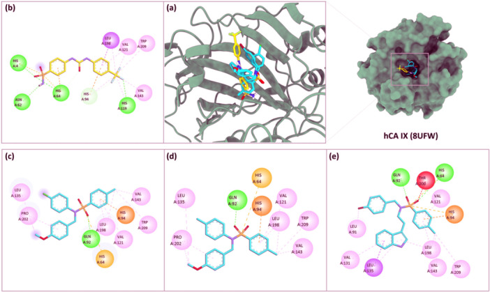
Binding mode analysis of the most active sulfonamide derivatives within the hCA IX active site. (a) Superposition of the docked poses of compounds4g–4iwith the co‐crystallized ligand inside the binding pocket of hCA IX (PDB: 8UFW). (b) Two‐dimensional interaction diagram of the native ligand. (c–e) Two‐dimensional interaction diagrams of compounds4g, 4h,and4i, respectively.
Finally, for hCA XII (Figure5), the docked sulfonamide derivatives retained several of the key contacts observed for the native ligand, indicating a closely related binding mode within the active site. The most consistently shared interactions involved Gln92, His94, His96, Leu198, Val121, and Ala131, which were preserved by the three tested compounds. In addition,4gand4halso maintained common contacts with His119 and Trp209, whereas4ireproduced the native binding pattern through shared interactions with Thr199 in addition to the conserved residues. Overall, the interaction profiles indicate that compounds4g–4ioccupy the hCA XII binding pocket through the same principal anchoring residues involved in recognition of the co‐crystallized ligand.
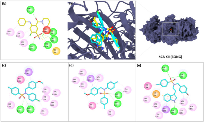
Binding mode analysis of the most active sulfonamide derivatives within the hCA XII active site. (a) Superposition of the docked poses of compounds4g–4iwith the co‐crystallized ligand inside the binding pocket of hCA XII (PDB: 6QNG). (b) Two‐dimensional interaction diagram of the native ligand. (c–e) Two‐dimensional interaction diagrams of compounds4g, 4h,and4i, respectively.
Collectively, the docking results support a clearer preference of the selected sulfonamide derivatives for the carbonic anhydrase binding sites over VEGFR‐2. This conclusion is supported by both the docking scores and the interaction analysis. In the carbonic anhydrase isoforms, the docked compounds reproduced a larger portion of the native binding pattern and showed better spatial accommodation within the catalytic cavity, consistent with the known compatibility of sulfonamide derivatives with carbonic anhydrase active sites. In addition, part of this stabilization appears to arise from directional polar contacts consistent with hydrogen‐bonding interactions, together with hydrophobic fitting within the pocket. By contrast, although some relevant contacts were observed in VEGFR‐2, the compounds showed weaker binding energies and a more limited overlap with the interaction network of the co‐crystallized ligand, indicating less efficient occupation of the kinase active site under the present docking conditions. Taken together, these findings suggest that carbonic anhydrases, particularly hCA II, hCA IX, and hCA XII, are more plausible molecular targets than VEGFR‐2 for the most active sulfonamide derivatives, with compound4ishowing the most favorable overall docking profile among the tested ligands.
Conclusion
In summary, a series of novel sulfonamide derivatives (4a–i) was successfully synthesized via a straightforward three‐step sequence involving Schiff base formation, sodium borohydride reduction, andN‐sulfonylation with methylphenylsulfonyl chloride. All target compounds were thoroughly characterized by FT‐IR,1H NMR,13C NMR, and HRMS analyses, which unequivocally confirmed their structures. Biological evaluation revealed that compound4dpossesses outstanding antioxidant activity, surpassing the reference standard BHT at specific concentrations, attributable to the synergistic electronic effects of its chloro and methyl substituents. Cytotoxicity was screening against A2780 and LNCaP cancer cell lines and compounds4g,4h, and4iwas identified as the most potent candidates, with4hexhibiting the lowest log IC50value (1.343) against LNCaP cells. The methoxy substituent was identified as a key structural determinant of enhanced cytotoxic activity, likely through increased lipophilicity and electron donation facilitating target engagement. These findings highlight the potential of sulfonamide‐based scaffolds as dual antioxidant–anticancer agents and provide a foundation for further structural optimization and mechanistic investigation. Docking studies revealed that sulfonamide derivatives4g,4h, and4iare potential inhibitor against VEGFR‐2, hCA II, hCA IX‐mimic, and hCA XII.
Author Contributions
Ouissal Bouraoui:investigation, writing – original draft.Güldeniz Şekerci:investigation.Khaled Mesbah:writing – review and editing.James A. Ezugwu:investigation, methodology, writing – review and editing.Rachid Benkiniouar:writing – review and editing.Suat Tekin:investigation, methodology.Fatümetüzzehra Küçükbay:investigation, methodology.Houssem Boulebd:data curation, software, methodology.Hasan Küçükbay:conceptualization, investigation, methodology, writing – review and editing, project administration, supervision.
Conflicts of Interest
The authors declare no conflicts of interest.
Acknowledgments
The authors thank TUBİTAK (The Scientific and Technological Research Council of Turkey) Project No: E‐21514107‐115.02‐612434/2221 Program 2024/1 and İnönü University, Turkey (BAPB‐ Grand No‐FBG‐ 2024‐3240). The supercomputing resources used in this work were supported by the HPC of UCI‐UFMC (Unité de Calcul Intesif of the University Fréres Mentouri Constantine 1).
Data Availability Statement
The data that supports the findings of this study are available in the supporting material of this article.
Associated Data
Data Availability Statement
The data that supports the findings of this study are available in the supporting material of this article.
References
- Rubab L., Afroz S., Ahmad S., et al., “An Update on Synthesis of Coumarin Sulfonamides as Enzyme Inhibitors and Anticancer Agents,” Molecules 27, no. 1604 (2022): 2–1631, 10.3390/molecules27051604. doi.org/10.3390/molecules27051604
- Scozzafava A., Owa T., Mastrolorenzo A., and Supuran C., “Anticancer and Antiviral Sulfonamides,” Current Medicinal Chemistry 10, no. 11 (2003): 925–953, 10.2174/0929867033457647. doi.org/10.2174/0929867033457647
- Khan F. A., Mushtaq S., Naz S., et al., “Sulfonamides as Potential Bioactive Scaffolds,” Current Organic Chemistry 22, no. 8 (2018): Ç 818–Ç 830, 10.2174/1385272822666180122153839. doi.org/10.2174/1385272822666180122153839
- Zerbib S., Cruz‐Martins N., and Bouissane L., “Exploring Sulfonamides,” Archiv der Pharmazie 359 (2026): e70180, 10.1002/ardp.70180. doi.org/10.1002/ardp.70180
- Buğday N., Küçükbay F. Z., Küçükbay H., et al., “Synthesis of Novel Dipeptide Sulfonamide Conjugates with Effective Carbonic Anhydrase I, II, IX, and XII Inhibitory Properties,” Bioorganic Chemistry 81, no. August (2018): 311–318, 10.1016/j.bioorg.2018.08.032. doi.org/10.1016/j.bioorg.2018.08.032
- Ezugwu J. A., Küçükbay F., Küçükbay H., Tatlıcı E., Touil N., and Yeşilada Ö., “Synthesis, Molecular Docking Study of Benzoylated Sulfonamide and Benzamide Conjugates of 7‐Chloroquinoline with Antioxidant and Antimicrobial Activity,” ChemSelect 11 (2026): e06242, 10.1002/slct.2025062421of12. doi.org/10.1002/slct.2025062421of12
- Tantawy A. H., Farag S. M., Abdel‐Haleem D. R., and Mohamed H. I., “Facile Synthesis, Larvicidal Activity, Biological Effects, and Molecular Docking of Sulfonamide‐Incorporating Quaternary Ammonium Iodides as Acetylcholinesterase Inhibitors Against Culex Pipiens L,” Bioorganic Chemistry 128, no. July (2022): 106098, 10.1016/j.bioorg.2022.106098. doi.org/10.1016/j.bioorg.2022.106098
- Qu B., Xu Y., Lu Y., et al., “Design, Synthesis and Biological Evaluation of Sulfonamides Inhibitors of XPO1 Displaying Activity Against Multiple Myeloma Cells,” European Journal of Medicinal Chemistry 235 (2022): 114257, 10.1016/j.ejmech.2022.114257. doi.org/10.1016/j.ejmech.2022.114257
- Boussaker M., Berredjem M., Grib I., et al., “In Silico Study, X‐Ray Characterization, Antimicrobial and Antioxidant Activities of Sulfonylphthalimide: Interaction with Dihydropteroate Synthase (DHPS),” Journal of Molecular Structure 1327, no. December 2024 (2025): 141111, 10.1016/j.molstruc.2024.141111. doi.org/10.1016/j.molstruc.2024.141111
- Mohamed N. M., El Rabeeb S. I., El Deeb M. A., Mahfoz A. M., and Abdulrahman F. G., “Design, Synthesis and Anti‐Tumor Evaluation of Novel Thiohydantoin Congeners as Androgen Receptor Antagonists with In Vivo Study,” Journal of Molecular Structure 1312, no. P2 (2024): 138565, 10.1016/j.molstruc.2024.138565. doi.org/10.1016/j.molstruc.2024.138565
- Mishra V. R., Ghanavatkar C. W., Mali S. N., Chaudhari H. K., and Sekar N., “Schiff Base Clubbed Benzothiazole: Synthesis, Potent Antimicrobial and MCF‐7 Anticancer Activity, DNA Cleavage and Computational Study,” Journal of Biomolecular Structure & Dynamics 38, no. 6 (2020): 1772–1785, 10.1080/07391102.2019.1621213. doi.org/10.1080/07391102.2019.1621213
- Tarasiuk T. M., Volovnenko T. A., Volovenko Y. M., Medviediev V. V., and Shishkin O. V., “Synthesis of N ‐Carboxyalkyl‐1, 4‐ Benzothiazepine‐3 (2 H) ‐One Derivatives Using Esters of N‐(2‐Chloro‐5‐Nitrobenzyl) Amino Acids,” Monatshefte für Chemie—Chemical Monthly 145 (2014): 483–489, 10.1007/s00706-013-1133-1. doi.org/10.1007/s00706-013-1133-1
- Peerzada M. N., Khan P., Ahmad K., Hassan M. I., and Azam A., “European Journal of Medicinal Chemistry Synthesis, Characterization and Biological Evaluation of Tertiary Sulfonamide Derivatives of Pyridyl‐Indole Based Heteroaryl Chalcone as Potential Carbonic Anhydrase IX Inhibitors and Anticancer Agents,” European Journal of Medicinal Chemistry 155 (2018): 13–23, 10.1016/j.ejmech.2018.05.034. doi.org/10.1016/j.ejmech.2018.05.034
- Yang J., Guo J., and Yuan J., “In Vitro Antioxidant Properties of Rutin,” LWT—Food Science and Technology 41, no. 6 (2008): 1060–1066, 10.1016/j.lwt.2007.06.010. doi.org/10.1016/j.lwt.2007.06.010
- Küçükbay H., Gönül Z., Küçükbay F. Z., et al., “Synthesis of New 7‐Amino‐3,4‐dihydroquinolin‐2(1H)‐one‐peptide Derivatives and Their Carbonic Anhydrase Enzyme Inhibition, Antioxidant, and Cytotoxic Activities,” Archiv der Pharmazie 354, no. 11 (2021): 1–13, 10.1002/ardp.202100122. doi.org/10.1002/ardp.202100122
- Doğan H., Bahar M. R., Çalışkan E., et al., “Synthesis and Spectroscopic Characterizations of Hexakis[(1‐(4′‐oxyphenyl)−3‐(substituted‐phenyl)prop‐2‐en‐1‐one)]cyclotriphosphazenes: Theirin Vitrocytotoxic Activity, Theoretical Analysis and Molecular Docking Studies,” Journal of Biomolecular Structure and Dynamics 40, no. 7 (2022): 3258–3272, 10.1080/07391102.2020.1846621. doi.org/10.1080/07391102.2020.1846621
- Küçükbay H., Mumcu A., Tekin S., and Sandal S., “Synthesis and Evaluation of Novel N,N'‐Disubstituted Benzimidazolium Bromides Salts as Antitumor Agents,” Turkish Journal of Chemistry 40, no. 3 (2016): 393–401, 10.3906/kim-1510-15. doi.org/10.3906/kim-1510-15
- Özen F., Tekin S., Koran K., Sandal S., and Görgülü A. O., “Synthesis, Structural Characterization, and in Vitro Anti‐Cancer Activities of New Phenylacrylonitrile Derivatives,” Applied Biological Chemistry 59, no. 2 (2016): 239–248, 10.1007/s13765-016-0163-x. doi.org/10.1007/s13765-016-0163-x
- Koran K., Tekin Ç., Çalışkan E., Tekin S., Sandal S., and Görgülü A. O., “Synthesis, Structural and Thermal Characterizations and in Vitro Cytotoxic Activities of New Cyclotriphosphazene Derivatives,” Phosphorus, Sulfur and Silicon and the Related Elements 192, no. 9 (2017): 1002–1011, 10.1080/10426507.2017.1315420. doi.org/10.1080/10426507.2017.1315420
- Koran K., Çalıs E., Çapan D.A., Tekin I., and Oztürk S., “The First Peptide Derivatives of Dioxybiphenyl‐Bridged Spiro Cyclotriphosphazenes: In Vitro Cytotoxicity Activities and DNA Damage Studies,” Bioorganic Chemistry 132, no. October 2022 (2023): 239–248, 10.1016/j.bioorg.2022.106338. doi.org/10.1016/j.bioorg.2022.106338
- Morris G. M., Huey R., Lindstrom W., et al., “AutoDock4 and AutoDockTools4: Automated Docking with Selective Receptor Flexibility,” Journal of Computational Chemistry 30, no. 16 (2009): 2785–2791, 10.1002/jcc.21256. doi.org/10.1002/jcc.21256
- Trott O. and Olson A. J., “AutoDock Vina: Improving the Speed and Accuracy of Docking with a New Scoring Function, Efficient Optimization, and Multithreading,” Journal of Computational Chemistry 31, no. 2 (2010): 455–461, 10.1002/jcc.21334. doi.org/10.1002/jcc.21334
- Pettersen E. F., Goddard T. D., Huang C. C., et al., “UCSF Chimera—A Visualization System for Exploratory Research and Analysis,” Journal of Computational Chemistry 25, no. 13 (2004): 1605–1612, 10.1002/jcc.20084. doi.org/10.1002/jcc.20084
- Dastyafteh N., Negahdaripour M., Sayahi M. H., et al., “Design, Synthesis, Biological Evaluation, and in,” RSC Advances 14 (2024): 35323–35335, 10.1039/d4ra04492d. doi.org/10.1039/d4ra04492d
- Charissopoulos E. and Pontiki E., “Sulfonamides a Promising Hit for Cancer Therapy Through VEGFR‐2 Inhibition,” Biomedicines 13, no. 4 (2025): 772, 10.3390/biomedicines13040772. doi.org/10.3390/biomedicines13040772
- Shaldam M. A., Hadia, Angeli Andrea, Badi Rehab Mustafa, et al., “Sulfonamides a Promising Hit for Cancer Therapy Through VEGFR‐2 Inhibition,” Journal of Enzyme Inhibition and Medicinal Chemistry 38, no. 1 (2023): 2203389, 10.1080/14756366.2023.2203389. doi.org/10.1080/14756366.2023.2203389
- McDonald P. C., Chia S., Bedard P. L., et al., “A Phase 1 Study of SLC‐0111, a Novel Inhibitor of Carbonic Anhydrase IX, in Patients with Advanced Solid Tumors,” American Journal of Clinical Oncology 43, no. 7 (2020): 484–490, 10.1097/COC.0000000000000691. doi.org/10.1097/COC.0000000000000691
- Gazzaroli G., Tavani C., Filiberti S., et al., “An Ureido‐Substituted Benzenesulfonamide Carbonic Anhydrase Inhibitor Exerts a Potent Antitumor Effect In Vitro and In Vivo,” Experimental Hematology & Oncology 14, no. 104 (2025): 1–5, 10.1186/s40164-025-00690-z. doi.org/10.1186/s40164-025-00690-z
Republished from the open web under CC-BY. Authors: Bouraoui O, Şekerci G, Mesbah K, Ezugwu JA, Benkiniouar R, Tekin S, Küçükbay F, Boulebd H, Küçükbay H. Read the original.