Systematic Review of Intravenous Ceftriaxone Administration in Animals.
Background Ceftriaxone is an antibiotic frequently prescribed in veterinary medical practice due to its effectiveness against several pathogenic microorganisms responsible for diseases in different animal species. Objective To select scientific articles from the academic databases PubMed and Web of Science that analysed the pharmacokinetic aspects of intravenously administered ceftriaxone in animals. Methods In accordance with the principles of scientific transparency, the systematic review protocol and all research stages were previously registered on the Open Science Framework (OSF) platform. The scientific articles that met the inclusion criteria were subjected to a checklist comprising 24 minimum assessment items designed for reporting pharmacokinetic studies. Results A total of 52 pharmacokinetic profiles were identified from the 29 selected scientific articles, including records involving 13 different animal species used in ceftriaxone experimental studies. Conclusion The limitations and inconsistencies identified in this review highlight the importance of detailed reporting of pharmacokinetic data derived from experimental trials in animals.
Introduction
Ceftriaxone is listed by the World Health Organization as one of the essential medicines recommended for the treatment of infections caused by microorganisms prone to resistance against other antibiotics (World Health Organization2023). From a pharmacodynamic perspective, ceftriaxone exerts its action by binding to bacterial cell wall proteins, thereby disrupting the third and final stage of the bacterial cell wall (Mora‐Ochomogo and Lohans2021). Its effectiveness stems from its broad‐spectrum activity against gram‐positive bacteria (Steptococcussspp. andStaphylococcusspp.) (Nazir et al.2025), gram‐negative bacteria (Escherichia coli,Proteusspp.,Klebsiellaspp.,Enterobacterspp.,Salmonellaspp. andPasteurellaspp.) (Rodríguez‐Baño et al.2018; Tamma et al.2023) and certain anaerobic bacteria (Bacteroides fragilis,Peptostreptococcusspp., Clostridium spp.) (Ligero‐López et al.2023).
Experimental evidence has demonstrated that ceftriaxone promotes a reduction in inflammation and airway resistance in septic rats, although it is associated with pharmacokinetic alterations characterized by decreased bioavailability and increased clearance (Simões et al.2024). Additionally, the investigation of the kinetic disposition and efficacy of ceftriaxone in cows with staphylococcal mastitis allowed the identification of therapeutic concentrations of the drug and its circulating metabolite, which were associated with the elimination ofStaphylococcus aureusand the clinical resolution of the disease (Buragohain et al.2021). However, the pharmacokinetic evaluation of ceftriaxone and meropenem in dogs indicated efficacy against susceptible bacteria, with meropenem demonstrating superior effectiveness compared to ceftriaxone (Orooba and Shwaish2024).
The pharmacokinetics of ceftriaxone provide crucial information regarding its disposition in the animal body, encompassing routes of administration, distribution, metabolism, excretion and potential toxicity (Orooba and Shwaish2024). Among the available administration routes, intravenous injection is recognized for achieving high therapeutic efficacy and extensive distribution across most tissues and body fluids in animals (Al Nasr et al.2025). Additionally, the affinity of ceftriaxone for plasma protein binding indicates that higher binding reduces the rate of drug transfer between plasma and extravascular compartments, contributing to its prolonged elimination half‐life (Ahmed et al.2025). Moreover, the free fraction of the drug requires sufficiently high systemic concentrations to attain the desired therapeutic effect (Gonzalez et al.2013).
Accordingly, the administration of ceftriaxone demands a rational, scientifically grounded approach to dosing regimens across different species, as improper management may result in toxicity or therapeutic failure (Poapolathep et al.2020). Therefore, the objective of this systematic review was to identify and analyse scientific articles from academic databases that reported pharmacokinetic (PK) parameters of intravenously administered ceftriaxone in animals.
Materials and Methods
Investigation Method
The first methodological procedure consists of identifying articles available on digital scientific publication platforms, such as PubMed and Web of Science. The search descriptors used in English were pharmacokinetics, ceftriaxone and animal, combined using the operators “AND” and “All Field” (Table1).
Table: Complete search plan in the digital platform databases.
During this exploration phase, platform‐specific filtering tools, such as publication date or type, were not applied. In line with scientific transparency principles, the protocols for this systematic review were preregistered on the Open Science Framework (OSF) and are available at: osf.io/bsn74.
Selection Criteria
The selection of scientific articles from PubMed and Web of Science included in this systematic review followed established methodological criteria (Okoli and Duarte2019). Based on this approach, the titles and abstracts retrieved from the databases were initially screened for eligibility. To enhance efficiency, the virtual platform Rayyan was used to manage and coordinate study selection, allowing collaborative review among team members.
Screening was conducted independently and first by two reviewers, while a third reviewer was consulted in case of discrepancies. This phase was developed by applying exclusion criteria that omitted articles written in non‐Roman scripts, languages other than English or Portuguese, studies on the pharmacological associations of ceftriaxone, literature reviews, non‐intravenous administration routes and studies that did not provide comprehensible pharmacokinetic (PK) parameters of ceftriaxone obtained from animal experiments.
Following initial screening, the remaining articles were fully assessed to select studies reporting the pharmacokinetics of intravenously administered ceftriaxone in healthy and diseased animals, irrespective of sex. Selected studies were required to report at least key PK parameters, including elimination half‐life, elimination rate constant, clearance, area under the curve (AUC) and plasma free fraction or concentration. All selection steps were documented in a PRISMA 2020 flowchart (Galvão et al.2015) to ensure full traceability of decisions throughout the review.
Analysis of the Integrity of Selected Scientific Articles
The scientific articles that met the inclusion criteria of this review were evaluated using the ClinPK questionnaire (Kanji et al.2015), which consists of a 24‐item checklist designed to verify the minimum essential criteria for conducting pharmacokinetic studies, in line with PRISMA guidelines for systematic reviews. During the application of this method, items 12, 14, 20 and 21 were disregarded, as they were not applicable to animal study designs.
The presentation of results from the checklist was systematized using an adaptation of the Evidence Gap Maps (EGM) methodology (Campbell et al.2023). The procedure enhanced transparency and reliability by identifying reporting gaps, classifying studies by checklist adherence (100%, 50%, 0%), and ensuring clear and consistent analysis of ceftriaxone data across animal species.
Systematic Assessment of Pharmacokinetic Parameters
The data extraction procedure comprised the identification of PK parameters reported in the studies included in the review, followed by verification of the availability of corresponding mean values and standard deviation (SD) indicators associated with these parameters. The standardization of measurement units for ceftriaxone PK data was conducted using unit conversion established in the literature (Ogden and Fluharty2015; Ansel et al.2016; Bauer2014; Maradiya et al.2010), ensuring consistency with the intrinsic mathematical relationships governing each extracted PK parameter.
Among the mean values and corresponding SDs of the ceftriaxone PK parameter reported in the selected articles, the following were included in this review: AUC from zero to the last observation (AUC0‐t), AUC from zero to infinity (AUC0‐∞), clearance (Cl), elimination rate constant (Kel or β), free fraction (FU), elimination half‐life (T1/2) and volume of distribution (Vd). Furthermore, compartmental modelling data reported in the studies were organized according to the number of compartments, parameter type and animal species investigated.
The final methodological step for PK parameter extraction consisted of systematically compiling an electronic spreadsheet, which included additional information on the selected experimental trials, such as administered doses (mg kg−1), animal species, sex (female or male), age (years), weight (kg), number of animals and pharmacokinetic profile. Additionally, this information verified whether the studies monitored ceftriaxone administration alone or in combination with another drug, as well as the use of different doses and the clinical status of the animals. Thus, the systematization of parameters considering these informative elements was implemented to support the interpretative analysis of the experimental design of ceftriaxone administration.
Results and Discussion
Investigation and Selection
The investigation conducted in the databases resulted in a total of 398 scientific articles, of which 271 studies were registered in PubMed and 127 studies in Web of Science. The subsequent phase consisted of the initial screening of 330 articles using the digital platform Rayyan, leading to the identification of 55 studies that met the selection criteria established at this screening stage. Ultimately, the full‐text evaluation performed on 51 articles accessible in the databases identified 29 studies that adequately fulfilled all previously defined selection criteria (Figure1).
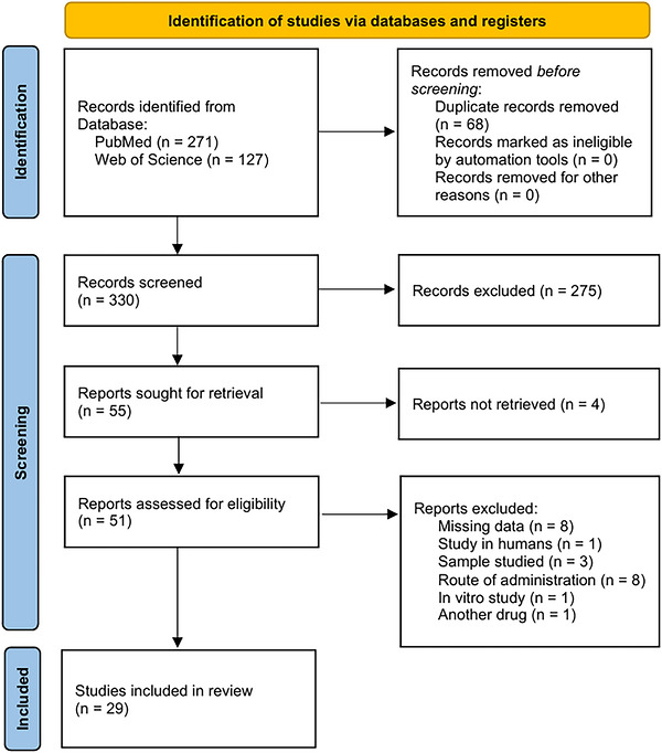
Flowchart of the systematic review of ceftriaxone.
Considering the commitment to accuracy of the results obtained during the stages of applying the selection criteria to the scientific articles identified in the previously established platforms, it is important to note that the incorporation of the digital tool Rayyan was decisive for the identification and removal of 68 duplicate records, followed by the elimination of a total of 275 publications during the initial screening that met the exclusion criteria. In the final screening stage, four studies were found to have no full‐text access available, while full‐text assessment resulted in the exclusion of 22 articles for not meeting the eligibility criteria, yielding a total of 29 studies included in this review on the pharmacokinetics of intravenously administered ceftriaxone in animals.
Study Characteristics
The systematic survey of information provided by the selected articles contributed to the identification of 52 pharmacokinetic profiles from 13 different animal species that received ceftriaxone in their experiments (Table2). A detailed evaluation of these studies revealed 42 profiles (80.77%) from healthy animals, whereas the remaining 10 profiles (19.23%) were derived from animals with specific clinical conditions, such as prematurity, experimentally induced febrile state, meningitis, endometritis, hepatopathy and nephropathy in the species investigated. Regarding the temporal distribution of the selected articles, 6 studies were published in the 1980s, followed by 7 studies in the 1990s, and from the 2000s onwards, 16 studies were made available presenting PK data related to ceftriaxone administered in animals.
Table: Baseline information extracted from selected articles.
The compiled data demonstrated ceftriaxone pharmacokinetic parameters across several species, totalling 284 animals (Table3): 44 (15.49%) cattle, 11 (3.87%) buffaloes (Bubalus bubalis), 28 (9.86%) goats, 9 (3.17%) dogs, 5 (1.76%) camels (Camelus dromedarius), 16 (5.63%) horses, 46 (16.20%) white rabbits, 5 (1.76%) cats, 2 (0.70%) monkeys, 3 (1.06%) miniature pigs (Yucatan miniature pigs), 40 (14.08%) sheep, 70 (24.65%) rats and 5 (1.76%) green turtles (Chelonia mydas). Profile analysis of these animals revealed particularities among individuals of the same species, such as differences in body proportions between horses and ponies. Some studies also showed divergences regarding the breeds investigated, as observed in sheep (Akkaraman sheep;Merino sheep;Chhotanagpuri sheep) (Cetin et al.2021; Dhanani et al.2020; Ranjan et al.2011) and calves (Crossbred calves;Holstein calves;Israeli‐Friesian calves) (Corum et al.2019; Johal and Srivastava1999; Soback and Ziv1988), whereas other studies included mixed‐breed animals, specifically in cats, dogs and mares. Additionally, lineage diversity was observed in laboratory animals, including regional lineages in white rabbits (Australian original white rabbitsandNew Zealand white rabbits) (Kwon et al.1985; Lutsar et al.1997) and laboratory strains in rats (Sprague Dawley ratsandWistar rats) (Hakim et al.1989; Zhang et al.2019).
Table: Data extracted using standardized units.
Regarding the sex variable, the evaluated articles reported a predominance of male animals (n= 155) compared to females (n= 95). In this context, the study conducted by Dhanani et al. (2020) stood out for obtaining PK data of ceftriaxone through an experimental model using castrated males. The experiment conducted by Ismail (2005) presented results related to lactating goats to explore ceftriaxone concentrations in blood and its elimination in milk. Additionally, research by Rebuelto et al. (2002) adopted a comparative methodology between both sexes in the canine species.
Analysis of age data reported in 20 articles revealed significant variability, with only the studies conducted by Guerrini et al. (1985) and Goudah (2008) presenting animals of similar age (2–3 years). In some studies, terminology allowed inference of age range, particularly in calves (Maradiya et al.2010; Corum et al.2019; Johal and Srivastava1999; Soback and Ziv1988) and foals (Ringger et al.1998). However, some experimental models did not detail animal age, describing only adult (Hakim et al.1989) or mature animals (Rebuelto et al.2002), premature animals or those registered by days of life (Corum et al.2019).
With respect to body weight, discrepancies were identified in 27 analysed articles, while the experiments conducted by Kumar et al. (2010) and Soback and Ziv (1988) did not report the weight of animals used for PK parameter acquisition. Additionally, data demonstrated ceftriaxone administration both alone and in combination with other drugs, compared across 10 PK profiles, including experiments involving ketoprofen or tolfenamic acid (Cetin et al.2021), Danhong (Zhang et al.2019), itraconazole (Cavalier et al.1997) and caffeine (Kwon et al.1985; Kwon and Bourne1986). The identification of experimental protocols involving different dosing regimens within the same study, as well as the induction of distinct clinical conditions, broadened the understanding of ceftriaxone PK parameters across various scenarios.
The rigorous analysis developed during the methodological selection stage sought to prioritize studies aligned with the scope of this review, particularly the PK and PD properties of isolated ceftriaxone administered alternative routes of administration constituted the first group of works excluded during screening, such as intraperitoneal application in horses (Campos et al.2017) and inhalational administration in mice (Valiulin et al.2022). According to Torrent Rodríguez et al.2024(Ofokansi et al.2007), drug interactions represent a recurrent challenge in clinical practice, particularly due to their capacity to interfere with drug metabolism or treatment safety, so that appropriate management of compound coadministration must consider aspects such as the intensity and duration of these interactions. In terms of the therapeutic efficacy of ceftriaxone, scientific articles that addressed clinical scenarios unrelated to the treatment of infections had to be disregarded from the review.
Assessment of Qualitative Attributes
The interpretation of the information evaluated from the investigated article, considering the checklist guiding PK studies, demonstrated results consistent with methodological guidelines, such that they were suitable to be systematized in the EGM, which substantially contributed to the objective analysis of reported evidence (Figure2). In continuity with the previous stage, the scientific articles submitted to the ClinPK checklist were organized according to the findings evidenced in each analysed item, favouring a clear and consistent interpretation of the respective evidence contained in the studies.
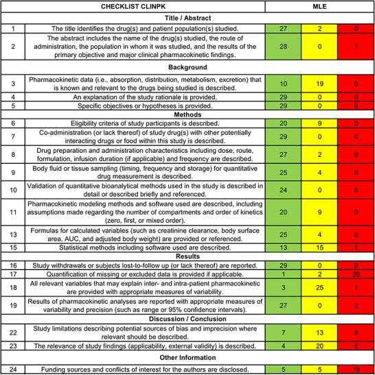
ClinPK analysis of the reviewed reports.
In accordance with the findings evidenced in the checklist of the articles included in the review, important information linked to specific questions was assessed, such as the understanding of the justifications and specific objectives of the studies (Questions 4 and 5), which aimed at the efficacy of ceftriaxone against microorganisms causing injuries in different animal species, as well as how this performance could be achieved through the perception of PK of the compound alone or under coadministration. Additionally, aspects related to the methodology addressed in the articles indicated that single or combined administration of ceftriaxone occurred in all studies (Question 7), and no potential interactions with food provided to the animals were identified, since compound administration was performed intravenously. Subsequently, the evaluation of animal eligibility criteria (Question 6) revealed substantial information for baseline data analysis; however, the findings identified 9 studies that demonstrated partial availability of information regarding the investigated species, particularly data concerning the number of animals, sex, age and weight.
The findings regarding the protocols for handling and administration of ceftriaxone (Question 8) were clearly delineated in the publications, except for the studies by Kovar et al. (1997) and Lutsar et al. (1997), which provided incomplete data due to the absence of specification of the ceftriaxone formulation administered. In addition, the description of blood collection and storage procedures (Question 9) indicates limitations in 4 studies, particularly regarding the detailing of the preservation techniques and the frequency at which collections were performed, whether daily or weekly. Meanwhile, findings related to PK modelling and analysis software (Question 11) revealed omissions of information in 9 studies, including the research by Cavalier et al. (1997) and Ringger et al. (1996), which showed the absence of a description regarding kinetic order and the number of compartments, a condition that may be attributed to the fact that such data were not recurrent requirements among scientific journals at the time. Subsequently, the results concerning mathematical procedures (Question 13) demonstrated that PK parameter modelling in the studies was conducted with samples limited to small groups of animals, thus promoting the adoption of mean and SD values of PK parameters without exploring sources of interindividual variability through covariates.
Regarding the evaluation of the results reported in the articles, it became evident that the PK variables explored (Question 18) indicated relevant contributions to this review, especially in studies reporting the influence of the animals’ clinical conditions on ceftriaxone parameters, such as the studies by Sar et al. (2008) and Ranjan et al. (2011). In addition, the analysis of precision measures (Question 19) found that only the studies by Kwon and Bourne (1986), Rebuelto et al. (2002) and Ringger et al. (1996) reported only mean values of certain PK parameters and presented an absence of SD.
Pharmacokinetic Parameters of Ceftriaxone in Animals
In alignment with the previously proposed methodological approach, the evaluation of the extracted parameters demonstrated consistency among the included studies regarding the adoption of dosing protocols for intravenously administered ceftriaxone. Among the observed findings, 19 studies implemented similar dosage regimens, such as 10 mg kg−1(27.59%), 25 mg kg−1(10.34%), 50 mg kg−1(17.54%) and 100 mg kg−1(10.34%). The study by Lutsar et al. (1997), however, investigated high doses of the compound in guinea pigs (150, 200, 300, 400 mg kg−1), aiming to determine the appropriate therapeutic regimen for the treatment of pneumococcal meningitis induced byS. pneumoniae.
Continuing the evaluation of the protocols recorded in the articles selected for this review, the studies by Cetin et al. (2021) and Dhanani et al. (2020) presented data related to isolated ceftriaxone at a dose of 40 mg kg−1, compared with coadministration with 3 mg kg−1of ketoprofen, 2 mg kg−1of tolfenamic acid (88 mg) and 15 mg kg−1of ertapenem (645.75 mg). Meanwhile, the study by Cavalier et al. (1997) investigated a 36‐day protocol consisting of oral administration of isolated ceftriaxone (1 g) at 12‐h intervals for a period of 5 days, a consecutive 10‐day interval without providing medications to the animals, and finally a bolus administration of ceftriaxone (1 g). The studies by Kwon et al. (1985) and Kwon and Bourne (1986) reported results referring to doses of the single compound (30 and 100 mg kg−1) and in association with caffeine (5, 10 and 20 mg kg−1). In contrast, Zhang et al. (2019) reported results referring to the isolated dose of ceftriaxone (90 mg kg−1day−1), prepared in 0.9% saline solution, as well as its association with different volumes of Danhong (2.19 mL; 15.33 and 30.66 mL), administered at three distinct time windows in aged rats.
In accordance with the previously processed results, the availability of PK parameters of interest across 52 PK profiles in the articles presented substantial data, although in certain evaluated studies, it proved to be limited, particularly regarding values associated with the parameters AUC0‐t(23 articles), AUC0‐∞(5 articles), Cl (Lutsar et al.1997), Kel or β (11 articles), FU (21 articles), T1/2(Dhanani et al.2020), Vd(3 articles) and α (18 articles). Within this same context, limitations were observed concerning studies that documented SD values of PK parameters, with only mean values identified in some reports, as in the findings described by Kwon and Bourne (1986), Ringger et al. (1996) and Rebuelto et al. (2002).
The PK characteristics of ceftriaxone enabled the development of studies in different compartmental models, and the findings identified 2 studies using a one‐compartment model, 13 studies using a two‐compartment model, and 3 studies using a three‐compartment model (Table4). Although this evaluation of the articles included such variations in experimental design, it was observed that 6 studies employed noncompartmental models, while 4 studies did not report the modelling structure. In the study conducted by Kwon and Bourne (1986), estimates compatible with a multicompartmental model were described, although the exact number of compartments addressed was not detailed. On the other hand, the study provided a physiologically based schematic representation, highlighting the trajectories of elimination constants and the transfer of ceftriaxone from plasma to peripheral tissue compartments of each organ weighted in the experiment.
Table: Systematization of the compartmental models from the articles selected.
Following the aspects discussed, experiments that addressed multicompartmental models exhibited limitations regarding the availability of parameters referring to the volume of distribution of ceftriaxone for the central compartment (V1) and the peripheral compartment (V2). Although the findings revealed that 7 studies recorded values related to the V1parameter, the results for V2were limited to the articles, which reported the following: 1.3516 ± 0.124 in dogs and 0.11424 ± 0.2184 in monkeys (Matsui et al.1984), 0.4205 ± 0.3074 for isolated ceftriaxone, 0.0899 ± 0.0232 for ceftriaxone coadministration with caffeine (5 mg) and 0.1044 ± 0.0203 for ceftriaxone coadministration with caffeine (10 mg) (Kwon et al.1985).
The physicochemical factors inherent to the behaviour of antimicrobials, with emphasis on cephalosporins, combined with the biological characteristics of patients, play a determining role in modulating the ADMET processes (absorption, distribution, metabolism, excretion and toxicity) of the compound (Kauss et al.2019; Kar et al.2025). In light of this theoretical framework, the data synthesized in this review demonstrated pharmacokinetic aspects of intravenous administration of ceftriaxone that differed significantly among the species investigated in the selected articles (Table3).
In the conceptual exposition by Curry and Whelpton2022(Torrent Rodríguez et al.2024), the parameters AUC0‐tor AUC0‐∞are recognized as assuming a central role in the interpretation of systematic kinetics, as they constitute a metric capable of quantifying drug entry and elimination, with applicability in evaluating patient exposure to the compound of interest. From this perspective, the synthesis of the articles included in this review revealed that fever induced in calves reflected lower AUC0‐∞values (25.2 ± 1.97 µg h mL−1), as reported by Dardi et al. (2005), whereas higher values (32.63 ± 1.47 µg h mL−1) of this parameter were described by Johal and Srivastava (1999) in healthy calves, despite certain similarities in the pharmacokinetic design of both studies, particularly the three‐compartment modelling. Although different cephalosporins and species were administered, the study by Sagar et al.2021(Alikhani et al.2025) demonstrated that fever induction cannot promote significant alterations in the pharmacokinetic parameters of cefquinome, a third‐generation cephalosporin administered intravenously in goats. Meanwhile, the research by Kwon et al. (1985) associated with the increase in AUC0‐∞(949 ± 229 µg h mL−1) of ceftriaxone in rabbits with concomitant administration of caffeine, an interpretation later discussed by Woziwodzka et al.2022(Taverne et al.2016) in the context of the potential adjuvant role of caffeine for third‐generation cephalosporins and its influence on rapid drug elimination. However, the results presented in the study by Kwon and Bourne (1986) revealed a significant reduction in Cmax(36.6%) and AUC (54.5%) of ceftriaxone in the rat brain due to cerebral hemodynamic alterations attributed to caffeine. Complementarily, compiled results referring to AUC0‐tdemonstrated wide variations according to the clinical condition of the animals, reaching minimum values (1.393 ± 273 µg h mL−1) in rats with meningitis (Lutsar et al.1997) and maximum values (570,000 ± 144,000 µg h mL−1) in healthy rats (Hakim et al.1989). Nevertheless, the study by Hertzsch and Richter2022(Curry and Whelpton2022) demonstrated greater ceftriaxone exposure in the cerebrospinal fluid (CSF) of dogs with meningitis, reflected by a higher CSF AUC/plasma AUC ratio (0.224) in neurologically compromised dogs, contrasting with the low ratio value (0.01) recorded in healthy dogs.
The combined analysis of ceftriaxone Vdvalues extracted from the studies included in this review demonstrated that the lowest value of the parameter (0.096 ± 0.012 L kg−1) was recorded in rats (Hakim et al.1989), whereas the highest Vdvalue (1.55 ± 0.52 L kg−1) was associated with cows (Kumar et al.2010). As reported in the literature and exploratory research, physiological factors associated with animal body size may influence drug distribution kinetics, presenting an allometric relationship with blood flow and compound circulation time, culminating in faster distribution rates in smaller species (Sagar et al.2021; Woziwodzka et al.2022; Hertzsch and Richter2022). In addition, the study by Kumar et al. (2010) indicated that inflammatory conditions may influence drug disposition, as elevated Vdvalues of ceftriaxone were recorded. Properties of ceftriaxone are relevant in limiting compound distribution through lipophilic tissues due to its high polarity, elevated molecular weight and strong plasma protein binding within the vascular compartment (Li et al.2015; Odival Cezar2011).
Within the discussions raised by Ahmed et al.2025(Sharma and McNeill2009), the impact of analysing plasma protein binding for β‐lactam antibiotics was emphasized, as this parameter has proven to be less predictable and highly variable among species. Although the availability of this information was limited in the articles included in the review, synthesis of the results demonstrated relevant differences among the evaluated models. It was verified that the lowest percentages of minimum FU of ceftriaxone (5.6%–32.8%) were associated with a study conducted in rats (Kwon and Bourne1986), whereas the highest values of this parameter (80%–71%) were reported in turtles (Mapongpeng et al.2019). In parallel, exploratory research has already reported broad variability of ceftriaxone FU in different animal species, encompassing percentages of 20%–30% in cattle (Johal and Srivastava1999; Soback and Ziv1988), 25% in dogs (Santos et al.2006), 39%–45% in goats (Ismail2005) and 84%–96% in rats and rabbits (Santos et al.2006; Wanat2020). Complementarily, the work by Ahmed et al. (2025) described the analytical relevance of the distinct interaction level of ceftriaxone with types of plasma proteins in animals, presenting a comparison between the intermediate affinity of the compound with α1‐acid glycoprotein (AAG) in bovine and rat plasma, common in infection and inflammatory states, with the high drug binding to plasma albumin in both animal models.
As explored in the studies by Ahmed et al.2022(Sharma and McNeill2009) and Di2021(Ahmed et al.2022), understanding plasma protein binding of antimicrobials is fundamental for interpreting compound disposition in patients, as it modulates central pharmacokinetic processes, directly reflecting on parameters such as T1/2, Kel (or β) and Cl. From this perspective, the T1/2data recorded in the selected scientific articles indicated differences between mammals and reptiles, requiring particular attention during parameter evaluation. In this context, the study by Sar et al. (2008) reported minimum T1/2values (0.19 ± 0.002 h) in the experimental goat model, whereas the research by Mapongpeng et al. (2019) recorded prolonged T1/2(5.81 ± 0.7 h) in turtles. As expected, analysis of Kel results indicated inverse behaviour, with low values of 0.12 ± 0.02 h kg−1associated with turtle modelling (Mapongpeng et al.2019) and higher records of parameters (3.57 ± 0.04 h kg−1) in research conducted in goats (Sar et al.2008). In general, physiological characteristics specific to reptiles, especially the relationship between temperature and metabolic rate, influence the low transfer of body fluids between vascular and extravascular compartments and reduce the compound elimination rate in these species (Mapongpeng et al.2019; Popick et al.1987; Granero et al.1995).
In the final evaluation of the pharmacokinetic profiles of the reviewed studies, wide interspecies variability regarding Cl was identified, with minimum values of 0.0181 ± 0.0351 L h−1kg originating from the experimental design in monkeys (Matsui et al.1984), whereas maximum values of 1.41 ± 0.24 L h−1kg were observed in goats (Sar et al.2008). Additionally, the study by Li et al.2015(Sagar et al.2021) pointed out that in comparative pharmacokinetics, drug behaviour among different species encompasses a range of aspects to be considered, such as the impact of feeding habits, promoting low clearance values of an investigated drug in carnivores, intermediate results in omnivores, and elevated rates in herbivores. Finally, the research by Ahmed et al.2022(Sharma and McNeill2009) reinforced that extrapolation of pharmacokinetic results of antimicrobials from animal models remains limited, such as those related to compound elimination in monkeys, due to methodological restrictions associated with low sample representativeness, despite the analytical model approach incorporating body weight and other interspecies physiological covariates.
Conclusions
The present systematic review demonstrated that intravenously administered ceftriaxone exhibits a pharmacokinetic profile consistent with rapid systemic availability, wide distribution, and a relatively prolonged elimination half‐life across different animal species. However, parameters such as clearance, volume of distribution, and AUC showed high interspecies variability, influenced by physiological factors, clinical status, and coadministration of drugs. Additionally, high plasma protein binding was shown to be a determining factor for systemic persistence of the drug, highlighting the relevance of the free fraction for therapeutic efficacy.
Author Contributions
Michel Leandro de Campos: conceptualization, investigation, funding acquisition, writing – original draft, writing – review and editing, formal analysis, supervision.Gabriel de Brito Rodrigues: investigation, writing – original draft.Paula Maria Fernandes de Vasconcelos: investigation, writing – original draft, writing – review and editing, methodology.
Conflicts of Interest
The authors declare no conflicts of interest.
Acknowledgements
The research was supported by the Coordenação de Aperfeiçoamento de Pessoal de Nível Superior (CAPES) under the Social Demand Program (Process number 88887.14400/2025‐00), Fundação de Amparo À Pesquisa do Estado de Mato Grosso (FAPEMAT‐PRO.000513/2023) and Universidade Federal de Mato Grosso (UFMT).
Data Availability Statement
The data that support the findings of this study are available from the corresponding author upon reasonable request.
Associated Data
Data Availability Statement
The data that support the findings of this study are available from the corresponding author upon reasonable request.
References
- Ahmed, H. , Bergmann F., and Zeitlinger M.. 2022. “Protein Binding in Translational Antimicrobial Development‐Focus on Interspecies Differences.” Antibiotics 11, no. 7: 923. 10.3390/antibiotics11070923. doi.org/10.3390/antibiotics11070923
- Ahmed, H. , Dorn C., and Zeitlinger M.. 2025. “Interspecies Differences in Plasma Protein Binding of Beta‐lactam Antibiotics.” International Journal of Antimicrobial Agents 65, no. 5: 107476. 10.1016/j.ijantimicag.2025.107476. doi.org/10.1016/j.ijantimicag.2025.107476
- Albarellos, G. A. , Kreil V. E., and Landoni M. F.. 2007. “Pharmacokinetics of Ceftriaxone After Intravenous, Intramuscular and Subcutaneous Administration to Domestic Cats.” Journal of Veterinary Pharmacology and Therapeutics 30, no. 4: 345–352. 10.1111/j.1365-2885.2007.00871.x. doi.org/10.1111/j.1365-2885.2007.00871.x
- Alikhani, M. S. , Nazari M., and Hatamkhani S.. 2025. “Enhancing Antibiotic Therapy Through Comprehensive Pharmacokinetic/Pharmacodynamic Principles.” Frontiers in Cellular and Infection Microbiology 15: 1–13. 10.3389/fcimb.2025.1521091. doi.org/10.3389/fcimb.2025.1521091
- Al Nasr, I. S. , Ma J., Khan T. A., et al. 2025. “Antiparasitic and Antifungal Activities of Cetyl‐Maritima, a New N‐Cetyl‐Modified Maritima Derivative.” Antibiotics 14, no. 3: 321. 10.3390/antibiotics14030321. doi.org/10.3390/antibiotics14030321
- Ansel, H. C. , Stockton S. J. P., and Stockton S. J.. 2016. Pharmaceutical Calculations. Wolters Kluwer Health.https://books.google.com.br/books?id=aWj3jgEACAAJ.
- Bauer, L. A. 2014. Applied Clinical Pharmacokinetics 3/E. McGraw Hill LLC.https://books.google.com.br/books?id=aHFAAwAAQBAJ.
- Buragohain, R. , Sar T. K., Biswas U., Samanta I., and Mandal T. K.. 2021. “Pharmacokinetics and Efficacy of Ceftriaxone in Staphylococcal Mastitis in Crossbred Cows Following Single Intravenous Administration.” Current Drug Metabolism 22, no. 5: 383–390. 10.2174/1389200222666210210113641. doi.org/10.2174/1389200222666210210113641
- Campbell, F. , Tricco A. C., Munn Z., et al. 2023. “Mapping Reviews, Scoping Reviews, and Evidence and Gap Maps (EGMs): The Same but Different—The “Big Picture” Review Family.” Systematic Reviews 12, no. 1: 45. 10.1186/s13643-023-02178-5. doi.org/10.1186/s13643-023-02178-5
- Campos, M. L. , de Moura Alonso J., dos Santos Martins E., Oliveira J. A., Hussni C. A., and Peccinini R. G.. 2017. “Ceftriaxone Pharmacokinetics by New Simple and Sensitive Ultrahigh‐Performance Liquid Chromatography Method.” Diagnostic Microbiology and Infectious Disease 88, no. 1: 95–99. 10.1016/j.diagmicrobio.2017.02.012. doi.org/10.1016/j.diagmicrobio.2017.02.012
- Cavalier, A. , Levêque D., Peter J. D., et al. 1997. “Pharmacokinetic Interaction Between Itraconazole and Ceftriaxone in Yucatan Miniature Pigs.” Antimicrobial Agents and Chemotherapy 41, no. 9: 2029–2032. 10.1128/aac.41.9.2029. doi.org/10.1128/aac.41.9.2029
- Cetin, G. , Durna Corum D., Corum O., Atik O., Coskun D., and Uney K.. 2021. “Effect of Ketoprofen and Tolfenamic Acid on Intravenous Pharmacokinetics of Ceftriaxone in Sheep.” Journal of Veterinary Pharmacology and Therapeutics 44, no. 6: 945–951. 10.1111/jvp.13001. doi.org/10.1111/jvp.13001
- Corum, O. , Yildiz R., Ider M., Altan F., Ok M., and Uney K.. 2019. “Pharmacokinetics and Bioavailability of Cefquinome and Ceftriaxone in Premature Calves.” Journal of Veterinary Pharmacology and Therapeutics 42, no. 6: 632–639. 10.1111/jvp.12789. doi.org/10.1111/jvp.12789
- Curry, S. H. , and Whelpton R.. 2022. Drug Disposition and Pharmacokinetics: Principles and Applications for Medicine, Toxicology and Biotechnology. John Wiley & Sons.
- Dardi, M. S. , Sharma S. K., and Srivastava A. K.. 2005. “Pharmacokinetics and Dosage Regimen of Ceftriaxone inE. coliLipopolysaccharide Induced Fever in Buffalo Calves.” Journal of Veterinary Science 6, no. 2: 147–150. 10.4142/jvs.2005.6.2.147. doi.org/10.4142/jvs.2005.6.2.147
- Dhanani, J. A. , Ahern B., Lupinsky L., et al. 2020. “Comparative Plasma Pharmacokinetics of Ceftriaxone and Ertapenem in Normoalbuminemia, Hypoalbuminemia, and Albumin Replacement in a Sheep Model.” Antimicrobial Agents and Chemotherapy 64, no. 7: 1–8. 10.1128/AAC.02584-19. doi.org/10.1128/AAC.02584-19
- Di, L. 2021. “An Update on the Importance of Plasma Protein Binding in Drug Discovery and Development.” Expert Opinion on Drug Discovery 16, no. 12: 1453–1465. 10.1080/17460441.2021.1961741. doi.org/10.1080/17460441.2021.1961741
- Galvão, T. F. , de Pansani T. S. A., and Harrad D.. 2015. “Principais Itens para Relatar Revisões Sistemáticas e Meta‐análises: A Recomendação PRISMA.” Epidemiologia e serviços de saúde 24: 335–342.
- Gardner, S. Y. , and Aucoin D. P.. 1994. “Pharmacokinetics of Ceftriaxone in Mares.” Journal of Veterinary Pharmacology and Therapeutics 17, no. 2: 155–156. doi.org/10.1111/j.1365-2885.1994.tb00226.x
- Gohil, P. V. , Patel U. D., Bhavsar S. K., and Thaker A. M.. 2009. “Pharmacokinetics of Ceftriaxone in Buffalo Calves (Bubalus bubalis) Following Intravenous and Intramuscular Administration.” Iranian Journal of Veterinary Research 10, no. 1: 33–37.
- Gonzalez, D. , Schmidt S., and Derendorf H.. 2013. “Importance of Relating Efficacy Measures to Unbound Drug Concentrations for Anti‐Infective Agents.” Clinical Microbiology Reviews 26, no. 2: 274–288. 10.1128/CMR.00092-12. doi.org/10.1128/CMR.00092-12
- Goudah, A. 2008. “Pharmacokinetic Parameters of Ceftriaxone After Single Intravenous and Intramuscular Administration in Camels (Camelus Dromedarius).” Research in Veterinary Science 84, no. 3: 483–489. 10.1016/j.rvsc.2007.07.005. doi.org/10.1016/j.rvsc.2007.07.005
- Goudah, A. , Shin H. C., Shim J. H., and Abd El‐Aty A. M.. 2006. “Characterization of the Relationship Between Serum and Milk Residue Disposition of Ceftriaxone in Lactating Ewes.” Journal of Veterinary Pharmacology and Therapeutics 29, no. 4: 307–312. 10.1111/j.1365-2885.2006.00749.x. doi.org/10.1111/j.1365-2885.2006.00749.x
- Granero, L. , Santiago M., Cano J., Machado A., and Peris J. E.. 1995. “Analysis of Ceftriaxone and Ceftazidime Distribution in Cerebrospinal Fluid of and Cerebral Extracellular Space in Awake Rats by In Vivo Microdialysis.” Antimicrobial Agents and Chemotherapy 39, no. 12: 2728–2731. 10.1128/AAC.39.12.2728. doi.org/10.1128/AAC.39.12.2728
- Guerrini, V. H. , Filippich L. J., Cao G. R., English P. B., and Bourne D. W. A.. 1985. “Pharmacokinetics of Cefaronide, Ceftriaxone and Cefoperazone in Sheep.” Journal of Veterinary Pharmacology and Therapeutics 8, no. 2: 120–127. doi.org/10.1111/j.1365-2885.1985.tb00935.x
- Hakim, L. , Bourne D. W. A., and Triggs E. J.. 1989. “Disposition of ceftriaxone in Rat: Application of a Pharmacokinetic‐protein Binding Model and Comparison With cefotaxime.” Xenobiotica 19, no. 8: 815–822. 10.3109/00498258909043142. doi.org/10.3109/00498258909043142
- Hertzsch, R. , and Richter A.. 2022. “Systematic Review of the Pharmacological Evidence for the Selection of Antimicrobials in Bacterial Infections of the Central Nervous System in Dogs and Cats.” Frontiers in Veterinary Science 8: 769588. 10.3389/fvets.2021.769588. doi.org/10.3389/fvets.2021.769588
- Ismail, M. M. 2005. “Pharmacokinetics, Urinary and Mammary Excretion of Ceftriaxone in Lactating Goats.” Journal of Veterinary Medicine Series A: Physiology Pathology Clinical Medicine 52, no. 7: 354–358. 10.1111/j.1439-0442.2005.00744.x. doi.org/10.1111/j.1439-0442.2005.00744.x
- Johal, B. , and Srivastava A. K.. 1999. “Disposition Kinetics and Dosage Regimen of ceftriaxone in Crossbred Calves (Short Communication).” Acta Veterinaria Hungarica 47, no. 2: 243–246. 10.1556/004.47.1999.2.9. doi.org/10.1556/004.47.1999.2.9
- Kanji, S. , Hayes M., Ling A., et al. 2015. “Reporting Guidelines for Clinical Pharmacokinetic Studies: The ClinPK Statement.” Clinical Pharmacokinetics 54, no. 7: 783–795. doi.org/10.1007/s40262-015-0236-8
- Kar, P. , Rajesh J. B., Das M., et al. 2025. “Drug Administration Routes and Therapeutic Strategies in Animals: A Comprehensive Review.” Journal of Scientific Research and Reports 31, no. 10: 992–1003. 10.9734/jsrr/2025/v31i103642. doi.org/10.9734/jsrr/2025/v31i103642
- Kauss, T. , Langlois M. H., Guyonnet‐Dupérat A., et al. 2019. “Development of Rectodispersible Tablets and Granulate Capsules for the Treatment of Serious Neonatal Sepsis in Developing Countries.” Journal of Pharmaceutical Sciences 108, no. 8: 2805–2813. 10.1016/j.xphs.2019.03.004. doi.org/10.1016/j.xphs.2019.03.004
- Kovar, A. , Costa T. D., and Derendorf H.. 1997. “Comparison of Plasma and Free Tissue Levels of Ceftriaxone in Rats by Microdialysis.” Journal of Pharmaceutical Sciences 86, no. 1: 52–56. 10.1021/js960244a. doi.org/10.1021/js960244a
- Kumar, S. , Srivastava A. K., Dumka V. K., Kumar N., and Raina R. K.. 2010. “Plasma Pharmacokinetics and Milk Levels of Ceftriaxone Following Single Intravenous Administration in Healthy and Endometritic Cows.” Veterinary Research Communications 34, no. 6: 503–510. 10.1007/s11259-010-9421-2. doi.org/10.1007/s11259-010-9421-2
- Kwon, K. I. , and Bourne D. W. A.. 1986. “Effect of Caffeine on Ceftriaxone Disposition and Plasma Protein Binding in the Rat.” Journal of Pharmacokinetics and Biopharmaceutics 14, no. 4: 397–408. 10.1007/BF01059199. doi.org/10.1007/BF01059199
- Kwon, K. I. , Bourne D. W. A., and Ho P. C.. 1985. “Effect of Caffeine on the Plasma Protein Binding and the Disposition of Ceftriaxone.” Journal of Pharmacy and Pharmacology 37, no. 11: 836–839. 10.1111/j.2042-7158.1985.tb04983.x. doi.org/10.1111/j.2042-7158.1985.tb04983.x
- Li, M. , Gehring R., Lin Z., and Riviere J.. 2015. “A Framework for Meta‐Analysis of Veterinary Drug Pharmacokinetic Data Using Mixed Effect Modelling.” Journal of Pharmaceutical Sciences 104, no. 4: 1230–1239. 10.1002/jps.24341. doi.org/10.1002/jps.24341
- Ligero‐López, J. , Rubio‐Mora E., Ruiz‐Bastián M. D., Quiles‐Melero M. I., Cacho‐Calvo J., and Cendejas‐Bueno E.. 2023. “Antimicrobial Susceptibility Testing of Anaerobic Bacteria Causing Bacteremia: A 13‐Year (2010–2022) Retrospective Study in a Tertiary Hospital.” Anaerobe 84: 102803. 10.1016/j.anaerobe.2023.102803. doi.org/10.1016/j.anaerobe.2023.102803
- Lutsar, I. , Ahmed A., Friedland I. R., et al. 1997. “Pharmacodynamics and Bactericidal Activity of Ceftriaxone Therapy in Experimental Cephalosporin‐resistant Pneumococcal Meningitis.” Antimicrobial Agents and Chemotherapy 41, no. 11: 2414–2417. 10.1128/aac.41.11.2414. doi.org/10.1128/aac.41.11.2414
- Mapongpeng, R. , Laovechprasit W., Poapolathep A., et al. 2019. “Pharmacokinetics of ceftriaxone in Green sea Turtles (Chelonia mydas) Following Intravenous and Intramuscular Administration at Two Dosages.” Journal of Veterinary Pharmacology and Therapeutics 42, no. 1: 104–110. 10.1111/jvp.12723. doi.org/10.1111/jvp.12723
- Maradiya, J. J. , Goriya H. V., Bhavsar S. K., Patel U. D., and Thaker A. M.. 2010. “Pharmacokinetics of Ceftriaxone in Calves.” Veterinarski Arhiv 80, no. 1: 1–9.
- Matsui, H. , Komiya M., Ikeda C., and Tachibana A.. 1984. “Comparative Pharmacokinetics of YM‐13115, Ceftriaxone, and Ceftazidime in Rats, Dogs, and rhesus Monkeys.” Antimicrobial Agents and Chemotherapy 26, no. 2: 204–207. 10.1128/AAC.26.2.204. doi.org/10.1128/AAC.26.2.204
- Mora‐Ochomogo, M. , and Lohans C. T.. 2021. “β‐Lactam Antibiotic Targets and Resistance Mechanisms: From Covalent Inhibitors to Substrates.” RSC Med Chem 12, no. 10: 1623–1639. 10.1039/D1MD00200G. doi.org/10.1039/D1MD00200G
- Nazir, A. , Nazir A., Zuhair V., et al. 2025. “The Global Challenge of Antimicrobial Resistance: Mechanisms, Case Studies, and Mitigation Approaches.” Health Sci Rep 8, no. 7: e71077. 10.1002/hsr2.71077. doi.org/10.1002/hsr2.71077
- Odival Cezar, G. 2011. Fisiologia Animal Comparada. UFSC.
- Ofokansi, K. C. , Adikwu M. U., and Okore V. C.. 2007. “Preparation and Evaluation of Mucin‐Gelatin Mucoadhesive Microspheres for Rectal Delivery of Ceftriaxone Sodium.” Drug Development and Industrial Pharmacy 33, no. 6: 691–700. 10.1080/03639040701360876. doi.org/10.1080/03639040701360876
- Ogden, S. J. , and Fluharty L.. 2015. Calculation of Drug Dosages—E‐Book: A Work Text. Elsevier Health Sciences.https://books.google.com.br/books?id=RzFTBwAAQBAJ.
- Okoli, C. , and Duarte D.. 2019. Revisão técnica e introdução:João Mattar . “Guia Para Realizar Uma Revisão Sistemática de Literatura.” EaD em Foco 9, no. 1: 1–40. 10.18264/eadf.v9i1.748. doi.org/10.18264/eadf.v9i1.748
- Orooba, M. S. I. , and Shwaish M. M.. 2024. “Pharmacokinetic Profile of Ceftriaxone and Meropenem in Dogs.” Academic International Journal of Veterinary Medicine 2, no. 2: 34–42. 10.59675/V224. doi.org/10.59675/V224
- Poapolathep, S. , Giorgi M., Chaiyabutr N., et al. 2020. “Pharmacokinetics of Ceftriaxone in Freshwater Crocodiles (Crocodylus siamensis) After Intramuscular Administration at Two Dosages.” Journal of Veterinary Pharmacology and Therapeutics 43, no. 2: 141–146. 10.1111/jvp.12801. doi.org/10.1111/jvp.12801
- Popick, A. C. , Crouthamel W. G., and Bekersky I.. 1987. “Plasma Protein Binding of Ceftriaxone.” Xenobiotica 17, no. 10: 1139–1145. 10.3109/00498258709167406. doi.org/10.3109/00498258709167406
- Ranjan, R. , Roy B. K., Ranjan A., et al. 2011. “Effect ofE. coliEndotoxin Induced Fever on the Pharmacokinetic Endotoxin Induced Fever on the Pharmacokinetic Profi Le and Dosage Regimen of ceftriaxone in Sheep (profi le and dosage regimen of ceftriaxone in sheep (Ovis aries Ovis aries)).” Veterinarski Arhiv 81, no. 4: 423–432.
- Rebuelto, M. , Albarellos G., Ambros L., et al. 2002. “Pharmacokinetics of ceftriaxone Administered by the Intravenous, Intramuscular or Subcutaneous Routes to Dogs.” Journal of Veterinary Pharmacology and Therapeutics 25, no. 1: 73–76. 10.1046/j.1365-2885.2002.00389.x. doi.org/10.1046/j.1365-2885.2002.00389.x
- Ringger, N. C. , Brown M. P., Kohlepp S. J., Gronwall R. R., and Merritt K.. 1998. “Pharmacokinetics of Ceftriaxone in Neonatal Foals.” Equine Veterinary Journal 30, no. 2: 163–165. 10.1111/j.2042-3306.1998.tb04477.x. doi.org/10.1111/j.2042-3306.1998.tb04477.x
- Ringger, N. C. , Pearson E. G., Gronwall R., and Kohlepp S. J.. 1996. “Pharmacokinetics of Ceftriaxone in Healthy Horses.” Equine Veterinary Journal 28, no. 6: 476–479. 10.1111/j.2042-3306.1996.tb01620.x. doi.org/10.1111/j.2042-3306.1996.tb01620.x
- Rodríguez‐Baño, J. , Gutiérrez‐Gutiérrez B., Machuca I., and Pascual A.. 2018. “Treatment of Infections Caused by Extended‐Spectrum‐Beta‐Lactamase‐, AmpC‐, and Carbapenemase‐Producing Enterobacteriaceae.” Clinical Microbiology Reviews 31, no. 2: 1–42. 10.1128/CMR.00079-17. doi.org/10.1128/CMR.00079-17
- Sagar, R. , Sultana M., Dumka V. K., and Sidhu P. K.. 2021. “Pharmacokinetic Evaluation of Cefquinome in Febrile Goats Following Intravenous Administration.” Indian Journal of Animal Sciences 90, no. 10: 1340–1345. 10.56093/ijans.v90i10.111249. doi.org/10.56093/ijans.v90i10.111249
- Santos, I. C. , Sousa R. V., and Santana G. C.. 2006. “Princípios da Antibioticoterapia em Medicina Veterinária.” Boletim Agropecuário, Lavras 38: 1–38.
- Sar, T. K. , Mandal T. K., Das S. K., and Chakraborty A. K.. 2008. “Pharmacokinetics of Ceftriaxone in Carbontetrachloride‐Induced Hepatopathic and Uranyl Nitrate‐Induced Nephropathic Goats Following Single Dose Intravenous Administration.” Drug Metabolism Letters 2, no. 1: 23–28. 10.2174/187231208783478515. doi.org/10.2174/187231208783478515
- Sharma, V. , and McNeill J. H.. 2009. “To Scale or Not to Scale: The Principles of Dose Extrapolation.” British Journal of Pharmacology 157, no. 6: 907–921. doi.org/10.1111/j.1476-5381.2009.00267.x
- Simões, J. S. , Rodrigues R. F., Zavan B., Emídio R. M. P., Soncini R., and Boralli V. B.. 2024. “Endotoxin‐Induced Sepsis on Ceftriaxone‐Treated Rats' Ventilatory Mechanics and Pharmacokinetics.” Antibiotics 13, no. 1: 83. 10.3390/antibiotics13010083. doi.org/10.3390/antibiotics13010083
- Soback, S. , and Ziv G.. 1988. “Pharmacokinetics and Bioavailability of Ceftriaxone Administered Intravenously and Intramuscularly to Calves.” American Journal of Veterinary Research 49, no. 4: 535–538. 10.2460/ajvr.1988.49.04.535. doi.org/10.2460/ajvr.1988.49.04.535
- Tamma, P. D. , Aitken S. L., Bonomo R. A., Mathers A. J., van Duin D., and Clancy C. J.. 2023. “Infectious Diseases Society of America 2023 Guidance on the Treatment of Antimicrobial Resistant Gram‐Negative Infections.” Clinical Infectious Diseases 77: 1–53. 10.1093/cid/ciad428. doi.org/10.1093/cid/ciad428
- Taverne, F. J. , van Geijlswijk I. M., Heederik D. J. J., Wagenaar J. A., and Mouton J. W.. 2016. “Modelling Concentrations of Antimicrobial Drugs: Comparative Pharmacokinetics of Cephalosporin Antimicrobials and Accuracy of Allometric Scaling in Food‐Producing and Companion Animals.” BMC Veterinary Research 12, no. 1: 185. 10.1186/s12917-016-0817-2. doi.org/10.1186/s12917-016-0817-2
- Torrent Rodríguez, A. , Font i Barceló A., Barrantes González M., et al. 2024. “Clinically Important Pharmacokinetic Drug‒Drug Interactions With Antibacterial Agents.” Revista Española de Quimioterapia 37, no. 4: 299–322. 10.37201/req/037.2024. doi.org/10.37201/req/037.2024
- Valiulin, S. V. , Onischuk A. A., Baklanov A. M., et al. 2022. “Aerosol Inhalation Delivery of Ceftriaxone in Mice: Generation Procedure, Pharmacokinetics, and Therapeutic Outcome.” Antibiotics 11, no. 10: 1305. 10.3390/antibiotics11101305. doi.org/10.3390/antibiotics11101305
- Wanat, K. 2020. “Biological Barriers, and the Influence of Protein Binding on the Passage of Drugs Across Them.” Molecular Biology Reports 47, no. 4: 3221–3231. doi.org/10.1007/s11033-020-05361-2
- World Health Organization . 2023. The Selection and Use of Essential Medicines: WHO Model List of Essential Medicines –23rd List.
- Woziwodzka, A. , Krychowiak‐Maśnicka M., Gołuński G., et al. 2022. “New Life of an Old Drug: Caffeine as a Modulator of Antibacterial Activity of Commonly Used Antibiotics.” Pharmaceuticals 15, no. 7: 872. 10.3390/ph15070872. doi.org/10.3390/ph15070872
- Zhang, Q. , Guo J., Dai G., et al. 2019. “Comparison of the Pharmacokinetic Profiles of Ceftriaxone Used Alone and Combined With Danhong Injection in Old Rats.” European Journal of Drug Metabolism and Pharmacokinetics 44, no. 4: 505–517. 10.1007/s13318-018-0530-6. doi.org/10.1007/s13318-018-0530-6
Republished from the open web under CC-BY. Authors: de Vasconcelos PMF, de Brito Rodrigues G, de Campos ML. Read the original.