The structural chemistry and biosynthesis of chlorophylls.
Chlorophylls (Chls) harvest the solar energy that drives photosynthesis, which underpins most of the food chains on our planet. Starting from protoporphyrin IX, just seven biosynthetic reactions culminate in the synthesis of Chl a , the major light-absorbing pigment on Earth. Other such pigments, Chls b , c , d and f , widen the absorption range in the visible and red regions of the spectrum, and several bacteriochlorophylls (BChls), BChls a , b and g in particular, open new spectral windows allowing organisms to harvest near infra-red light. This perspective surveys the structural features of porphyrins, chlorins and bacteriochlorins that impart their characteristic absorption features, then presents a similar analysis of the biosynthetic intermediates leading to Chls a , b , c , d and f . The interlinked Chl and BChl biosynthetic pathways are summarised, then the rest of the perspective focusses on the enzymes that synthesise Chls a , b , c , d and f . AlphaFold 3 was used to model a complete set of structures for Chl biosynthesis enzymes, predicting intersubunit associations and the arrangements of cofactors and bound substrates, and providing insights into catalytic mechanisms. A new scheme for binding substrates and transferring products between pathway enzymes suggests how synthetic biology approaches can assemble hybrid Chl and BChl pathways to expand the spectral range for harvesting and using solar energy.
Introduction
Chlorophylls (Chls) harvest the sunlight that provides the main source of energy for the biosphere. Photosynthetic metabolism uses absorbed solar energy to drive a cascade of excitation energy, electron and proton transfers, transiently storing this energy as a proton motive force, and subsequently in a chemical form, adenosine triphosphate (ATP), which fuels cell growth and division. Photoautotrophic metabolism fixes carbon dioxide, forming reduced carbon compounds such as carbohydrates on a massive scale, and global primary productivity, estimated as 119 ± 12 Pg carbon yr−1,1represents a vital, long-term store of solar energy for all life on Earth. Heterotrophs extract this energy from reduced carbon compounds by employing a series of catabolic reactions that also culminate in transfers of electrons and protons, formation of a proton-motive force, and the synthesis of ATP. Chl makes all this possible as “…the pigment that negotiates the cosmic gap between the light of the sun and life on Earth.”2
The Earth's Chl metabolism includes the annual terrestrial cycles of vernal Chl biosynthesis then autumnal breakdown, as well as the production of Chlain the oceans. Although largely unseen, marine bacteria and algae synthesise Chl on a vast scale, with their rapid turnover measured on timescales of hours and days, and with each cell synthesising millions of Chl pigments. The huge accumulation of biomass on Earth,3,4is attributable to the global production of Chls, mainly Chla. It has been remarked that Chl biosynthesis is the only product of a biochemical pathway visible from outer space,5and one can estimate that around 1033Chl molecules are present on the land and in the seas, equivalent to an annual production of ∼109tonnes. Thus, global primary productivity rests on the seven enzymes that convert protoporphyrin IX (PPIX) to Chla.
The purpose of this review is to conduct a comparative survey of the Chls and BChls, to summarise the catalytic steps leading to Chlsa,b,c,dandf, and to present the structures of the enzymes of Chl biosynthesis. The functions of Chls, as essential pigments in photosynthetic light-harvesting and reaction center (RC) complexes, are outside the scope of this review, as is the pathway leading to the synthesis of PPIX. Some valuable articles on Chl biosynthesis were consulted for this review, for example.6–9Also very useful are reviews on natural and synthetic Chls,10a review of symmetry and chirality in Chls,11and one wide-ranging review that includes rhodopsin-based energy transduction in marine bacteria.12The regulation of Chl biosynthesis, reviewed in ref.13, is crucial for the physiological role of these pigments and their assembly into photosynthetic complexes, but is not considered here. The motivation for this article is to bring together information on the structural chemistry and absorption properties of Chls and BChls. Furthermore, it aims to update the Chl biosynthesis field by including recent progress towards the understanding of mechanistic and structural aspects of Chl biosynthesis enzymes, some of which had eluded biochemical and structural analyses for decades. Specifically, we provide a complete structural overview of the Chl biosynthesis pathway, using the AlphaFold family of computational tools to augment existing structures of Chl biosynthesis enzymes. Finally, we consider what it means to have a biochemical pathway, how substrates and products might move between enzymes, and the future directions that research on Chl and BChl biosynthesis might take, by thinking of these pathways as biosynthetic modules. As such, they can be re-routed and combined to provide engineered photosynthetic organisms with a wider palette of pigments than those furnished by evolution. Such modules could be transplanted into heterotrophs with no previous history of making Chl,14,15providing a foundation for designing and building new photosynthetic organisms.
The structures of porphyrins, chlorins and bacteriochlorins in relation to their biological function
This group of pigments consists of cyclic arrangements of aromatic bonds that form an extended π–electron system, all derived from a porphyrin macrocycle consisting of four pyrrole units connected to one another by methine bridges. Depending on the extent of oxidation of the macrocycle, these pigments can be classified as porphyrins, which are fully unsaturated; chlorins (17,18-dihydroporphyrins) in which ring D is reduced; and bacteriochlorins (7,8-17,18-tetrahydroporphyrins) with two reduced rings, B and D. These three types of tetrapyrrole are depicted inFig. 1, which shows (in red outlines) how the reduction of one, then two rings progressively modifies the extent of conjugation of these π systems. One anomalous feature of the chlorin group is the presence of BChlsc,d,eandf, which, despite their name, are actually chlorins, hence their listing inFig. 1along with Chls. The accompanying tables inFig. 1catalogue the wide variety of exocyclic groups that can further modify the optical properties of Chls and BChls.
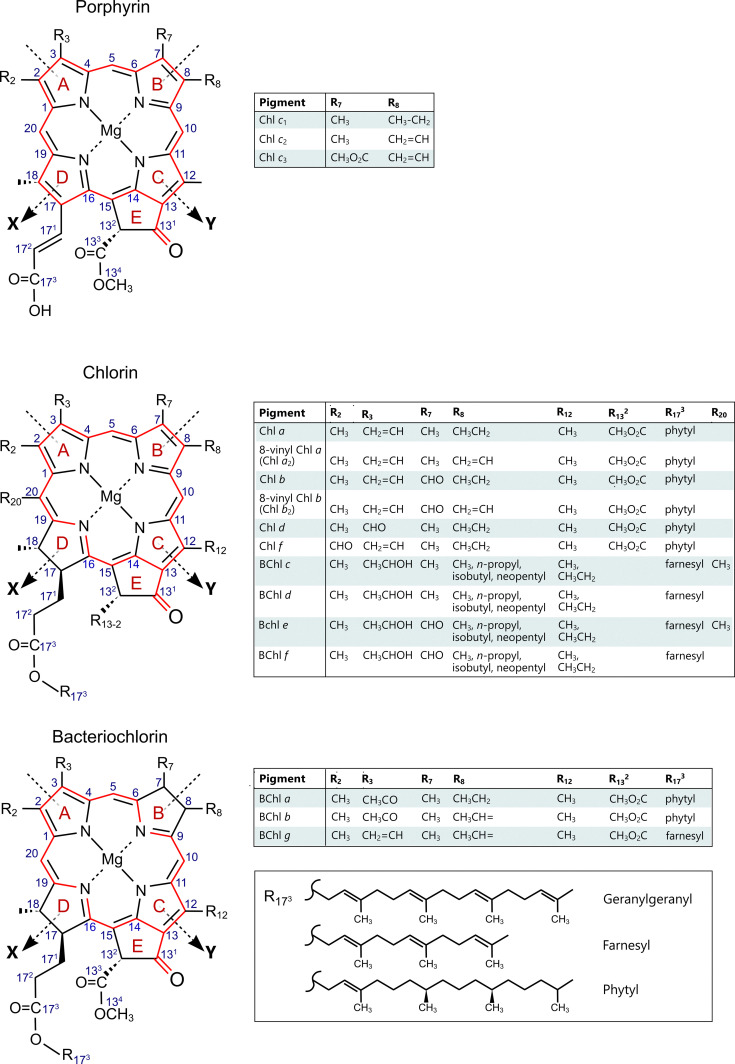
Structures of porphyrin-, chlorin-, and bacteriochlorin-type Chls and BChls. The red outlines indicate the extent of conjugation for each macrocycle. The arrows andX,Ylabels denote the molecular axes that run between central pyrrole nitrogen atoms. Carbon atoms are numbered (in blue) according to standard IUPAC-IUB nomenclature. Exocyclic R groups for each pigment appear in the respective tables. Esterification at C173generally involves attachment of a phytyl group, with farnesyl used for some of the BChls. Geranylgeranyl (GG) is also included in the bottom table, because this is the esterifying group in the BChlafound in the purple phototrophRhodospirillum rubrum.
π-conjugated molecules of this size would be expected to absorb visible light and indeed porphyrins, Chls and BChls are coloured, with a strong B-band (Soret) absorption around 380–450 nm.Fig. 2shows the absorption spectra of the porphyrin-type Chlsc1, c2andc3; the chlorins, Chlsa,b,dandfand BChlsc,d,eandf; and the bacteriochlorins, BChlsa,bandg, all in solvent. The choice of solvent affects the absorption wavelength maxima, as shown by the spectra in PhotochemCAD,16and diethyl ether was arbitrarily selected for the spectra inFig. 2. Each spectrum is accompanied by a pigment structure, with diffuse red outlines used to provide a purely graphic representation of the extent of conjugation. The absorption spectra, which are arranged in two equal columns according to their Chl/BChl nomenclature, fall unevenly into two main spectroscopic groups – eleven with their lowest energy bands in the 630–700 nm region, and the three true BChlsa,bandgfurther redshifted above 760 nm. Within the former group, only Chlsc1,c2andc3retain some porphyrin symmetry. The spectra are stacked to show the progressive intensification and redshifting of the lowest energy absorption band, for the Chls (left) and BChls (right). The following sections briefly explain why the state of reduction of the macrocyle, and modification of conjugation by exocyclic conjugated groups, are functionally important for various classes of photosynthetic organisms, allowing them access to different spectral regions of the solar energy that reaches the Earth.
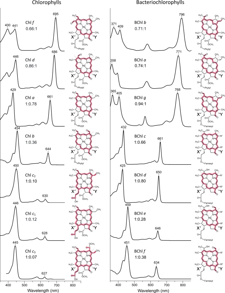
The absorption spectra of some Chls and BChls, with their respective chemical structures. The spectra of pigments in diethylether were downloaded from PhotochemCAD (https://www.photochemcad.com/databases/natural-chlorophylls;16). Ref.246also contains valuable data on the optical and structural properties of Chls and BChls. Chl and BChl spectra were replotted to facilitate stacking according to the position of the QYabsorption maximum, with the lowest energy QYbands at the top of each stack. Diffuse red outlines illustrate macrocyle conjugation and its extension to exocyclic groups. The ratio of B-band : QY-band amplitudes at their respective absorption maxima are shown for each pigment. In nearly all cases this ratio correlates with the extent of the red shift of the QYband. The 8V derivatives of Chlsaandbare not included.
Porphyrins
The four-orbital model of Gouterman17(also discussed in ref.11,18and19) can account for porphyrin absorption bands; in this model the two highest occupied molecular orbitals (HOMO) and two lowest unoccupied π-molecular orbitals (LUMO) result in four possible HOMO to LUMO π–π* transitions. Excited state orbitals are intermixed, but also polarised along theXandYaxes shown inFig. 1 and 2, which run between the pyrrole nitrogen atoms on rings A and C (Y), and rings B and D (X). For a completely symmetrical conjugated system, theX- andY-polarised transitions are degenerate and the main absorption feature for such a molecule is a strong B-band (Soret band) in the 380–420 nm region. In practice, the symmetry of porphyrin structures found in nature, such as PPIX inFig. 3and Chlsc1,c2andc3inFig. 2, is weakened, in the former case by the opposing central hydrogens on two of the four pyrrole rings. In the latter, insertion of Mg in thec-type Chls removes the asymmetry arising from the pyrrole hydrogens, but addition of the 131-keto group conjugated to the macrocycle provides another source of asymmetry. The consequence in each case is some loss of degeneracy in the excited states associated with theXandYtransitions and a splitting of the absorption, yielding B (BX, BY) bands in the UV/blue region of the spectrum and Q (QX, QY) bands in the visible region.
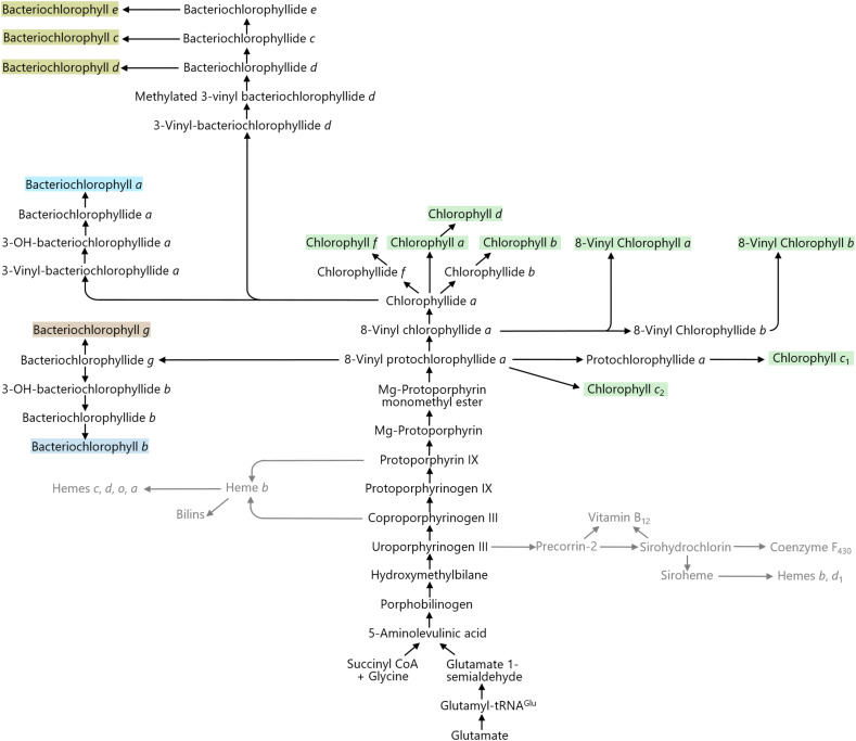
The biosynthetic pathways for Chls and BChls, including the routes (in grey) from uroporphyrinogen III leading to vitamin B12, siroheme, cofactor F430, hemeband hemed1. The steps from uroporphyrinogen III then lead to PPIX, the precursor of hemesa,b,c,dando, and the bilins (also in grey). Insertion of Mg into the protoporphyrin macrocycle commits this part of tetrapyrrole metabolism to the biosynthesis of BChls and Chls.
Chlorophyllsc1,c2andc3
Thetrans-acrylate group at C17, which is unique to these pigments, extends macrocycle conjugation along the QXaxis (Fig. 2). The absorption of Chlc1, with strong B-band absorption at 439 nm and weak absorption around 630 nm, closely resembles the absorption of the Chl biosynthesis intermediate 8-vinyl protochlorophyllide (8V-PChlide;Fig. 4), also called 3,8 divinyl protochlorophyllide (DV-PChlide). Further small B-band shifts are imparted by the 8V (Chlc2) and 7-methoxycarbonyl (Chlc3) groups. A full discussion of all aspects of Chlsc1, c2andc3can be found in ref.20, which details several more Chlcvariants, as well as discussing their evolution, biosynthesis and function in the light-harvesting complexes of brown algae and diatoms.Fig. 2shows that Chlsc1,c2andc3have the highest B/Q band intensity ratio among all the Chls and BChls; the strong B-band absorption is functionally important because organisms withc-type Chls are particularly suited to harvest the blue light that selectively penetrates into deep water.12,20,21Chlscperform an auxiliary role in light harvesting and they transfer absorbed energy to the more abundant Chla.20
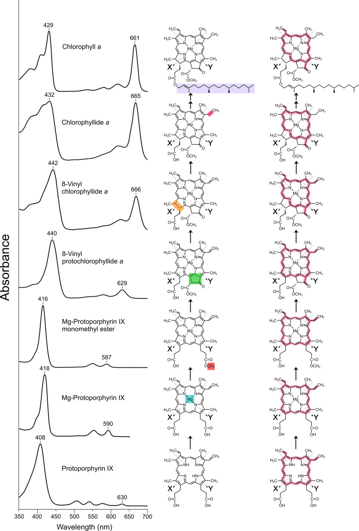
The biosynthetic pathway leading to Chla, depicted in three ways. (Left) The progression towards Chlavisualised as a series of absorption spectra, for the pigments solvated in methanol (but with PPIX in chloroform), showing the wavelength positions of major absorption maxima. Two arrows represent the successive addition of GG, then reduction to phytyl; both pigments have the same absorption spectrum. (Middle) The chemical structures of the biosynthetic intermediates are shown, with coloured boxes used to indicate the various modifications, according to the colour scheme used in.14(Right) The chemical structures of the biosynthetic intermediates, with diffuse red outlines used as inFig. 2to illustrate the successive changes in macrocycle conjugation, as the pathway progresses towards the final product, Chla.
Chlorins
Ring D, through which theX-axis runs, is reduced in all these pigments. As a result of removing the C17–C18 π-bond, orbitals associated with excited states along theYaxis gain oscillator strength,11redshifting and intensifying QYabsorption as seen with Chlsa,b,dandf(Fig. 2). Thus, QYabsorption for Chlaat 661 nm is much stronger than the ∼630 nm QYband for Chlsc1,c2andc3(Fig. 2). The biosynthetic pathway depicted inFig. 4also illustrates the consequences reducing the C17 Created by potrace 1.16, written by Peter Selinger 2001-2019 C18 double bond; the conversion of 8V-PChlide to 8V-chlorophyllide (8V-Chlide) redshifts the QYband from 629 nm to 666 nm, while also increasing its amplitude. Relative to porphyrins, this basic pattern of reconfigured conjugation and redshifted, intensified QYabsorption is found in all the chlorin pigments, namely Chlsa,b,dandfand BChlsc,d,eandf(Fig. 2). Exocyclic conjugated double bonds impart further spectral changes, and account for differences between Chlsa,b,dandf.
Chlorophyllsa,b,dandf
These are the most globally abundant pigments by virtue of Chla, which is the main light absorber on Earth for oxygenic photosynthesis.19The ubiquity and abundance of Chlaare apparent from satellite imaging, which shows how variations in temperature and nutrient availability affect the distribution of Chlain the oceans as well as displaying the huge seasonal fluctuations in terrestrial Chla.22–24The absorption spectrum of Chla(Fig. 2) leaves a ‘green gap’ between 450–600 nm, filled in photosynthetic organisms by a range of accessory light-absorbing pigments such as carotenoids and bilins, which transfer energy to the QYabsorption band of Chla.
One such accessory pigment is Chlb, which is found in light-harvesting complexes of algae and plants.25Unlike Chla, Chlbhas a 7-formyl group that affects conjugation along theX-axis, redshifting the B-band from 429 to 454 nm while also blueshifting and attenuating QYabsorption. These attributes narrow the ‘green gap’ (Fig. 2) and the complementary absorption properties of Chlsaandb, together with carotenoids, underpin the essential role of Chla/b(LHCII) complexes as the major light-harvesting complexes in plants and algae.26–28Indeed, the LHCII complex is likely the most abundant membrane protein on Earth.29Chlsaandbare closely packed within the LHCII complex, and energy absorbed by the ChlbB band can transfer rapidly and efficiently to the ChlaQY,viathe ChlbQYband at 644 nm.30LHCII donates harvested energy to Photosystem II (PSII) and Photosystem I (PSI) complexes,26–28but it is more than just an absorber and transmitter of energy; LHCII can be considered as a ‘smart antenna’ that can regulate its light-harvesting function by dealing with large fluctuations in solar irradiance. Instead of feeding excitation energy to photosystem complexes LHCII can switch to a quenching mode, sparing its acceptor partners the damaging consequences of excessive energy input.31
The 8V derivatives of Chlsaandb(also known as Chlsa2andb2) are not shown inFig. 2, but the effect of converting the 8V group to an 8-ethyl (8E) group can be seen in the biosynthetic pathway (Fig. 4). The B-band absorption of 8V-Chlidea(often called DV-Chlidea) is redshifted and relatively stronger than for the monovinyl pigment (ChlideainFig. 4, but also called 3V, 8E-Chlidea, 3V-Chlidea, or MV-Chlidea). Similarly, the 8V group in 8V-Chlashifts the B-band maximum from 429 nm in Chlato 436 nm (for pigments in Et2O), with no significant effect on the position of the QYband; there is also a small (∼7%) increase in the B-band amplitude relative to QY(not shown). These small alterations in B-band absorption are important for some marine cyanobacteria, conferring marginal gains in their ability to harvest blue light, the only wavelengths that can penetrate down to 100 m or more in the oceans.12,21This group of bacteria includes the globally abundant picocyanobacteriumProchlorococcus, so the 8V-Chls ofProchlorococcusrepresent a substantial proportion of the mass of marine Chls.32–34
Chldprovides another example of an exocyclic conjugated double bond affecting absorption; in this case the 3-vinyl group on ring A of Chlais replaced with the more electron-withdrawing 3-formyl group. This change selectively modifies electron density along theY-axis, increasing QYintensity and redshifting the B and QYbands relative to Chlaby 15 nm and 25 nm, respectively. Thus, the 3-formyl group enablesAcaryochloris marina, a cyanobacterium that makes Chld, to use light for photosynthesis that its Chla-utilising neighbours cannot.A. marinawas originally found in a biofilm that lies underneath the didemnid ascidianLissoclinum patella,35but it is now known to be widespread in environments enriched in far-red light.36
Chlfis the most redshifted of the Chl pigments, a consequence of being the only Chl with two exocyclic conjugated groups on ring A. The additive, electron withdrawing effects of the 2-formyl and 3-vinyl groups extend electron density along the QYaxis relative to Chlsa,bandd, and continuing the trend (Chlsb–a–d–f) of increasing QY: B band intensities and redshifted QYmaxima (Fig. 2). Chlf, which was discovered only recently in 2010,37has a QYabsorption maximum at 695 nm, and the assembly of this pigment within the photosystems of some cyanobacteria has allowed adaptation to life in spectrally filtered light environments, which are enhanced in far-red wavelengths above 700 nm.
Bacteriochlorophyllsc,d,eandf
These BChls form a distinct grouping, with modifications at C31, C82, C121, C132, and C20. Another distinguishing feature is C173esterification with farnesyl rather than the longer phytyl chain found in most of the Chls and BChls. In the green filamentous bacteriumChloroflexus aurantiacusC173is primarily esterified with stearyl alcohol.38These are the only (B)Chls with a 31-hydroxyl group, with a range of C82and C121substituents or with one at C20, and the only (B)Chls with no 132-methoxycarbonyl group. Despite this range of distinctive modifications, they have minimal effects on the absorption spectra of BChlsc,d,eandfin solvent, and their ∼420–460 nm B bands and 634–661 nm QYbands have similarities with Chlsaandb(Fig. 2). The C7 formyls in BChlseandfrelative to BChlscanddhave analogous effects to the C7 formyl of Chlbin relation to Chla; altered conjugation along theX-axis redshifts the B-band, and blue-shifts and attenuates QYabsorption. It should be noted here that BChlfis not found in nature, and it is synthesised by a genetically engineered strain of the photosynthetic bacteriumChlorobaculum(Cba.)limnaeum.39
The modifications seen in BChlsc,dandereflect their deployment in unique light-harvesting arrays; all the other Chls and BChls inFig. 2rely on binding to protein scaffolds in order to function, whereas BChlsc,dandecan self-assemble to form tightly packed, nanotubular, supramolecular structures, enclosed by a thin, protein-stabilized glycolipid membrane. Up to 250 000 of these BChls form ovoid assemblies called chlorosomes, which in bacteria such asCba. tepidumcan be as large as 133 × 57 × 36 nm.40,41
Chlorosomes can vary in size and shape, and their overall absorption and fluorescence properties depend on whether they are assembled from BChlsc,d, ore, but in general the C31, C82, C121, C132, C173and C20 modifications promote the serial, self-organising stacking of the macrocycles. The requirements for stacking BChl macrocyclesin vitrohave been extensively researched by the Tamiaki group and are now well understood.42The 31-hydroxyl is important because it ligates the central Mg atom of an adjacent BChl, so the stacking process is cooperatively extended and enhanced.40The other modifications, such as the absence of the 132-methoxycarbonyl group and methylation at C8 and C12, affect the packing between adjacent BChls. For example, methylation of the C8 ethyl to produce propyl, isobutyl, and neopentyl side chains, and methylation of the C12 methyl to produce an ethyl side chain, are used to fine-tune packing of the BChl macrocycles, thereby modulating the absorption of the chlorosomes.40,43As a result of forming extended, excitonically coupled arrays, the absorption of chlorosomes is greatly redshifted relative to the spectra inFig. 2so BChlc, with a QYabsorption maximum of 661 nm, is shifted as far as 750 nm inCba. tepidum.43Thus, the chemical organisation of this group of BChls determine macroscopic organisation and light-harvesting function.
Bacteriochlorins
Large bathochromic shifts are also found for protein-bound assemblies of BChlsa,bandg. This small group of pigments exhibits the most redshifted QYabsorption maxima (Fig. 2), which arises from the combined effects of reducing the C7 Created by potrace 1.16, written by Peter Selinger 2001-2019 C8 and C17 Created by potrace 1.16, written by Peter Selinger 2001-2019 C18 double bonds. The lower degeneracy of excited states associated with theXandYtransitions leads to further splitting of B and Q absorption, with BX, and BYbands in the UV/blue region 30–40 nm apart and QXand QYbands separated by as much as 170 nm in the case of BChlb. For comparison, the QX, and QYbands for Chlaare only 35 nm apart (Fig. 2). Compared to the chlorins, reduction of the opposing rings B and D further limits the extent of macrocycle conjugation along theXaxis, and QYabsorption for BChlsa,bandgshifts even more to the red, almost to 800 nm. Thus, photosynthetic organisms with BChlsa,b, orgas antenna and RC pigments can occupy spectral niches that are inaccessible to Chl-producers.21
An overview of the chlorophyll and bacteriochlorophyll biosynthesis pathways
This perspective will focus on the biosynthetic pathway for Chla, given its importance as the main light-absorbing pigment on Earth. The seven biosynthetic reactions from PPIX to Chlaare just a small subset of the metabolic network that synthesises the tetrapyrrole cofactors required for respiration, nitrogen and sulfur metabolism, photosynthesis, mammalian metabolism, and methanogenesis.44The ∼90 interrelated reactions that form these ‘molecules of life’, namely hemes, bilins, Chls, vitamin B12and the F430cofactor, have been compiled into an interlinked tetrapyrrole roadmap,44a simplified version of which is depicted inFig. 3. The central spine of the Chl/BChl pathways leads from 5-aminolevulinic acid to 8V-PChlidea,viaa series of intermediates with central importance to the rest of the tetrapyrrole network. Thus, vitamin B12, cofactor F430, siroheme and hemes are derived from uroporphyrinogen III, and PPIX is the precursor of hemesa,b,c,d,o, and the bilins (Fig. 3). Importantly, PPIX also acts as the substrate for the Mg chelatase (MgCh) enzyme complex that catalyzes insertion of Mg into the protoporphyrin macrocycle, committing this part of tetrapyrrole metabolism to the biosynthesis of (B)Chls. The BChl/Chl biosynthetic steps then proceed until the formation of 8V-PChlidea, the last common intermediate for the family of BChls and Chls. Here, 8V-PChlideacan form the substrate for synthesising BChlsbandg, or Chlsc1andc2(Fig. 3), or it can be converted into 8V-Chlidea, which can be considered as a hub for biosynthesis of nearly all Chls and BChls.44One set of reactions from 8V-Chlideayields the 8V versions of Chlsaandb, which play an important role as the major pigments in the globally abundant picocyanobacteriumProchlorococcus.32–34Continuation of the main biosynthetic route leads next to Chlidea, from which most Chls and BChls originate. Thus, Chlsa,b,dandf, as well as BChlsa,c,dande, are formed from Chlidea(Fig. 3).
The structures and absorption properties of biosynthetic intermediates leading to chlorophylla
The biosynthetic steps outlined inFig. 4introduce a series of structural and energetic properties that enable Chlato participate not only in absorbing solar energy in antenna complexes, but also in converting and transiently storing this energy in the form of a charge-separated state in RC complexes.19These steps are mediated by a series of enzyme-catalysed reactions that progressively strengthen and redshift the absorption band associated with the QYtransition dipole.
At the start of the pathway, PPIX has strong B-band absorption in the 380–420 nm region, but only minimal QYabsorption between 590–630 nm. Then, Mg is inserted, with only small effects on absorption, but with pervasive and powerful effects on function. Several metals, Fe for example, could also confer the ability to form ligands on protein side chains, but nature has selected Mg as the central metal. Mg has a coordination number of six, strongly preferring oxygen-containing ligands such as water, but also able to form stable ligands to the pyrrole nitrogen atoms of Chls (and their biosynthetic intermediates), as well as to protein side chains such as histidine residues. However, in most cases the central Mg of Chls and BChls in photosynthetic complexes is pentacoordinated.45Thus, single or paired Chls, ligated to proteins, are held in place for energy transfer or photochemical functions, while also contributing decisively to the overall stability of Chl–protein complexes. Similarly, stacked BChlsc,d, orein chlorosomes are stabilised in part by their 31-hydroxyls ligating to the central Mg of an adjacent BChl.40Mg in chlorins and bacteriochlorins also maximises the excited state lifetimes of the pigments, which enables participation in energy transfer and redox reactions, while minimising formation of potential toxic triplet states due to intersystem crossing from the singlet state.9,19The strong ligands formed by the central Mg within protein binding sites allow proteins to exert control over the aggregation state and electrostatic environment of Chls and BChls, contributing to their remarkable and contrasting ability within RCs to generate either strongly oxidising or reducing Chl species.19
The next pigment in the pathway, Mg-PPIX monomethyl ester (MgPME), is also very similar in absorption to the preceding intermediates (Fig. 4), but addition of a methyl group to the C13 propionic acid group is a prerequisite for the subsequent and crucial formation of the isocyclic ring E.11This fifth ring is found in all Chls and BChls, so it imparts essential structural and functional properties. The presence of the isocyclic pentanone ring E braces the macrocycle and as a result the C Created by potrace 1.16, written by Peter Selinger 2001-2019 O group at C-131of ring E is coplanar with the rest of the macrocycle,9which promotes delocalisation of the conjugated π-system along the QYaxis and redshifting of absorption from 587 nm to 629 nm (Fig. 4). Lowered macrocycle flexibility due to ring E is suggested to help prevent dissipation of excited states due to internal conversion.9Another functional aspect of the 131-keto group is forming important hydrogen bonding interactions within antenna complexes, for example.46,47The other major peripheral group on ring E is the methoxycarbonyl at C132, which is one of the three stereochemical locations in Chla(aside from those in the phytyl chain), the others being at C17 and C18, and withR,S,Sconfigurations, respectively.10,11Enolisation then reprotonation of Chlaat C132forms theS-stereoisomer, termed Chla′,9,10,48which forms part of the charge-separating core of Type 1 RCs.49A pair of BChlg′ epimers forms the primary electron donor in RCs of the anoxygenic phototrophHeliomicrobium modesticaldum.50,51The C132methoxycarbonyl is sterically significant and although it is an invariant feature of all Chls and the ‘true’ BChlsa,bandg(Fig. 2), it is absent from BChlsc,dande, where it would otherwise impede the serial stacking of the macrocycles within chlorosomes.40,43
The next biosynthetic step forms a chlorin, and reduction of ring D at C17–C18 provokes a major absorption change, as outlined earlier, because of the altered macrocycle conjugation (Fig. 4). The balance between the oscillator strengths associated with the QXand QYaxes changes, increasing and redshifting QYabsorption from 629 nm to 666 nm, which is more or less the final absorption position for Chla(seeFig. 4). Thus, the final three biosynthetic steps do not exert much effect on the wavelength maximum of QYabsorption, although reduction of the 8V group to 8E blue-shifts the B-band from 442 nm to 432 nm. As mentioned earlier, oceanic bacteria such asProchlorococcusbenefit from retaining the vinyl at C8 and the attendant 442 nm absorption, because only blue light is available at depths of 100 m or more. Conversely, a C8 ethyl group increases the amplitude of QYabsorption relative to the B-band maximum by ∼60% (Fig. 4), which allows cyanobacteria growing in shallow water, or terrestrial photosynthesisers, to absorb more red light.
The next biosynthetic step esterifies the C17 propionate group with a long-chain alcohol, usually geranylgeraniol, which is followed by the stepwise reduction of geranylgeranyl Chl (ChlGG) to phytyl Chl,viadihydrogeranylgeranyl Chl (ChlDHGG) and tetrahydrogeranylgeranyl Chl (ChlTHGG). In principle, the esterification and reduction steps could proceed in a different order, with prior reduction of geranylgeranyl pyrophosphate (GGPP) to phytyl PP (PPP), followed by its attachment to Chlideaforming Chla(phytyl Chla). Although the final esterifying moiety is generally phytyl, there are examples where GG is found, in the case of BChlainR. rubrum,52and farnesyl in the case of BChlsc,d,e,fandg.40The green bacteria that synthesise BChlsc,d,eandf, can esterify the C17 propionate with geranylgeraniol, phytol, 2,6-phytadienol, hexadecanol and octadecanol.11,44
The absorption and excited state properties of the pigments in solvent are not affected significantly by C173esterification, but the addition of the long-chain (C20H40O) isoprenoid phytol to monovinyl Chlidea, for example, represents a major modification that accounts for a third of the molecular mass of Chla, while also increasing its hydrophobicity with important consequences for membrane location, the packing of Chls, and their attachment to proteins. The addition of phytol is a therefore decisive and essential step in photosystem assembly.53–55The disposition of the phytyl tails has been revealed by numerous high-resolution structures of photosynthetic complexes, which show that the phytyls influence pigment–pigment and pigment–protein distances and orientations, sometimes intertwining or associating with carotenoids (for example45,56,57).
The enzymes of chlorophyllabiosynthesis
The following sections outline the enzymes that catalyse the biosynthesis of Chlsa,b,candf, but more depth and detail covering historical, regulatory and physiological aspects can be found in reviews such as,6–11,13,20,58and we do not cover the steps unique to the BChl pathways. Indeed, each Chl biosynthesis step merits its own review, but here the focus is on the structures and mechanisms of the Chl pathway enzymes. We have used AlphaFold 3 (AF3)59to augment the existing experimentally determined structures, permitting a complete structural overview of the Chl biosynthesis enzymes. The performance of AF2 and AF3 has been tested previously using critical assessment of structure prediction (CASP),60,61and their value demonstrated by providing starting models for X-ray crystallographic and cryogenic electron microscopy (cryo-EM) model reconstruction,62–65and in the design of structurally validatedde novo-designed proteins.66,67A set of useful parameters has been compiled for the modelling of Chl pathway enzymes, and can be found in Table S1 in supplementary information (SI). This includes the ipTM (interface predicted template modelling) confidence scores for all AF3 models and, where appropriate, the root mean square deviation (RMSD) values for AF3vs.experimentally determined structures in the RCSB PDB (research collaboratory for structural bioinformatics protein data base,https://www.rcsb.org/). The zipped structure files for all models are also available athttps://doi.org/10.1039/d6cb00082g.
Step 1 – magnesium chelatase
Insertion of magnesium into porphyrins
The chemical mechanisms of metalation of porphyrins have been studied for decades (reviewed in ref.68), and several steps have been identified, including deformation of the porphyrin ring, association of the solvated outer-sphere of the metal ion and the porphyrin, then exchange of an (unprotonated) porphyrin nitrogen atom with a solvent molecule in the coordination sphere. Further desolvation of the metal ion allows formation of the second metal–porphyrin bond, with the metal ion now coordinating both of the unprotonated pyrrole nitrogen atoms. The presence of hydrogen atoms on the other two pyrrole nitrogen atoms obliges the bound metal to sit above the porphyrin plane, forming a sitting atop (SAT) complex, as originally proposed by Fleischer and Wang.69The rate of insertion of Mg into porphyrins follows the order Cu > Zn > Mn, Co, Fe > Ni > Cd > >Mg.70,71Relative to other metals, Mg2+has a lower affinity for porphyrin and a stronger affinity for water, so the six water molecules in the first coordination sphere are strongly polarised, creating a second hydration shell. Valuable insights into Mg insertion have been provided by density functional calculations, which followed the stepwise displacement of water molecules from the first hydration shell through to Mg–porphyrin formationviaMg–porphyrin SAT complexes.72,73A ten-step reaction mechanism was proposed, starting with formation of an outer-sphere complex between the hydrated metal and the porphyrin, followed by exchange of one water ligand leading to formation of the first bond from Mg to a central pyrrole nitrogen atom. After two more such exchanges the second Mg–pyrrole N bond is formed, then the fourth and fifth water molecules are moved, modelled as transferring to the second coordination sphere,73but this outer sphere might not be present in an enzyme-catalysed mechanism. At this stage there are four Mg–N bonds, with two of the pyrrole nitrogens still protonated, and with Mg retaining one of its original water ligands. Increasing the number of metal–porphyrin bonds is accompanied by progressive distortion of the porphyrin, with the mounting strain released when the two pyrrole NH groups are deprotonated and the metal moves from its SAT position into the ring plane.73
These theoretical studies highlight the problems that confront a Mg-chelating enzyme, which must remove five water molecules from the strongly bound hexacoordinate hydration shell and deprotonate two pyrrole nitrogens. Thus, the enzyme-catalysed insertion of Mg2+into the PPIX macrocycle is energetically demanding; this requirement, and the need to regulate this first step in Chl biosynthesis, necessitate an ATP-fuelled, multi-subunit, allosterically controlled enzyme. Early experiments with recombinant proteins produced inEscherichia coliusingbchH,IandDgenes fromRhodobacter(Rba.)sphaeroides, or withchlH,I, andDgenes fromSynechocystissp. PCC 6803 (hereafterSynechocystis), showed that magnesium chelatase (MgCh) is a three-subunit complex that catalyses the ATP-dependent conversion of PPIX to Mg-protoporphyrin IX (MgPIX).74,75The ATP-driven catalytic cycle involves the ChlI (∼35 kDa) and ChlD (∼75 kDa) subunits, which are members of the AAA+(ATPases associated with various cellular activities) superfamily.76–79As many as 14 ATP are required to drive each catalytic cycle.80ChlH (∼150 kDa) binds the PPIX substrate.74,75,81
Structural, kinetic and mutagenesis studies of magnesium chelatase
Although there are currently no structures of the MgCh complex, early crystallographic work determined the structure of BchI fromRba. capsulatusand showed that it could form hexamers,82subsequently also investigated by cryo-EM.83,84A 2.9 Å resolution crystallographic structure showed that ChlI purified fromSynechocystisforms hexamers,85while a more recent cryo-EM study of ChlI from the nitrogen-fixing cyanobacteriumNostocsp. PCC 7120 revealed pentamers and hexamers.86An earlier, low resolution study revealed heptamers of BchI,79so it is likely that the oligomerisation of I subunits is poorly controlled in the absence of the D subunit. The cryo-EM resolutions for the hexamers (3.8 Å and 4.0 Å) and for the pentamer (4.9 Å) were sufficient to show five ATP molecules and one ADP bound in hexamer conformation A, with four ATPs and two ADPs in conformation B. ATP hydrolysis was proposed to rearrange the hexamer ring, providing the basis for a motor function that couples ATP hydrolysis to Mg insertion.86
The 2.5 Å resolution crystal structure of ChlH fromSynechocystisshowed the overall architecture of this subunit and that the active site is buried within the protein interior.87Subsequently, a combination of X-ray crystallography, computational modelling, mutagenesis and enzymology was used to identify the PPIX binding site, which is close to the catalytically essential E660 residue.88The active site of ChlH is connected to the exterior by a solvent-filled channel, and the strictly conserved residue E625 sits at the interface with bulk solvent. E625 was suggested to play a role in delivering Mg to the active site, but this channel could also provide a route for abstracting protons from pyrrole N atoms during the insertion of Mg into PPIX.88
Unravelling the roles of ChlD is key for understanding MgCh, and the functionally crucial interaction between ChlD and ChlH (Kd∼ 330 nM) was mapped using chemical cross-linking coupled with mass spectrometry, microscale thermophoresis, and by modifications that either truncate ChlD or modify single residues.89The C-terminal integrin I domain of ChlD has a metal ion-dependent adhesion site (MIDAS) motif90that mediates the Mg2+-dependent ChlD–H interaction. Five Glu residues in the C-terminal domain ofSynechocystisChlD, E510, E513, E600, E603, and E605, are important for the cooperative response of MgCh to Mg2+.91ChlD bindsviathe integrin I domain to the body region of ChlH,89and the AAA+N-terminal domain of ChlD, which shares ∼40% sequence identity with ChlI, binds tightly to ChlI with aKd≈ 7 nM,52,92which would close an arc of five ChlI subunits86to form a six-membered I5D ring.
A series of kinetic studies on the cyanobacterial MgCh formed from recombinant enzyme subunits79,80,91,93–96suggests a model where a heterogeneous ChlI5D ring interacts with the body region of the ChlH proteinviathe C-terminal integrin domain of ChlD.89Hydrolysis of ATP by ChlI79,80drives a conformational change transmitted initially to ChlD then to the ChlH–porphyrin complex, promoting the insertion of the Mg2+ion into the PPIX ring.88Thus, MgCh is a molecular machine that chemomechanically couples the free energy from ATP hydrolysis by the curved array of AAA+ChlI subunits to the metal ion insertion site on ChlH, through the bridging subunit, ChlD.
Computational modelling of the magnesium chelatase complex
A structure of the three-subunit MgCh enzyme would reveal essential mechanistic information, so new computational tools have been used to build a model of this complex, shown inFig. 5. AF3 was used to calculate a hypothetical structure of the ChlHI5D complex. The model incorporates existing data for the ChlD–H interaction89and for PPIX binding within ChlH.88The HI5D stoichiometry in the model inFig. 5is consistent with native mass spectrometry analyses, and migration on native gel electrophoresis (P. J. Jackson and A. A. Brindley, personal communication). This model provides only a single snapshot of the complex, but the central position of ChlD shows how it might transmit free energy from ATP hydrolysis by the ring of ChlI subunits to ChlH, where it is used for metal ion insertion.
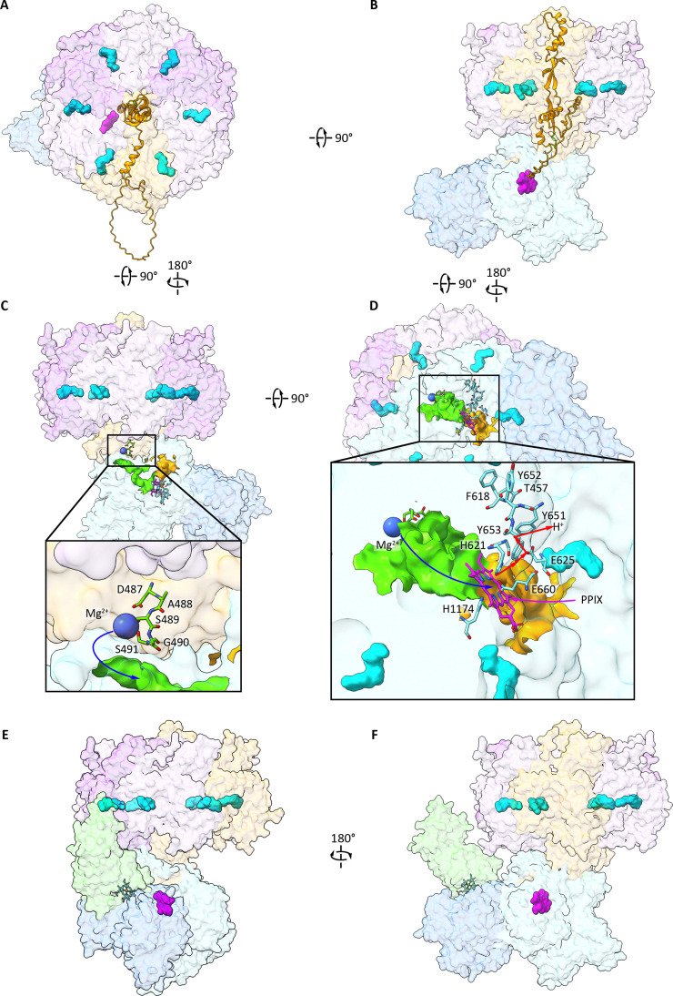
AF3 model of a putative magnesium chelatase complex highlighting the ChlD (pastel orange), ChlI (light mauve and light lavender) and ChlH (light blue – body; baby blue – head) subunits with the stoichiometry ChlHI5D. The PPIX substrate (magenta) sits in the body of ChlH and the ATP cofactors (cyan) bind at the ChlI–ChlI and ChlI–ChlD interfaces within the HI5D AAA+complex. (A) and (B) Two surface views of the overall structure of the complex. The intrinsically disordered portion of ChlD (pastel orange ribbon) connects the ChlI-like portion of ChlD to the integrin, ChlH-interacting portionviathe energy transducing subdomain (shown in cartoon representation) that sits within the AAA+pore of the ChlI5D ring. (C) and (D) Insets showing detail of the putative Mg2+(bright green) and PPIX substrate (orange) pores along with a Mg2+ion (blue sphere) bound to the MIDAS motif at the entry to the putative Mg2+channel. The AF3 modelled substrate, PPIX, is shown in stick representation (heteroatom colours: blue – nitrogen, red – oxygen, magenta – carbon) at the confluence of the PPIX and Mg2+channels. Previously identified essential catalytic residues H1174, E660 and H621 (cyan, stick representation) point directly to the center of Mg2+-chelating tetrapyrrole nitrogen atoms. Blue and magenta arrows indicate the direction of substrate entry to the active site for Mg2+and PPIX respectively, along with a putative proton conductance channel that proceeds along H621, Y653, E625 and Y651, highlighted with a red arrow. (E) and (F) Model of the MgCh–Gun4 complex with Gun4 (pastel green) sitting on the head group of ChlH, where the Gun4 tetrapyrrole binding site forms part of the binding interface. See Table S1 in the SI for ipTM scores for AF3 models and RMSD values for the correspondence of AF3 models with experimentally determined structures in the RCSB PDB.
The crystallographic and cryo-EM structures of rings formed by the AAA+protein ChlI show a central pore,85,86which is a common feature of other AAA+complexes.97,98The AAA+family uses the free energy from hydrolysing ATP to perform mechanical work, acting as molecular screws, unwinders, or threaders of a substrate within this central cavity.98The model inFig. 5depicts a heterogeneous I5D ring, where the N-terminal half of D, which has homology with ChlI, replaces a 6th I subunit while the central pore encloses D-subunit residues 413–478. This extended segment of ChlD forms a right-handed pseudo-helical fold, which was predicted independently by AF3 and also ESMFold;99near to the point of entry into the I5D pore D413–D459 wrap around a central core formed by residues D459–D478. The end of this central core directly connects to a beta-sheet (D479–D487), at the end of which is the MIDAS motif (D487–D491).
By analogy with other AAA+complexes, and in line with the molecular machine concept, hydrolysis of ATP by the ChlI components performs mechanical work that is transmitted to a spindle formed by the ring-enclosed residues D413–D478 (coloured in orange inFig. 5A and B), which represent the AAA+substrate. In our model, chemomechanical forces are transmitted sequentially from the ChlI5D ring to residues D459–D478, subsequently to residues D413–D459, then to the MIDAS motif (D487–491). Extrusion of the MIDAS-bound Mg2+ion along the pore in ChlH (blue arrow inFig. 5C; pore coloured green), is proposed to be accompanied by progressive removal of at least some waters from the hydration shell of Mg2+, which enters the active site within ChlH. PPIX also enters this site (Fig. 5Dinset, magenta arrow)viaanother channel (Fig. 5Dinset; shown in orange). Large patches of basic residues (not shown) in ChlH regions 105–184 (body domain) and 852–888 (head domain), are proposed to associate with the outer leaflet of the thylakoid membrane. The acquisition of PPIX is likely to take place near to or at the membrane surface, and the initial PPIX binding site environment must change to become an enclosed chamber where no external waters are admitted, likely requiring large domain movements in ChlH. The chemistry of Mg2+insertion into PPIX within the active is likely to follow some of steps identified by density functional calculations.72,73The red arrow (Fig. 5D, inset), indicates a possible pathway for conducting protons removed from the central pyrrole nitrogen atoms of PPIX, which is part of the chelation process (see earlier section). All of these events, from movement of PPIX and the initially hydrated Mg2+into the catalytic site, the insertion of Mg2+into the porphyrin macrocycle and deprotonation of the pyrrole nitrogens, then product release, are all somehow driven by conformational changes in ChlH fuelled by ATP hydrolysis, and these processes need to be resolved by structures of catalytic intermediates in the coming years.
Structure and function of the Gun4 subunit
The multi-subunit nature of MgCh, and its slow, energetically costly catalytic cycle,80,93,95sharply contrasts with ferrochelatase (FeCh), which is a single polypeptide of ∼40 kDa that catalyses the rapid and energetically favourable insertion of Fe into PPIX.73The Fe and Mg chelation steps launch porphyrins down the heme and Chl branches of tetrapyrrole metabolism, respectively (Fig. 3), so it is strategically and physiologically important to regulate this branchpoint, while taking into account the widely differing catalytic capacities of the FeCh and MgCh enzymes. Allocating the correct levels of flux down these branches must also respond to environmental variations such as changes in light and temperature, matching the supply of Chls with the assembly and repair of photosystems while avoiding the accumulation of potentially phototoxic Chl intermediates. ChlD is the regulatory hub for the MgCh enzyme complex,89,94and the AAA+site in ChlD is involved in the allosteric and cooperative responses of MgCh to both Mg2+and MgATP2;94it has been shown that ChlI2 ofChlamydomonas reinhardtiican stimulate chelatase activity by phosphorylating the integrin-I domain of ChlD.100The Mg-dependent binding between ChlD and ChlH, involving the C-terminal integrin I domain of ChlD, mediates the cooperative response of theSynechocystischelatase to Mg.89,91
Another important aspect of regulating MgCh involves the auxiliary porphyrin-binding subunit, Gun4.101High-resolution structures of this small, soluble, 25 kDa protein from cyanobacterial sources revealed non-conserved N-terminal and conserved Gun4 domains linked by a 12–15 residue loop.102,103Extensive kinetic analyses showed that Gun4 dramatically enhances the sensitivity of MgCh to Mg2+, so although there is almost no activity at 2 mM Mg2+the complex is fully active in the presence of Gun4.102TheOryza sativaGUN4 increases the maximum reaction rate of Mg chelation 16-fold;104lower Chl levels, PPIX accumulation, and lowered MgCh and FeCh activities were found ingun4mutants ofSynechocystis.105,106A subsequent crystallographic study of Gun4-porphyrin complexes revealed ‘half-open’ binding sites for deuteroporphyrin IX (DIX; a more water-soluble analogue of PPIX) or magnesium deuteroporphyrin IX (MgDIX), compatible with a role for Gun4 in transferring porphyrins to biosynthetic enzymes.107Gun4 from the green algaC. reinhardtiibinds bilins,108further indicating its role as a regulator of tetrapyrrole biosynthesis.109
The panels inFig. 5E and Fshow a computational model in which the tetrapyrrole-binding face of Gun4 (heme coloured in green) binds to the head domain of ChlH. This binding site could be partially mediated by a tetrapyrrole, consistent with a regulatory role for Gun4 that could involve responding to flux down the heme/bilin and Chl branches of tetrapyrrole metabolism.
Step 2 – magnesium-protoporphyrin IX O-methyltransferase
The second step in Chl biosynthesis involves addition of a methyl group fromS-adenosyl-l-methionine (SAM) to the C13 propionate of MgPIX, catalysed by Mg–protoporphyrin IX O-methyltransferase. This modification prepares the porphyrin for the next reaction, which forms the isocyclic ring E.11Early steady-state kinetic studies on recombinant ChlM fromSynechocystisusing the MgDIX substrate showed that MgDIX and SAM bind to ChlMviaa random ternary mechanism, with parametersKSAMM= 38 µM,KMgDIXM= 2.37 µM,kcat/KSAMM= 1500 M−1s−1.110A subsequent transient kinetics study showed that rapid binding of MgDIX to ChlM (>600 s−1) is followed by a slower (70 s−1) isomerization of the enzyme.111Pre-steady-state catalysis, monitored using quenched-flow and high-performance liquid chromatography, showed evolution of a catalytic intermediate (rate constant of 11.9 ± 0.5 s−1), the decay of which (11.8 ± 0.5 s−1) coincides with the evolution of the Mg–deuteroporphyrin IX monomethylester (MgDME) product; given thatkcatis 0.057 s−1release of the MgDME and S-adenosylhomocysteine (SAH) products is likely rate-limiting under the conditions of this assay.111
Crystal structures of ChlM fromSynechocystiswere obtained with either SAM or SAH bound, at resolutions of 1.6 and 1.7 Å, respectively.112This monomeric, soluble protein has a central seven-stranded β-sheet, with the predicted strand order and conformation,110flanked by 8 α-helices; structural differences were noted for ChlM–SAH and ChlM–SAM.112In the absence of a structurally defined bound MgPIX substrate, it was dockedin silicoand the binding pocket was tested using mutagenesis, isothermal calorimetry and functional assays. These analyses showed that Tyr-28 and His-139 were necessary for catalysis, and modelling showed the C13 propionate group of MgPIX lies close to the hydroxyl of Tyr-28, the Nε nitrogen of His-139, and theS-methyl group of SAM.112It was suggested that the C13 propionate carboxyl, positioned by Tyr-28, is deprotonated by His-139, creating the conditions for nucleophilic attack by the carboxyl on the SAMS-methyl group.112Furthermore, the identification of two flexible arms of ChlM could explain earlier observations of random binding and enzyme isomerisation.110,111The AF3 model of the SAM–MgPIX–ChlM ternary complex inFig. 6agrees with this previous crystallographic andin silicomodelling study, placing the C13 propionate of MgPIX within 3.8 Å of the SAM methyl group (Fig. 6D). No obvious Mg coordinating residues were identified in our model although a water, mediated by the backbone carbonyl of Phe219 or the hydroxyl oxygen of Ser185, could provide a coordination bond, but it is not modelled here.Fig. 6Cdepicts the surface hydrophobicity of the methyltransferase, with an open substrate cleft flanked by hydrophobic residues (in gold), providing a possible hydrophobic interface with the underlying membrane bilayer, and a path for substrate and product tetrapyrroles to enter and leave the enzyme active siteviathe membrane bilayer. This topic is covered in more detail in Section 6 andFig. 15.
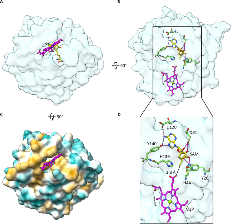
AF3 model of Mg-Protoporphyrin IX O-methyltransferase. (A) and (B) Two surface views of the overall topology of the enzyme with MgPIX and SAM shown in magenta and yellow, respectively (heteroatom colours: blue – nitrogen, red – oxygen). (C) View of the open substrate binding cleft of the enzyme highlighting the surface hydrophobicity (gold – hydrophobic, blue – hydrophilic) of the protein and indicating the potential for this face of the enzyme to dock onto the photosynthetic membrane where the substrate resides. (D) Enlarged view of the box in (B) highlighting important residues involved in SAM binding – the H44 pyrrole nitrogen interacts with the carboxyl group of the SAM methionine, the D91 carboxyl group interacts with the alcohol groups of the ribose moiety, and the Y140 hydroxyl and D120 carboxyl groups interact with the adenine moiety of SAM. Y28 and H139 are highlighted due to their importance for catalysis and are found either side of the axis running from the MgPIX propionate group to the methyl group of SAM. Aside from a hydrophobic cleft, no obvious interacting groups were found for the MgPIX substrate. The distance from the SAM methyl group to the substrate propionate carboxyl was modelled to be 3.8 Å. See Table S1 in the SI for ipTM scores for the AF3 model and RMSD values for the correspondence of the AF3 model with the experimentally determined structure in the RCSB PDB.
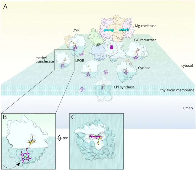
(A) Cartoon depiction of the membrane location and transfer of substrates and products between membrane-associated Chl biosynthesis enzymes. With the exception of MgCh, a slice through each enzyme structure shows that its active site cavity is flanked by hydrophobic residues that form a membrane interface, consistent with images of surface hydrophobicity inFig. 6C, 7C, 8C, 9C and 11D. The extent of penetration of the Chl enzymes into the bilayer is purely to illustrate the proposal that Chl intermediates, represented by generic macrocycles in magenta, could gain access to, and move between, pathway enzymesviathe membrane bilayer. (B) Zoomed view of the methyltransferase, used as an example of a Chl biosynthesis enzyme sitting in, but not through, the membrane. The direction of movement of the substrate into the active site cleft is shown with an arrow. (C) View of the methyltransferase from within the membrane, looking up at the membrane-embedded active site cleft.
Linkage between the magnesium chelation and methytransferase steps
There were indications from kinetic studies that the ChlH subunit of MgCh stimulates catalysis by ChlM, and a catalytic intermediate had been found that forms before the MgDME product.111Subsequently it was found that ChlH shortens the lag phase that precedes MgDME evolution, and rate constants for the accumulation of MgDME improved from 11.8 s−1to 31.7 s−1in the presence of 2 µM ChlH.113The ChlH concentration at half the maximal rate of product formation, 1.2 ± 0.3 µM, was suggested to represent the binding constant (KD) for the binding of ChlH to ChlM.113It is possible that ChlH directly accelerates the methytransferase reaction chemistry, and the ChlM structure indicated that ChlH could interact with the flexible N-terminal and G arms of ChlM,112thereby mediating the stimulatory effect of ChlH on the methytransferase reaction. Further control and regulation of the coupling between chelatase and methyltransferase steps could be exerted by Gun4, which preferentially binds MgDIX over DIX (KD0.3 ± 0.02 µMvs.2.29 ± 0.28 µM, respectively102). TheSynechocystisGun4–MgDIX structure shows an exposed ring C propionate, the methylation target for ChlM, pointing at a possible substrate delivery mechanism for the porphyrin.107
Step 3 – magnesium–protoporphyrin IX monomethyl ester [oxidative] cyclase
The formation of the isocyclic ring E is crucial for all Chls and BChls in several respects. As noted earlier, the stiffening of the macrocycle by this fifth ring enforces coplanarity of the C-131C Created by potrace 1.16, written by Peter Selinger 2001-2019 O group with the macrocycle,9selectively delocalising the conjugated π-system along the QYaxis and redshifting the associated absorption band. Thus, the red cyclase substrate, MgPME, is converted to a green product, 8V-PChlidea(Fig. 4)viaan oxygen-requiring reaction. Two types of MgPME cyclase have evolved, with oxygenic phototrophs such as cyanobacteria, algae and plants, as well as some purple bacteria, using molecular oxygen,114,115whereas most anoxygenic phototrophic bacteria employ a mechanistically different cyclase that forms the 131-oxo group using the oxygen atom from water.116Sourcing oxygen from water allows the cyclase reaction to proceed in anaerobic environments, catalysed by an oxygen-sensitive radical SAM enzyme with [4Fe–4S] and cobalamin cofactors.117
This section will focus on the O2-dependent cyclase, which falls into three classes118that all possess a core catalytic subunit, AcsF; this is the only cyclase component in the Betaproteobacteria, Gammaproteobacteria, Acidobacteria and Chloroflexi, whereas an auxiliary subunit is required by the Alphaproteobacteria (BciE)119and the oxygenic phototrophs (Ycf54).120–123AcsF was first identified inRubrivivax(Rvi.)gelatinosus,124followed by homologues inC. reinhardtii,125Synechocystis,126–128Arabidopsis thalianaand barley (Hordeum vulgare).129
Eventually, it became possible to measure the kinetics of the O2-dependent cyclase using the purified protein.130Recombinant AcsF was produced inE. colias a single, ∼44 kDa polypeptide containing 2.35 ± 0.04 iron atoms per monomer, which oligomerised to form dimers or trimers; absorption spectra indicated the presence of an µ-oxo-bridged di-iron cluster. A continuous assay was developed for the cyclase, by combining purified AcsF with NADPH, ferredoxin (Fd) and Fd:NADP+reductase (FNR); measurements of reaction kinetics revealed a turnover rate of 0.9 min−1, aKMfor MgPME of 7.0 µM and aKDfor MgPME of 0.16 µM.130Liquid chromatography–electrospray ionization–tandem mass spectrometry (LC–ESI–MS/MS) established that formation of the 8V-PChlideaproduct proceedsvia131-hydroxy–MgPME and 131-keto–MgPME, each of which successively evolved then decayed during the cyclase assay in the manner of reaction intermediates.130
The catalytic cycle consists of three sequential reactions, each involving the supply of two electrons by NADPHviaFNR and the Fd carrier.130The turnover rates of the chelatase (0.8 min−1) and cyclase (0.9 min−1) enzymes are similarly slow relative to the intervening methyltransferase step (3420 min−1), and both reflect the difficult chemistries involved. Cyclisation involves three, two-electron transfers coupled to three successive activations of molecular oxygen by active site iron atoms, in each caseviaa reactive diiron(iv)-bis–oxo intermediate.131Formation of the 131-hydroxy–Mg PME intermediate will consist of a multistep catalytic cycle requiring the concerted supply of electrons and molecular oxygen, followed by another cycle forming the 131-keto–Mg PME, then yet another that generates the final 8V-PChlideaproduct. It was possible to couple the cyclase assay to a reconstituted PSI electron transport system, so in oxygenic phototrophs the ultimate source of electrons for the cyclaseviareduced Fd is likely light-driven charge separation in this complex.130
In the absence of a structure for AcsF we used AF3 to generate a model, which has a core comprising a four-helix bundle incorporating a di-iron center held by H133, E198, E130, E211, H214 and E175 ligands (Fig. 7), while molecular oxygen forms the remaining two ligands to the di-iron cluster (not shown). Fd is also modelled, suggesting the [2Fe–2S] cluster is ∼17 Å from the di-iron center, within electron transfer distance. Of the nine Fds inSynechocystis,132the one modelled here is the [2Fe–2S] PetF (Ssl0020). The di-iron cluster is ∼5 Å from the substrate C131atom but an intervening molecular oxygen (Fig. 7D) would be 3.6 Å from C131. This arrangement closely aligns with the di-iron catalytic core of methane monooxygenase hydroxylase fromMethylococcus capsulatus.133Ser174 is ∼2 Å from the central Mg2+ion, which suggests it may provide a weak coordination bond.
![AF3 model of the Mg–protoporphyrin IX monomethyl ester cyclase complex formed from AcsF1 (light blue), Ycf54 (pastel orange) and Fd (light pink). (A) and (B) Two surface views of the overall topology of the cyclase complex with Mg–PME shown in magenta (heteroatom colours: blue – nitrogen, red – oxygen), a µ-oxo-bridged diiron cluster (heteroatom colours: red – oxygen, orange – iron) and the [2Fe–2S] cluster (heteroatom colours: yellow – sulfur, orange – iron). (C) Surface hydrophobicity (gold – hydrophobic, blue – hydrophilic) showing an open substrate binding cleft for AcsF1 flanked by hydrophobic residues. (D) Expanded view of the active site highlighting important residues involved in binding the µ-oxo-bridged diiron cluster – E98, E130, H133, E175, E211 and H214. A putative weak Mg–PME coordinating residue, S174, is also shown. The black dashed lines indicate distances from the [2Fe–2S] cluster to the diiron center and from the diiron center to the C131carbon of MgPME, which are 17.3 Å and 5.3 Å, respectively. See Table S1 in the SI for ipTM scores for the AF3 model and RMSD values for the correspondence of the AF3 model with any experimentally determined structure in the RCSB PDB.](https://pub-4f2af807d45642319f8e7f07a4c88770.r2.dev/PMC13297055/d6cb00082g-f7.jpg)
AF3 model of the Mg–protoporphyrin IX monomethyl ester cyclase complex formed from AcsF1 (light blue), Ycf54 (pastel orange) and Fd (light pink). (A) and (B) Two surface views of the overall topology of the cyclase complex with Mg–PME shown in magenta (heteroatom colours: blue – nitrogen, red – oxygen), a µ-oxo-bridged diiron cluster (heteroatom colours: red – oxygen, orange – iron) and the [2Fe–2S] cluster (heteroatom colours: yellow – sulfur, orange – iron). (C) Surface hydrophobicity (gold – hydrophobic, blue – hydrophilic) showing an open substrate binding cleft for AcsF1 flanked by hydrophobic residues. (D) Expanded view of the active site highlighting important residues involved in binding the µ-oxo-bridged diiron cluster – E98, E130, H133, E175, E211 and H214. A putative weak Mg–PME coordinating residue, S174, is also shown. The black dashed lines indicate distances from the [2Fe–2S] cluster to the diiron center and from the diiron center to the C131carbon of MgPME, which are 17.3 Å and 5.3 Å, respectively. See Table S1 in the SI for ipTM scores for the AF3 model and RMSD values for the correspondence of the AF3 model with any experimentally determined structure in the RCSB PDB.
Oxygenic phototrophs require an auxiliary cyclase subunit, which is Ycf54 inSynechocystis122,134and LCAA in tobacco.135This small, soluble protein of 12.5 kDa is essential for the assembly and function of the cyclase complex, and inactivatingslr1780encoding Ycf54 inSynechocystisgreatly lowers Chl and PChlide levels in the cell, while accumulating the cyclase substrate MgPME.122Although full segregation of aycf54deletion mutation could not be achieved, it was nevertheless clear that Ycf54 is required to form an active cyclase, although there were no bound cofactors or a likely catalytic site in structures of Ycf54 homologues deposited in the PDB (PDB 3HZE;Thermosynechococcus elongatus, and PDB 3JSR;Nostocsp. PCC 7120). A wider influence on the early stages of Chl biosynthesis was suggested because lowered amounts of Ycf54 in theycf54mutant were accompanied by decreases in the levels of the AcsF1 subunit, as well as the MgPIX methyltransferase and PChlide oxidoreductase.122
The Ycf54–AcsF1 interaction has been investigated using structural and functional methods. Sequence alignments of Ycf54 homologues from a wide range of oxygenic phototrophs revealed a core domain of 90 residues, seven of which, A9, F13, E22, E26, D39, F40 and R82 (Synechocystisnumbering) are particularly highly conserved. Mutating these residues showed that A9G decreases the amounts of Ycf54 and AcsF1, while R82A lowered Chl levels, abolished interaction with the AcsF1 catalytic subunit, and impaired photosynthetic growth.121D39A and F40A mutants also stopped the AcsF1–Ycf54 interaction. To investigate the Ycf54–AcsF1 interaction further, crystal structures were solved for Ycf54 fromSynechocystis(5M2P, 1.3 Å), and for the R82A (5M2U, 2.2 Å) and A9G (5M2R, 1.5 Å) mutants. Ycf54 comprises a single domain consisting of a central four-stranded antiparallel β-sheet (β1–β4) flanked by α1, α2 and α5 helices on one side and by helices α3 and α4 on the other, and these features were maintained in the R82A and A9G variants. However, R82A had lost stabilising hydrogen bonds with neighbouring W78, F20 and E17 residues, and the surface electrostatics on this part of the wild-type and R82A structures changed from being positive overall to predominantly negative. It was speculated that the flexibility and positive charge of R82 are required for docking of Ycf54 onto AcsF1.121AF3 modelling predicts that R82 interacts directly with AcsF1viaa salt bridge with D216. A9 lies at the end of the first β-strand and interacts internally with Ycf54 residues L14 and F15; the A9G mutant likely affects L14 and L15, which form part of the interface with AcsF1. The conserved residue D39 of Ycf54 is predicted to associate internally with Y31, which is found on a helix that directly interacts with AcsF1, and conserved residue F40 may form a π–cation interaction with R54 on the same helix. Thus, AF3 modelling shows how mutation of several conserved residues in Ycf54 might disrupt its interface with AcsF1.
Step 4 – light-dependent protochlorophyllide oxidoreductase
In this biosynthetic step reduction of the ring D double bond at C17–C18 forms a chlorin, which increases the relative amplitude of QYabsorption as well as redshifting the maximum from 629 nm to 666 nm, close to the absorption maximum for Chla(Fig. 4). Nature has found two contrasting ways of reducing this double bond: anoxygenic phototrophic bacteria use a PChlide oxidoreductase (POR) enzyme that is mechanistically and structurally related to nitrogenase, the multi-subunit, ATP-requiring metalloenzyme that reduces nitrogen to ammonia. As with nearly all enzymes, this type of POR does not require light to initiate its catalytic cycle, but it is called light-independent POR or DPOR (EC 1.3.7.7) to distinguish its operation in the dark from the wholly different light-dependent enzyme, (LPOR, E.C. 1.3.1.33). Crystal structures of DPOR have been reported,136–138and the following reviews summarise the structure, function and biological role of this enzyme.6,8,58,139,140
LPOR is used by cyanobacteria, algae and plants, all of which generate oxygen as a by-product of water splitting by PSII,141thus creating problems for a nitrogenase-based DPOR enzyme.142There are several isoforms of LPOR in plants, which have been studiedin vitro,58,143,144but the structural modelling in this section the focusses on the LPOR of cyanobacteria, specificallySynechocystis. The literature on POR exceeds that for any other Chl biosynthesis enzyme, partly because the POR photocycle is intertwined with the wider topic of photomorphogenesis in plants.58,145Thus, the development and assembly of photosynthetic membranes is held back in the dark so POR, together with its PChlide and NADPH substrates, accumulates in etiolated plant tissues within proplastids (etioplasts) forming prolamellar bodies (PLBs).146–149PLBs are remarkable, tubular paracrystalline structures, which rapidly dissemble when light-dependent turnover of POR converts PChlide to Chlide,150–152with the eventual formation of the lamellar thylakoid membranes that house photosynthetic complexes for light harvesting and charge separation.153
The other large body of research on POR relates to its value as a model system, because LPOR is one of a select group of enzymes that use light to trigger catalysis.154–158Thus, mechanistic research on POR has transcended the field of Chl biosynthesis, and kinetic and structural studies of LPOR have revealed new and generally important information on the rates and mechanisms of enzymatic hydride, proton and electron transfers.156,159,160As with the other Chl pathway enzymes, the availability of large quantities of pure, active recombinant protein, from a cyanobacterial source, was decisive.161Early cryo-trapping experiments with mesophilic and thermophilic LPORs fromSynechocystisandT. elongatus, respectively, showed that the initial light-driven reaction of the ternary NADPH–PChlide POR complex could occur even at 120 K, followed by a series of stepwise ‘dark’ reactions that could either proceed or be halted by careful temperature control, implying the involvement of domain movements and/or reorganization of the protein on release of the NADP+and Chlide products, then also for binding of another round of NADPH and PChlide substrates.154,155,162,163Early ultrafast absorption experiments with 50 fs laser pulses showed that product states appear on a picosecond timescale,164but more detailed ultrafast studies were required to dissect the hydride, proton and electron transfers that collectively reduce the C17–C18 double bond.156,159,160
Structures were clearly essential to formulate a coherent mechanistic view of the LPOR catalytic cycle, and an early model of LPOR fromSynechocystis, based on its homology to the family of short-chain dehydrogenase and reductase (SDR) enzymes, was a useful guide.165Then, crystal structures of cyanobacterial NADPH–LPOR complexes fromSynechocystisandT. elongatuswere determined;166,167although PChlide was absent, a series of mutagenesis and docking studies positioned PChlide within a binding pocket, and a LPOR–NADPH–PChlide ternary complex was proposed.167Currently, the only structural studies that include both the NADPH and PChlide substrates are cryo-EM analyses of tubular LPOR assemblies fromA. thaliana; these tubular filaments can be assembledin vitrofrom purified lipid, protein and substrate components, and are used as a convenient proxy for the more intricate PLBs foundin vivo, in which interconnected tubules form a 3D cubic lattice.152,168Thus, such work brings together the two notable features of LPOR, namely its crucial role in photomorphogenesis and thylakoid membrane formation, and its value as a structurally defined, kinetically accessible model enzyme system.
The structural model of the LPOR–NADPH–PChlide ternary complex fromSynechocystis(SynLPOR) inFig. 8, calculated using AF3, closely resembles the cryo-EM structure of the plant complex (AraLPOR) (7JK9;152), rather than the PChlide-free cyanobacterial structures (6L1G (91) and 6R48 (92)). There is close agreement for all structurally determined sites for binding NADPH,152,166,167but not for PChlide. In both the AF3 SynLPOR model and the AraLPOR structure PChlide sits in a cavity, free from the obstructing loop present in 6R48 and 6L1G; instead, this membrane-facing region of POR is rearranged, and the loop moves to one side to admit PChlide, with the α-10 helix on the distal side (not shown inFig. 8). The opening to the PChlide binding cleft of AraLPOR lies on the membrane,152effectively enclosing the PChlide, and a similar arrangement could apply to PChlide bound to SynLPOR. The AraLPOR lipid-binding residues are situated on the loop that forms the base of the PChlide binding site, opposite the α-10 helix, and the monogalactosyldiacylglycerol (MGDG) lipid head group largely bindsviaa combination of polar and non-polar residues. This suggests that the hydrophobic α-10 helix is important for interacting with the membrane bilayer, and this topic will be returned to in Section 6 because it illustrates a general point about the way Chl biosynthesis enzymes might interact with the underlying membrane and with one another.
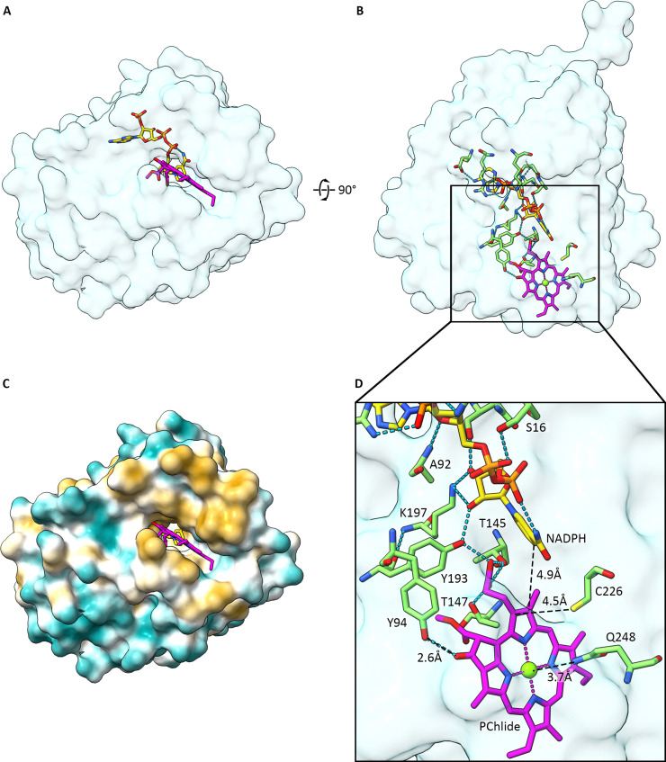
AF3 model of the light-dependent PChlide oxidoreductase. (A) and (B) Two surface views of the overall topology of the protein with 8V-PChlide and NADPH shown in magenta and yellow, respectively (heteroatom colours: blue – nitrogen, red – oxygen, orange – phosphate). (C) Surface hydrophobicity (gold – hydrophobic, blue – hydrophilic) showing an open substrate binding cleft for LPOR flanked by hydrophobic residues. (D) Expanded view of the active site highlighting important residues involved in binding NADPH and 8V-PChlide, and some residues implicated in catalysis. The backbone amino nitrogen of A92 interacts with the adenosine ribose ring oxygen of NADPH; the alcohol group of S16 interacts with one of the bridging phosphate groups closer to the adenosine moiety; K197 and Y193, previously implicated in reducing the C17 Created by potrace 1.16, written by Peter Selinger 2001-2019 C18 double bond, interact with the alcohol groups of the nicotinamide ribose. Y193, T145 and T147 appear to form interactions with the C17 propionate carboxyl of 8V-PChlide; Y94 interacts with the 131-keto group of 8V-PChlide; Q248 may provide a weak co-ordination bond with the central Mgviathe amide nitrogen. The distance from the C4 group of the hydride donor NADPH is 4.9 Å from the C18 carbon and C226 is 4.5 Å from the C17 carbon. See Table S1 in the SI for ipTM scores for the AF3 model and RMSD values for the correspondence of the AF3 model with experimentally determined structures in the RCSB PDB.
The differing positions of the loop and α-10 helix in the SynLPOR (AF3)/AraLPOR (7JK9) ternary complexes compared with the SynLPOR–NADPH (6R48/6L1G) structures have consequences for the PChlide binding site and reveal a new set of active site residues. Interestingly, the SynLPOR–NADPH structures (6R48/6L1G) may represent a state in the catalytic cycle where the α-10 helix points into the photosynthetic membrane to “fish” for substrates. In relation to the AF3 model, we note that Q248, found on the α-10 helix, is 3.7 Å from the central Mg, potentially acting as a transient ligand to the PChlide substrate. Two moieties are close to the C17–C18 double bond on ring D (Fig. 8D); the nicotinamide ring of NADPH is 5.4 Å from C17 and 4.9 Å from C18, and the corresponding distances for C226 are 4.5 Å and 3.9 Å, respectively. Thus, based on distance alone, and bearing in mind their modelled inaccuracies, the hydride from NADPH and the proton from C226 could attack either carbon. The C226S mutant has only 7% of wild-type activity,169and proton transfer for the C226A variant is > 200-fold slower than for wild-type LPOR.158Y193 and K197 were originally assigned as a catalytic motif, based on their conserved roles in the SDR family,154,165,167and are essential for activity,170–172but here Y193 and K197 interact with the NADPH ribose oxygens and Y193 with the C17 propionate group of PChlide. The model highlights other PChlide-interacting residues which may be important for ensuring correct orientation of the macrocycle and access to the C17–18 bond by the hydride. Y94 is predicted to interact with the C131keto group of PChlide, while T147 and T145 interact with the propionate group oxygens.
Once PChlide is established within the active site, transiently liganded by Q248, oriented by interactions with LPOR side chains such as Y94, Y193, T147 and T145, and with the C17 Created by potrace 1.16, written by Peter Selinger 2001-2019 C18 bond held adjacent to C226 and the NADPH nicotinamide ring, the ternary complex is primed for catalysis. The POR catalytic cycle is initiated when PChlide absorbs a photon, and a charge transfer state157polarises the C17 Created by potrace 1.16, written by Peter Selinger 2001-2019 C18 double bond, predisposing it to hydride transfer from the nearby NADPH. Several possibilities exist for hydride transfer, discussed in ref.173, and it appears that this multi-step process proceeds in about 500 nsviaan initial electron transfer followed by a hydrogen (proton plus electron) transfer.159,160The next step involves microsecond proton transfer from C226 to conclude reduction of the C17 Created by potrace 1.16, written by Peter Selinger 2001-2019 C18 bond. More detailed discussions are beyond the scope of this summary and can be found in ref.158,173and174. The product release steps have been investigated,162,163and in angiosperms they are likely associated with the rapid dissolution of PLBs,151proposed to be initiated when Chlide forms.152The geometry at C17 changes upon reduction, from trigonal to tetrahedral, which displaces the propionate out of plane, and in angiosperms this movement is proposed to push the α-10 helix, leading to dissociation of LPOR from the membrane.152
Step 5 – 8-vinyl reductase
The majority of phototrophs convert 8V-Chlideato 8E-Chlidea(ChlideainFig. 4, but also called MV-Chlidea), which enhances the absorption of red wavelengths by the QYband (Fig. 4), although some marine cyanobacteria retain the 8V group.12,21There are several types of 8V reductase (8VR, also called divinyl reductase, DVR); a gene encoding an 8VR, AT5G18660, was discovered inArabidopsis,175,176and 8VR activity was confirmed following recombinant production inE. coli.175Homologues of the protein encoded by AT5G18660, now called BciA, have been found in rice,177Rba. sphaeroides178and green sulfur bacteria,179for example. Whereas BciA is an NADPH-requiring enzyme,179there is another type of 8VR, BciB, that obtains electrons from reduced Fd. The recombinant BciB from the green sulfur bacteriumChloroherpeton thalassiumhas a flavin adenine dinucleotide (FAD) cofactor and two [4Fe–4S] clusters.180Among the cyanobacteria,A. marinais unusual in having both BciA and BciB,181and inSynechocystis, the main subject of this review, formation of the 8-ethyl group is catalysed only by BciB.182,183
The AF3 structure ofSynechocystisBciB (Slr1923) is shown inFig. 9. AF3 places an FAD and two [4Fe–4S] clusters within this enzyme, consistent with earlier experimental work,180and the model also shows a Fd boundviaa positively charged patch on BciB in an area formed by residues 9–40. Many of these interactions are between backbone atoms, but the H37 and K22 side chains are predicted to interact with Fd. We used the [2Fe–2S] PetF (Ssl0020), the same Fd modelled in the cyclase complex (Section 4.3), which docks with its [2Fe–2S] center 11.2 Å from the proximal [4Fe–4S] cluster of BciB. For comparison, the distance between the [2Fe–2S] center of Fd to the [4Fe–4S] FBacceptor of Photosystem I is 11.7 Å.184Subsequent electron transfers in BciB, between the [2Fe–2S] centers, and then to the redox active FADH2isoalloxazine nitrogen, are over distances of 7.9 Å and 7.0 Å, respectively. The final step transfers a hydride from FADH2which is 4.0 Å from the C8 vinyl group (Fig. 9D). The AF3 model also reveals two residues predicted to interact with the substrate macrocycle; D197 provides a putative weak coordination bond to the central Mg of 3,8-divinyl Chlideaand there is a hydrogen bond from S358 to the C131keto group. Features of the AF3 model of BciB are found in the FrhB subunit of the F420-reducing [NiFe]-hydrogenase fromMethanothermobacter marburgensis,185which has sequence homology with BciB.180FrhB also has a [4Fe–4S] cluster, an FAD, and a binding site for a tetrapyrrole substrate, coenzyme F420.
![AF3 model of the 8V reductase in complex with Ferredoxin, Fd, which provides the electrons for the reduction reaction, along with its cofactors, two [4Fe–4S] clusters and FADH2. (A) and (B) Two surface views of the overall topology of the protein with 8V-Chlide in magenta (heteroatoms coloured: blue – nitrogen, red – oxygen). Fe–S clusters are coloured by heteroatom: dark orange – iron, yellow – sulfur. FADH2is shown in yellow (heteroatoms coloured: blue – nitrogen, red – oxygen, orange – phosphate). These panels also highlight the relative location of the Fd [2Fe–2S] cluster, the 8VR [4Fe–4S] clusters, the FADH2cofactor and the 8V-Chlideasubstrate. (C) Surface hydrophobicity (gold – hydrophobic, blue – hydrophilic) showing an open substrate binding cleft for 8V reductase flanked by hydrophobic residues. (D) Expanded view of the active site highlighting the cysteine residues involved in coordinating the Fe–S clusters. In Fd, C40, C45, C48 and C78 coordinate a [2Fe–2S] cluster. In 8VR, the [4Fe–4S] cluster proximal to the Fd binding site is coordinated by C25, C28, C31 and C43, and the [4Fe–4S] cluster that reduces the vinyl group is coordinated by C171, C195, C259 and C262. Residues that interact with FADH2have been omitted for clarity. The black dashed lines indicate various distances: the Fd [2Fe–2S] cluster to the first 8VR [4Fe–4S] cluster (11.2 Å), the first 8VR [4Fe–4S] cluster to the second (7.9 Å), the second [4Fe–4S] cluster to the FADH2nitrogen (7.0 Å), and from the FADH2nitrogen to the 8V group of 8V-Chlidea(4.0 Å). Two residues interacting with the macrocycle substrate were also identified; S358 provides a hydrogen bond to the C131keto group and D197 may provide a coordination bond to the central Mg2+ion. See Table S1 in the SI for ipTM scores for the AF3 models and RMSD values for the correspondence of models with any experimentally determined structures in the RCSB PDB.](https://pub-4f2af807d45642319f8e7f07a4c88770.r2.dev/PMC13297055/d6cb00082g-f9.jpg)
AF3 model of the 8V reductase in complex with Ferredoxin, Fd, which provides the electrons for the reduction reaction, along with its cofactors, two [4Fe–4S] clusters and FADH2. (A) and (B) Two surface views of the overall topology of the protein with 8V-Chlide in magenta (heteroatoms coloured: blue – nitrogen, red – oxygen). Fe–S clusters are coloured by heteroatom: dark orange – iron, yellow – sulfur. FADH2is shown in yellow (heteroatoms coloured: blue – nitrogen, red – oxygen, orange – phosphate). These panels also highlight the relative location of the Fd [2Fe–2S] cluster, the 8VR [4Fe–4S] clusters, the FADH2cofactor and the 8V-Chlideasubstrate. (C) Surface hydrophobicity (gold – hydrophobic, blue – hydrophilic) showing an open substrate binding cleft for 8V reductase flanked by hydrophobic residues. (D) Expanded view of the active site highlighting the cysteine residues involved in coordinating the Fe–S clusters. In Fd, C40, C45, C48 and C78 coordinate a [2Fe–2S] cluster. In 8VR, the [4Fe–4S] cluster proximal to the Fd binding site is coordinated by C25, C28, C31 and C43, and the [4Fe–4S] cluster that reduces the vinyl group is coordinated by C171, C195, C259 and C262. Residues that interact with FADH2have been omitted for clarity. The black dashed lines indicate various distances: the Fd [2Fe–2S] cluster to the first 8VR [4Fe–4S] cluster (11.2 Å), the first 8VR [4Fe–4S] cluster to the second (7.9 Å), the second [4Fe–4S] cluster to the FADH2nitrogen (7.0 Å), and from the FADH2nitrogen to the 8V group of 8V-Chlidea(4.0 Å). Two residues interacting with the macrocycle substrate were also identified; S358 provides a hydrogen bond to the C131keto group and D197 may provide a coordination bond to the central Mg2+ion. See Table S1 in the SI for ipTM scores for the AF3 models and RMSD values for the correspondence of models with any experimentally determined structures in the RCSB PDB.
Step 6 – chlorophyll synthase
ChlG is the only a transmembrane enzyme in the Chl biosynthesis pathway, which reflects the large increase in hydrophobicity when the C17-propionate of the Chlide macrocycle is esterified by the C20 diterpenoid geranylgeraniol. Early studies had identified and assignedbchGgenes encoding the BChl synthase in purple bacteria,53,55and ChlG homologues followed, with overexpression inE. coliyielding active Chl synthase.186The hydrophobic nature of this enzyme and the inability to purify sufficient quantities for structural and kinetic studies delayed progress, but it was found that a ∼83 kDa ChlG2–HliD2complex accumulates in a mutant strain ofSynechocystis.187This complex could be purified and structure determination by cryo-EM yielded both apo (3.0 Å resolution) and GGPP-bound (3.2 Å) forms of the enzyme.188The overall structure comprises a central HliD dimer flanked on each side by a ChlG monomer consisting of nine TMHs that form a large substrate-binding cavity gated by cytoplasmic entry loops. These structural features have been found in other prenyltransferases,189,190and the similarities likely extend to their catalytic mechanisms. It was already known that ChlG associates with HliD, a high light-inducible protein (Hlip), and with the membrane protein insertion machinery,191but this work provides the first structure of the photoprotective ChlG2–HliD2complex. This advance also aids understanding of the HliD-mediated photoprotection of Chls delivered to the machinery for inserting nascent photosystem polypeptides into membranes.
The cryo-EM structure of the ChlG2–HliD2complex lacks Chlide bound within the active site, so AF3 was used to model the ChlG–GGPP–Chlide ternary complex188(Fig. 10). There is close agreement between the cryo-EM and AF3 models (RMSD = 1.69 Å), with the tetraprenyl chain of GGPP similarly positioned in a hydrophobic cleft in both cases, but in the AF3 model Chlidea(magenta), two catalytically important Mg2+ions (green) and GGPP (yellow) are all present.Fig. 10A and Bdisplays the overall semi-transparent shape of the ChlG monomer enclosing the transmembrane helices, andFig. 10Cshows the active site in more detail. Q218 interacts with the C132ester group, and Q305 coordinates the central Mg of Chlide, similar to the function of Q248 in POR; such coordination bonds help to position the macrocycle within an active site but are presumably weak enough to allow product release.
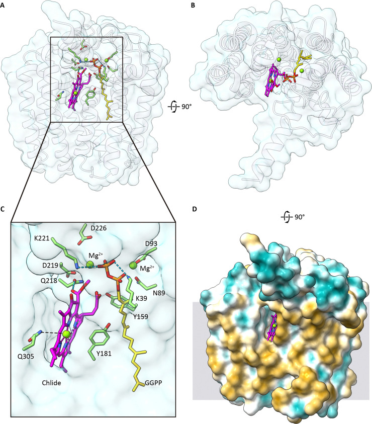
AF3 model of chlorophyll synthase in a ternary complex with its substrates Chlideaand GGPP. (A) and (B) Two surface views of the overall topology of the protein with Chlideaand GGPP in magenta and yellow, respectively (heteroatoms coloured: blue – nitrogen, red – oxygen, orange – phosphate). (C) Expanded view of the active site, showing several residues involved in substrate binding and activation. D93 and N89 coordinate a catalytic Mg2+, which interacts with the pyrophosphate group; D219 and D226 are hypothesised to play the same role with another Mg2+. K39 and K221 may also play an activating role, but at minimum they provide structural stability to the pyrophosphate group. Y159 and Y181 likely stabilise the ∂+terminal GG carbon. Q218 interacts with the C132ester group. Q305 provides a weak ligand to the central magnesium ion of Chlidea. (D) Surface hydrophobicity (gold – hydrophobic, blue – hydrophilic) showing the binding cleft for the Chlideasubstrate flanked by hydrophobic residues, and open to the membrane interior. The distribution of hydrophobic residues is consistent with indicating the transmembrane nature of ChlG – a cartoon representation of the membrane is shown in grey. See Table S1 in the SI for ipTM scores for the AF3 model and RMSD values for the correspondence of models with any experimentally determined structures in the RCSB PDB.
The esterifiable C17 carboxyl group of Chlideais positioned next to the pyrophosphate of GGPP (orange), which is stabilised by K39 and K221. Nearby, a catalytic Mg2+coordinated by D93 and the pyrophosphate group oxygens acts as a Lewis acid, drawing electron density away from the terminal GGPP carbon atom; D219 and D226 appear to play the same role with another Mg2+, which is also coordinated by pyrophosphate group oxygens on the other face of the molecule, adding to the electron withdrawing effect of the other Mg2+Collectively, these two Mg2+ions make the pyrophosphate a more potent leaving group.192We suggest that the terminal GG carbon is primed for a concerted SN2-type attack by the strongly nucleophilic C173carboxyl group oxygen, forming an ester bond between the GG moiety and the macrocycle. The SN2 mechanism proceeds efficiently if the directions of attack on one side of the ∂+terminal GG carbon and bond breakage on its distal side are linearly aligned, and the structural model188(Fig. 10) indicates a near-linear angle of 165°. The reaction can therefore proceed in a concerted manner, with no need for a two-stage SN1 mechanism involving a carbocation, as seen for other prenyltransferases.193Y159 and Y181 likely stabilise the ∂+terminal GG carbon and transition state intermediate, respectively.Fig. 10Dshows the substrate binding cleft of the enzyme with the hydrophobic surface facing out towards the membrane bilayer as expected. The membrane-intrinsic ChlG is therefore different from the other enzymes in the Chl pathway, which all have a hydrophobic face sitting on, rather than in, the membrane. This point will be discussed in section 6.
In terms of the substrate specificity of ChlG for Chlide rather than BChlide, it was shown that the I44F mutation enabled ChlG, heterologously produced inRba. sphaeroides, to participate in BChl biosynthesis.194We note that the AF3 model shows the B-ring of the Chlide macrocycle adjacent to I44, suggesting a link between the identity of the sidechain at position 44 and its compatibility with a chlorin with a C7 Created by potrace 1.16, written by Peter Selinger 2001-2019 C8 double bondversusa bacteriochlorin with a C7–C8 single bond.188Further mutagenesis, modelling and structural work should enhance our understanding of the selectivity of synthases for making either Chls or the redshifted BChls, accelerating progress towards the synthetic biology goal of engineering organisms to produce both Chls and BChls to expand the spectral range of photosynthesis;195see also Section 6.1.
Step 7 – geranylgeranyl diphosphate reductase
Early work on spinach chloroplasts had shown that ‘free’ GGPP could be converted to PPP in the chloroplast envelope, and a second pathway and location for reducing GG was found in the thylakoid membrane. This latter reaction initially esterified Chlideato form ChlGG, catalysed by Chl synthase, followed by reduction of the macrocycle-attached GG moiety, forming phytyl-Chla.196Both types of GG reduction required NADPH and proceeded in a stepwise mannerviathe DHGG and THGG intermediates. However, this work found a four-fold preference for PPP as the substrate for Chl synthase, in contrast to the synthase in etioplasts which favoured GGPP as a substrate, so the final step in Chl biosynthesis could be regarded as the attachment of PPP to Chlide. In support of this reaction sequence, PPP was found to be a better substrate for recombinant BChl and Chl synthases than GGPP.186A study of repair of damaged PSII inSynechocystisprovided evidence for de-esterification and re-esterification of Chl, with recycling of phytol and Chlide. Under these conditions at least, the substrates for Chl synthase are likely to be PPP and Chlide.197
Early genetic studies ofRba. capsulatusidentified mutations in the BChl biosynthesis genebchPwhich halted the pathway at the level of BChlGG,53and abchPmutant ofRba sphaeroidescould be partially complemented with a gene fromSynechocystis, assigned aschlP, providing evidence for strong similarities between the BchP and ChlP GGPP reductases.198RecombinantArabidopsisChlP produced inE. coliwas able to reduce GGPP to PPP as well as ChlGGto phytyl-Chl, suggesting that this enzyme can be recruited to serve both the prenylquinone and Chl pathways.199
We modelled the structure ofSynechocystisChlP using AF3.Fig. 11A and Bshow the macrocycle of ChlGG(magenta) within a cavity near a substrate entry site. Another feature seen for other Chl biosynthesis enzymes is a weak ligand to the central Mg, here formed by Q388 (Fig. 11C). The GG moiety extends away from the macrocycle into the interior of ChlP, along a pore (orange) that runs from the substrate entry site to the C4 atom of the nicotinamide group of NADPH, adjacent to a C41, which might play a catalytic role. However, there are three successive reductions along the GG tail, so the oxidised NADP+formed in converting GG to DHGG must be replaced with NADPH, and once more when THGG is finally reduced to phytyl. It is assumed that GGPP reductase possesses some processivity, allowing movement of the tail through the pore to enable these three reductions of the GG tail, namely the C6–7, C10–11 and C14–15 carbon double bonds, to take place. Our modelling does not provide any insights into any mechanistically relevant conformational movements. The predicted presence of a weak coordinating ligand, and a pore large enough to admit the GGPP tail and its attached macrocycle, are consistent with ChlGGas the substrate for GGPP reductase but do not exclude the same involvement for ‘free’ GGPP. So, although GGPP reductase is regarded as the final enzyme in the Chl pathway for the purpose of this review, another plausible sequence is the reduction of GGPP to PPP by GGPP reductase, and the final biosynthetic step would be esterification of Chlide by PPP, catalysed by Chl synthase.
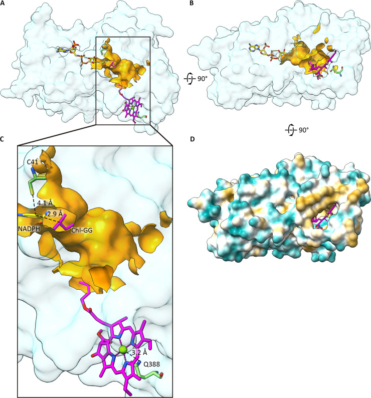
AF3 model of geranylgeranyl pyrophosphate reductase, ChlP. (A) and (B) Two surface views of the overall topology of the protein with ChlGGand NADPH shown in magenta and yellow, respectively (heteroatoms coloured: blue – nitrogen, red – oxygen, orange – phosphate). (C) Expanded view of the active site, showing several residues involved in substrate binding and activation, including a potential catalytic residue, C41, and a ChlGG-coordinating residue, Q388. Besides these two residues, the relatively well conserved NADPH binding residues and the residues which form the GGPP binding pore, no other obvious catalytic residues were identified. The C4 group of NADPH (where the hydride is located) is 2.9 Å from the GG tail and is 4.1 Å from C41. Panels A–C also highlight a pore (orange) that runs from the substrate entry site to the C4 atom of the nicotinamide group of NADPH. It is assumed the enzyme possesses some processivity, and the GG tail is likely to move through the pore as the reductions take place. (D) Surface hydrophobicity (gold – hydrophobic, blue – hydrophilic) showing the binding cleft for the ChlGGsubstrate flanked by hydrophobic residues. See Table S1 in the SI for ipTM scores for the AF3 model.
Finally,Fig. 11Dshows that the ChlGGsubstrate gains access to the active site through a pore flanked by hydrophobic ridges, which are proposed to form a membrane interface. Similar substrate pores/cavities, flanked by hydrophobic membrane interfaces, were predicted for the methytransferase, cyclase, LPOR, 8VR, Chlcsynthase and CAO enzymes (Fig. 6C, 7C, 8C, 9C, 13C and 14C), and this feature will be covered in more detail in Section 6.
![AF3 model of the trimeric chlorophyll(ide)aoxygenase (CAO) fromA. thaliana(subunits coloured in light blue, blue and violet), in a complex with its Chlideasubstrate, [2Fe–2S] and Fe2+cofactors, and Fd (pastel orange), which has its own [2Fe–2S] cofactor. (A, B) Two surface views of the overall topology of the CAO trimer with substrates and cofactors shown in stick representation. Chlideais in magenta (heteroatoms coloured: blue – nitrogen, red – oxygen); [2Fe–2S] clusters are coloured by heteroatom: dark orange – iron, yellow – sulfur; Fe2+is shown in dark orange. (C) Surface hydrophobicity (gold – hydrophobic, blue – hydrophilic) showing three open substrate-binding clefts for Chlideaflanked by hydrophobic residues. (D) Expanded view of the active site, highlighting the inter-subunit chain of redox-active cofactors, where electrons are transferred from the [2Fe–2S] cluster of the docked Fd to a [2Fe–2S] cluster bound within subunit A of CAO, and subsequently to an Fe2+ion held in CAO subunit B, where the Chlideasubstrate is also bound. Important substrate- and cofactor-interacting residues are shown, and distances are indicated by black dashed lines. See Table S1 in the SI for ipTM scores for the AF3 model.](https://pub-4f2af807d45642319f8e7f07a4c88770.r2.dev/PMC13297055/d6cb00082g-f13.jpg)
AF3 model of the trimeric chlorophyll(ide)aoxygenase (CAO) fromA. thaliana(subunits coloured in light blue, blue and violet), in a complex with its Chlideasubstrate, [2Fe–2S] and Fe2+cofactors, and Fd (pastel orange), which has its own [2Fe–2S] cofactor. (A, B) Two surface views of the overall topology of the CAO trimer with substrates and cofactors shown in stick representation. Chlideais in magenta (heteroatoms coloured: blue – nitrogen, red – oxygen); [2Fe–2S] clusters are coloured by heteroatom: dark orange – iron, yellow – sulfur; Fe2+is shown in dark orange. (C) Surface hydrophobicity (gold – hydrophobic, blue – hydrophilic) showing three open substrate-binding clefts for Chlideaflanked by hydrophobic residues. (D) Expanded view of the active site, highlighting the inter-subunit chain of redox-active cofactors, where electrons are transferred from the [2Fe–2S] cluster of the docked Fd to a [2Fe–2S] cluster bound within subunit A of CAO, and subsequently to an Fe2+ion held in CAO subunit B, where the Chlideasubstrate is also bound. Important substrate- and cofactor-interacting residues are shown, and distances are indicated by black dashed lines. See Table S1 in the SI for ipTM scores for the AF3 model.
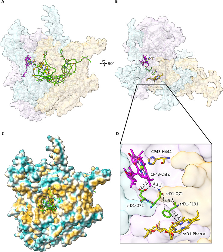
AF3 model of the srPSII Chlfsynthase composed of srD1 (light blue), D2 (pastel orange) and CP43 (light pink) with its co-factors P680, ChlD1, ChlD2, PheoD1, PheoD2, two PQ-9 molecules and beta-carotene (all shown in green, stick representation with heteroatoms coloured: blue – nitrogen, red – oxygen). Also shown is the proposed substrate, a CP43-bound Chla(magenta, stick representation with heteroatoms coloured: blue – nitrogen, red – oxygen). (A) and (B) Two surface views of the overall topology of the Chlfsynthase with transmembrane helixes shown in cartoon representation in light grey. (C) Illustration of the transmembrane nature of the synthase showing the surface hydrophobicity (gold – hydrophobic, blue – hydrophilic). (D) Residues relevant to the function of the srPSII Chlfsynthase enzyme, with distances indicated using black dashed lines. See Table S1 in the SI for ipTM scores for the AF3 model and RMSD values for the correspondence of models with any experimentally determined structures in the RCSB PDB.
Branches and extensions of the Chlapathway – the biosynthesis of Chlsb,c,dandf
Although Chlais the dominant such pigment on Earth, the extensions of the main pathway to form Chlsb,c,dandfare important because they allow plants, algae and cyanobacteria to colonise new ecological and spectral niches, by harvesting and using solar energy more effectively. Only one extra enzyme is apparently needed to create each Chl variant, and the structures of the respective synthases have been calculated using AF3.
Biosynthesis of Chlsc1andc2
Fig. 3shows that while Chlsa,b,dandfhave Chlideaas a common intermediate, Chlsc1andc2branch from the main pathway at the level of 8V-PChlidea. Brown algae, diatoms, and dioflagellates use these accessory Chls to absorb more of the blue light that filters through water columns, so they make a major contribution to marine productivity.20,200ACHLCgene encoding chlorophyllcsynthase was discovered in the marine diatomPhaeodactylum tricornutum, and recombinant CHLC protein converted 8V-PChlide and PChlide (MV–PChlide) to Chlc2and Chlc1, respectively,201in a reaction requiring Fe2+and 2-oxoglutarate (2OG). The synthase gene was also found in a dinoflagellate,Breviolum minutum, and its function was confirmed by heterologous expression inNicotiana benthamiana.202Here, the synthase has a predicted but dispensable Chla/bbinding domain, and an essential catalytic domain belonging to the superfamily of 2OG-Fe(ii) dioxygenases.203These catalytically flexible enzymes use 2OG and O2to oxidise a wide variety of substrates, most commonly hydroxylations but also desaturations,204as seen for CHLC, which forms an acrylate by introducing a C171–C172double bond into the C17 propionate.
In the absence of a Chlcsynthase structure, AF3 was used to calculate a model with its substrate and co-factors, 2-OG and Fe2+(Fig. 12). R167 interacts with 8V-PChlidea, providing hydrogen bonds to the C132methoxycarbonyl and the C17 propionate carbonyl oxygen, which presumably orients the C172carbon for attack by a water coordinated by the Fe2+ion. This Fe2+is coordinated by H216, R231, H298 and the 2-keto and 2-carbonyl groups of 2-OG, which leaves a single coordination site unoccupied and pointing towards the C172carbon where oxidation occurs, only 5.5 Å from the Fe2+. It is feasible that an activated, nucleophilic water is held in this final coordination site and abstracts a proton from the C172carbon initiating the oxidation. The C4 carbonyl group of 2-OG is also predicted to interact with Y162, R312 and W233, which presumably act to orient the 2-OG correctly and stabilise binding (Fig. 12D). As with most other enzyme structures of the Chl pathway, a hydrophobic patch surrounding the substrate binding cleft was identified, suggesting its docking to the membrane surface for substrate retrieval and product release (Fig. 12C).
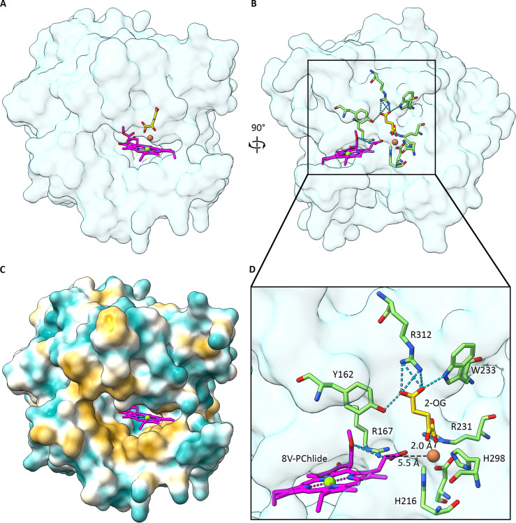
AF3 model of thePhaeodactylum tricornutumChlcsynthase, in a complex with its 8V-PChlideasubstrate and its cofactors, 2-oxoglutarate (2OG) and Fe2+. (A) and (B) Two surface views of the overall topology of the protein with substrates and cofactors shown in stick representation. 8V-PChlideais in magenta (heteroatoms coloured: blue – nitrogen, red – oxygen), 2-OG in yellow (heteroatom coloured: red – oxygen), and Fe2+is shown in dark orange. (C) Surface hydrophobicity (gold – hydrophobic, blue – hydrophilic) showing an open substrate binding cleft for 8V-PChlideaflanked by hydrophobic residues. (D) Expanded view of the active site highlighting important residues. R167 is predicted to form hydrogen bond interactions with the C132methoxycarbonyl and the C17 propionate group. 2-OG binding is stabilised by several interacting residues: the C4 carbonyl group is predicted to form interactions with Y162, R312 and W233, while the C2 carbonyl group interacts with R231. The 2-keto group of OG and one of the C2 carbonyl oxygens provide coordination bonds to the Fe2+, along with D218, R231 and H298. The remaining coordination site remains free and pointing at the C172carbon, where oxidation occurs. The distance between the Fe2+ion and the C172carbon is 5.5 Å. See Table S1 in the SI for ipTM scores for the AF3 model.
Biosynthesis of Chlb
Chlbhelps to fill the 450–600 nm ‘green gap’ between the B band and QYabsorption features of Chla(Fig. 2). This pigment differs from Chlain having a C-7 formyl group and it is highly abundant globally, by virtue of its essential role in the major light-harvesting complexes of algae and plants. Conversion of the 7-methyl group to a 7-formyl is catalysed by Chlaoxygenase (CAO), first discovered inC. reinhardtii.205This reaction requires molecular oxygen,206,207and CAO has consensus sequences for a Rieske-type [2Fe–2S] cluster and for a mononuclear non-heme Fe-binding site. A study of the recombinant enzyme fromA. thalianarequired reduced Fd, replenished by FNR and an NADPH-regenerating system. It was found that CAO was specific for Chlidearather than Chla, and that the formation of the 7-formyl group proceeded in two stepsviaa 71-OH intermediate,208the same transient metabolite found in the conversion of Chlbto Chla.209A later, detailed study with several recombinant enzymes showed they could catalyse the formation of Chlidebfrom Chlidea,210so the CAO acronym should refer to Chlideaoxygenase, as originally proposed.208This mechanistic work showed that CAO uses a novel Rieske chemistry to catalyse two successive monooxygenation reactions, and that CAO uses Rieske oxygenase chemistry to convert 71-OH–Chlideato Chlideb, establishing 71-OH–Chlideaas a true intermediate of the CAO reaction.210
Although there is no structure for CAO oxygenase, deep learning-based methods have been used to predict the tertiary structures of CAO fromA. thalianaand the PrasinophyteMicromonas pusilla, along with the Fd interaction site and the Chlabinding cavity.211Here, we used AF3 to calculate the trimeric structure of CAO fromA. thalianawith Fd, Chlideaand its [2Fe–2S] cluster and Fe2+cofactors (Fig. 13). The AF3 model shows that the Fd-binding site is formed by the interface between two neighbouring CAO subunits, the geometry of which necessitates a trimeric structure (Fig. 13B). Further indications of this constraint are found in the complex redox chain formed at the interface between two subunits, where Fd provides electrons to a [2Fe–2S] cluster in subunit A, after which electrons are transferred to a mononuclear Fe2+ion in subunit B, where the substrate binding site is located (Fig. 13D). The subunit A iron–sulfur cluster is located 16.2 Å from the Fd [2Fe–2S] cluster and is coordinated by A-C60, A-H62, A-C65 and A-C79. The mononuclear Fe2+ion is located 12 Å from the subunit A iron–sulfur cluster and is coordinated by B-N159, B-H165, B-H170 and B-D285; the Fe2+is ∼5 Å from the C7 methyl group of Chlidea, which is held transiently by a weak coordination bond with B-Q238. A further Fe2+coordination site is likely occupied by molecular oxygen, which would be ∼3 Å from the C7 methyl group of Chlidea. Progressive reductions of this oxygen likely activate the molecule for oxidation of the C7 methyl to a formyl group (Fig. 13D). As with most Chl biosynthesis enzymes, each CAO monomer has a hydrophobic patch surrounding the substrate binding cleft (Fig. 13C).
Biosynthesis of Chlsdandf
Although the extensive literature on these pigments cannot be covered here, there are many excellent reviews, such as,36that provide a detailed account of Chlsdandf. Briefly, ring A formyl groups in Chlsdandfredshift QYabsorption in Et2O to 686 and 695 nm, respectively (Fig. 2), and organisms able to make these pigments gain access to new spectral niches enriched in far-red light.8,36Chldis the major pigment in the cyanobacteriumA. marina35but, despite extensive efforts, no Chldsynthase has been found. Labelling experiments have shown that the 3-formyl is derived from molecular oxygen;212conversion of Chlato Chldhas been observed with thiol reagents213and a thiol protease,214and could involve cysteine-containing proteins such as allophycocyanins.215However, this part of the review on enzyme structures will concentrate on Chlf, as the Chlfsynthase has been identified.
Following the earlier isolation of Chldin 1943,216the discovery of Chlfin 201037was a landmark in Chl research. This most redshifted Chl, with two exocyclic conjugated groups on ring A, was discovered in a complex cyanobacterial community that forms mats that cover marine stromatolites. These layered, rock-like structures house complex consortia of cyanobacteria resulting in high levels of spectral shading deep within the stromatolite.217Culturing of samples from a stromatolite found in Shark Bay, Australia, under far-red light conditions, yielded a Chlf-containing filamentous cyanobacterium that was purified and named asHalomicronema hongdechloris.218Ganet al.219studied another cyanobacterium able to adapt to other niches enriched in far-red light,Leptolyngbyasp. strain JSC-1, and characterised the remodelling of the phycobilisome and photosystems as “far-red light photoacclimation” (FaRLiP). Far-red light activates a conserved cluster of 20 genes inLeptolyngbya, one of which,psbA4, encodes a functionally inactive (“super-rogue”) version of the PsbA subunit of PSII, identified previously in some diazotrophic bacteria,220and also found in two other cyanobacteria capable of FaRLiP, namelyChlorogloeopsis fritschiiPCC 9212 andSynechococcussp. PCC 7335. Heterologous expression of theC. fritschii psbA4gene inSynechococcussp. PCC 7002 conferred the ability to synthesise Chlf, showing thatpsbA4encodes at least part of the Chlfsynthase.221
Unlike the conversion of the C7 methyl to a formyl, catalysed by CAO (see Section 5.2), the oxidation of the ChlaC2 methyl group to a formyl in Chlfis light-induced, and the Chlfsynthase (ChlF) is a photo-oxidoreductase.221ChlF lacks most of the ligands for binding the water-oxidising Mn4Ca1O5cluster but it has Chla, pheophytina, tyrosine YZ, plastoquinone and binds carotenoids. Heterologous production and purification of [His]10-tagged ChlF fromFischerella thermalisPCC 7521 in a ΔpsbD1ΔpsbD2strain ofSynechococcussp. PCC 7002 yielded 3–4% Chlfrelative to total Chl content, about half the content of FaRLiP strains grown in FRL.222ChlF activity was inhibited by DCMU, consistent with plastoquinone as an electron acceptor, and alteration of the putative ChlZ, P680 and QAbinding sites inactivated the synthase but activity was unaffected by donor side electron transfer mutants.222The ChlZpigment was proposed as a candidate for oxidation to Chlf.223
Parallel studies were conducted onSynechocystisharbouring FLAG-tagged ChlF fromChroococcidiopsis thermalisPCC 7203 orF. thermalis.224,225An active PSII-like core complex was purified, termed the “super-rogue” PSII (srPSII) complex, which contained mainly ChlF (srD1), D2, CP47 and CP43, and enhanced expression of theC. thermalis chlfincreased production of Chlfpigment 30-fold, reaching 8.2% Chlf/Chla, a level comparable with that accumulated during FaRLiP acclimation.225M127–G128 in theSynechocystisPSII subunit D1 were replaced by the QD motif, which is conserved across ChlF srD1 proteins, and this small change enabled the production of 0.1% Chlf/Chla, demonstrating the likely role of this motif in Chlfproduction.224In one suggested mechanism for the oxidation of the ChlaC2 methyl group to a formyl, the QD motif lies 13 Å from the proposed site for generating reactive oxygen species (ROS), namely the pheophytinain the Chlfsynthase corresponding to the redox-active electron acceptor in PSII.224
Here, we used AF3 to generate a structural model of Chlfsynthase based on ChlF/srD1 fromSynechoccocussp. PCC 7335, which includes CP43 (light pink), srD1 and D2 (Fig. 14). All the Chls and carotenoids for CP43 were modelled but are not shown. The srPSII complex, comprising srD1 (light blue) and D2 (pastel orange), is shown with most of the usual PSII co-factors highlighted in green, except for the proposed catalytic srD1–pheophytina, which is shown in yellow (Fig. 14A). Following initial charge separation in the Chlfsynthase, the reduced pheophytin (srD1–PheoainFig. 14D) transfers an electron to a nearby oxygen generating a catalytically required ROS. SrD1–F191 is 3.2 Å from srD1–Pheoaand 4.9 Å from the QD motif, and may mediate electron transfer from the pheophytin to molecular oxygen (not shown) bound near the C2 methyl group of the Chlashown in magenta inFig. 14D. This Chl, the proposed substrate of the Chlfsynthase, forms a ligand to CP43 at the interface of the CP43–srD1 complex. The role of the QD motif is unclear but it may stabilise a transition state on the way to oxidation of the C2 methyl group to a formyl. Alternatively, it could stabilise the ROS such that oxidation of the C2 methyl group is favoured.
The discovery of Chlfhas had a significant impact on photosynthesis research, from its role in antenna complexes,226,227primary photochemistry;228,229and its possible future contribution to enhancing crop productivity.230,231Thus, there has been a lot of interest in conferring the ability to make Chlfon plants, so they can use more >700 nm light for photosynthesis, but this requires more knowledge of the Chlfsynthase. If Chlais the substrate, Chlfsynthase could lie in proximity to Chl synthase and the geranylgeranyl reductase ChlP, ready to accept their Chlaproduct. Another possibility is that the true substrate for the Chlfsynthase is Chlidea, and the Chlidefproduct would require coupling to ChlG and ChlP for attachment and reduction of GGPP. Co-location of the Chlfsynthase with other membrane-attached and membrane-embedded Chl biosynthesis enzymes is likely to be important, as would integration with the machinery for photosystem assembly.191Then, heterologously produced Chlfmust finally be incorporated into photosystems, replacing some native Chlapigments, as already observed for hybrid PSI complexes in a strain ofSynechococcussp. PCC 7002 that expresses the Chlfsynthase gene.232
Transfer of substrates and products between chlorophyll biosynthesis enzymes
The biosynthetic intermediates in the Chl pathway absorb light and are potentially photolabile, with the capacity to generate harmful ROS. Yet Chl biosynthesis and the assembly of Chls into active photosystems, as well as the repair of damaged complexes, must take place in the glare of the Sun. These considerations necessitate the rapid transfer of metabolites between enzymes, so the final Chl product can be efficiently assimilated into photosystem complexes where carotenoids can provide some measure of photoprotection. Perhaps the Chl synthase, coupled as it is to the membrane assembly machinery,191is especially vulnerable, hence the complex formed with carotenoid-binding Hlips.187One way to achieve efficient transfers of substrates and products between enzymes would be to ‘hardwire’ the whole pathway togetherviaa set of specific protein–protein associations to form a Chl biosynthesis ‘supercomplex’; this level of organisation would promote substrate channelling between enzymes and prevent the intermediates from diffusing randomly (and damagingly slowly) to find their cognate pathway enzymes.
The availability of a complete set of Chl enzyme structures, some of which are in the PDB but here are all predicted by AF3, suggests another possibility; as noted in the previous section, five of the seven pathway enzymes (methytransferase, cyclase, POR, 8VR, GG reductase;Fig. 6C, 7C, 8C, 9C and 11D) appear to have a pore or active site cleft flanked by hydrophobic residues, which would allow each enzyme to sit on the membrane. The same features were found for the Chlcsynthase (Fig. 12C) and CAO enzymes (Fig. 13C). Thus, Chl pathway intermediates could migrate between enzymes, and gain access to active sites,viathe underlying membrane, which acts as a quasi-two-dimensional solvent. In the case of the membrane-intrinsic Chl synthase (Fig. 10D) and Chlfsynthase (Fig. 14C), the active site clefts are accessed directly from within the bilayer. Thus, among our AF3 structures, MgCh is the only outlier, although a positively charged patch on ChlH could allow interaction of the MgCh complex with the negatively charged thylakoid membrane (see Section 4.1.3).
We note that bilayer-forming lipids comprise only about half of the thylakoid membrane, with the remainder consisting of the non-bilayer lipid monogalactosyldiacylglycerol (MGDG); however, dense packing of membrane-intrinsic proteins such as photosystems can enforce local bilayer formation.233It has been proposed that MGDG could create partially autonomous membrane domains,233and perhaps one of these regions could encompass or house a Chl biosynthesis pathway.Fig. 15depicts a cluster of membrane-associated Chl biosynthesis enzymes, but this model requires no particular organisation of enzymes, only their proximity. Communication between Chl biosynthesis components is proposed to rely on their shared location, forming a nanodomain on the membrane. If the pathway enzymes associate loosely to occupy 400 nm2of membrane surface, roughly equivalent to a membrane volume of 2000 nm3, then one molecule of a biosynthetic intermediate has an effective concentration of approximately 0.8 mM. The kinetic parameters for these enzymes have been measuredin vitroin the bulk phase, generally yieldingKdvalues in the 1–5 µM range,79–81,102,110,111,130,155so with the minimal assumptions used here the effective intramembrane concentrations of Chl biosynthesis intermediates are much higher than their binding site affinities, thereby promoting formation of enzyme–substrate complexes. Such concentrations also greatly exceed theKdvalues for enzyme–product complexes; for example, theKdvalues for the porphyrin substrate and Mg–porphyrin product binding to ChlH do not differ much and are 4.0 and 5.2 µM, respectively.81If this is the case, release of a product from the active site into the membrane bilayer will be more likely when the next enzyme in the pathway is ready to sequester this Chl intermediate within its own active site.
However, rather than considering a bulk parameter such asKdthe Chl pathway (and likely others), is best thought of as a series of single molecule encounters governed by a set of probabilities, where each Chl intermediate engages in a form of intermittent searching234for its target, namely the next enzyme in the pathway. Each product transfers from its active site to the membrane, where it could randomly encounter up to five pathway enzymes with mostly unsuccessful (less probable) attempts at binding before finding its correct (most probable) active site. This intermittent search strategy is made easier if the enzyme targets are clustered in patches, and if the search is confined to two dimensions,234which are both likely for the Chl pathway. Thus, the searching time is minimised, and although the diffusion rates for Chl intermediates in membranes are not known, a large molecule such as ubiquinone-10 (C59H90O4) can diffuse between photosystem and cytochrome complexes in milliseconds.235Given the slow catalytic rates for some of the Chl pathway enzymes, such as 0.9 min−1for the cyclase,130Chl intermediates engaged in rapid, local searching for active sites would likely not limit the operation of the Chl pathway. There are useful regulatory consequences for this mechanism: the chain of product release/searching/active site binding events would rapidly come to a halt if one or more active target sites are already occupied and so unavailable to bind Chl intermediates. Chl enzymes would be locked in a product binding state, highly unlikely to discharge their products into the membrane because a product molecule is already in the vicinity (see the earlier point regardingKdvalues for ‘free’ products exceeding those for enzyme–product complexes). Thus, a failure to complete Chl–protein assembly feeds back to Chl synthase because of the link between the synthase and the membrane insertase,191and then product inhibition is propagated back along the series of Chl enzymes in the nanodomainviaa series of single molecule encounters, shutting down the Chl pathway and preventing the accumulation of toxic Chl intermediates. Such a mechanism might explain why there is cessation of the BChl pathway in mutants ofRba. sphaeroideslacking genes encoding BChl–binding proteins,236and no accumulation of pathway intermediates.
Consequences of the membrane transfer model for engineering hybrid biosynthetic pathways
Given the proposed lack of specific contacts between the Chl enzymes, it should be possible to assemble hybrid (B)Chl pathways from components obtained from a variety of bacteria, or from plant and bacterial sources. As long as the enzymes can congregate at a membrane surface, metabolites should be able to diffuse between them through the membrane bilayer. There are several examples of engineering native (B)Chl pathways, and of installing pathwaysde novoin purely heterotrophic bacteria, all of which rely on metabolites moving between (B)Chl enzymes in non-native contexts.Rba. sphaeroideshas been used as a chassis to demonstrate the function of Chlide oxidoreductases (CORs) fromRhodopseudomonas palustris,Cba. tepidum, andRoseiflexus castenholzii.237The native BChlapathway ofRba. sphaeroideshas been modified to synthesise BChlbby replacing the native COR genes with those fromBlastochloris viridis;238further modification of theRba. sphaeroidesisoprenoid biosynthetic pathway, and replacing the native BChl synthase with its counterpart fromH. modesticaldum, yielded BChlgesterified with farnesol (BChlgF).239ExpressingchlPandchlGfromSynechocystisinRba. sphaeroides, re-routes the native BChlapathway to Chla.240The oxidative MgPME cyclases can be swapped betweenRvi. gelatinosusandSynechocystis, and they still function.118Extending this work, the MgPME cyclases fromSynechocystisandA. thalianaintegrate into the BChl pathway inRvi. gelatinosus.120An early example using anE. colichassis functionally linked the MgCh and methyltransferase enzymes fromSynechocystis,241and complete hybrid Chl and BChl pathways could be assembled inE. coliusing components fromSynechocystis,Rvi. gelatinosusandRba. sphaeroides.14,120Finally, cyanobacterial and plant chlorophyll synthases function inRba. sphaeroides,194,242,243and algal and plant Chl synthases can replace the native enzyme inSynechocystis, integrating into the Chl pathway and allowing normal assembly and function of photosystems.244
These many examples show that heterologous (B)Chl pathways can be assembled, and as long as there is only one foreign component a pathway can apparently function efficiently. However, pathways with diverse components often perform poorly when they are assembled in a heterotrophic host. For example, Chl or BChl production inE. coliis numbered in thousands of molecules per cell rather than the native millions in a photosynthetic bacterium, and efforts should be made to assemble these pathways with the normal stoichiometries and cellular levels of enzymes. Quantitative analysis of theSynechocystisChl biosynthesis enzymes, in terms of copies per cell (cpc), yielded numbers in the 1150–3550 cpc range, with ChlI, Ycf54 and LPOR somewhat higher and 8VR and ChlG in the 250–1000 range.245The stoichiometries of Chl biosynthesis enzymes revealed in that study, combined with their respectivekcatvalues (where available), highlight the potential for regulating various steps. For example, 500–1200 cpc of MgCh complexes might produce only 7–16 molecules of MgPIX s−1cell−1, based on akcatof 0.013 s−1,80,95whereas the methyltransferase has 105times more catalytic capacity on a cellular basis. As a result, there should be very little ‘free’ MgPIX. There are also mismatches between the very limited cyclase activity (1150–1700 cpc;kcatof 0.015 s−1; 7–16 molecules MgPIX s−1cell−1), and the 4–7-fold greater capacity of the next pathway enzyme, LPOR.245Natively, photosynthetic cells have used these stoichiometries and (presumed) enzyme clustering to overcome the limitations of the enzyme chemistries and enzyme assembly, but pathways newly installed in a heterotrophic host effectively must start again, and there is much work to do before they can operate efficiently.
Author contributions
CNH performed the literature search, preparedFig. 1–4 and 15, wrote and edited the manuscript; FSM-B preparedFig. 5–14, wrote and edited the manuscript.
Conflicts of interest
There are no conflicts to declare.
Acknowledgments
CNH and FSM-B gratefully acknowledge funding from European Research Council Synergy award 854126. We are indebted to Professor Jonathan Lindsey, Dr Masahiko Taniguchi, Professor Peter Nixon, Professor Nicholas Williams, Dr James Reid, and Dr Andrew Hitchcock for their helpful advice, and again to Dr Andrew Hitchcock for his careful reading of the manuscript.
Data availability
The data supporting this article have been included as part of the supplementary information (SI). Supplementary information: Table S1. Structure files for AF3 models are available for download. See DOI:https://doi.org/10.1039/d6cb00082g.
Associated Data
Data Availability Statement
The data supporting this article have been included as part of the supplementary information (SI). Supplementary information: Table S1. Structure files for AF3 models are available for download. See DOI:https://doi.org/10.1039/d6cb00082g.
References
- Li W. Ciais P. Wang Y. Yin Y. Peng S. Zhu Z. Bastos A. Yue C. Ballantyne A. P. Broquet G. Geophys. Res. Lett. 2018;45:1058–1068.
- Breidenbach R. Castelfranco A. Castelfranco J. Govindjee G. Smith K. Stemler A. Photosynth. Res. 2023;157:1–11. doi.org/10.1007/s11120-023-01017-x
- Bar-On Y. M. Milo R. Cell. 2019;179:1451–1454. doi.org/10.1016/j.cell.2019.11.018
- Bar-On Y. M. Phillips R. Milo R. Proc. Natl. Acad. Sci. U. S. A. 2018;115:6506–6511. doi.org/10.1073/pnas.1711842115
- Rüdiger W. Phytochemistry. 1997;46:1151–1167.
- Willows R. D., Advances in Botanical Research, Elsevier, 2019, vol. 90, pp. 141–182
- Willows R. D. Photosynthesis in algae: biochemical and physiological mechanisms. 2020:83–103. doi.org/10.1007/978-3-030-33397-3_5
- Chen M. Annu. Rev. Biochem. 2014;83:317–340. doi.org/10.1146/annurev-biochem-072711-162943
- Scheer H., Chlorophylls and bacteriochlorophylls: biochemistry, biophysics, functions and applications, 2006, pp. 1–26
- Tamiaki H. Kichishima S. Plant Cell Physiol. 2025;66:153–167. doi.org/10.1093/pcp/pcae094
- Senge M. O. Ryan A. A. Letchford K. A. MacGowan S. A. Mielke T. Symmetry. 2014;6:781–843.
- Larkum A. Ritchie R. Raven J. Photosynthetica. 2018;56:11–43.
- Brzezowski P. Richter A. S. Grimm B. Biochim. Biophys. Acta, Bioenerg. 1847;2015:968–985. doi.org/10.1016/j.bbabio.2015.05.007
- Chen G. E. Canniffe D. P. Barnett S. F. Hollingshead S. Brindley A. A. Vasilev C. Bryant D. A. Hunter C. N. Sci. Adv. 2018;4:eaaq1407. doi.org/10.1126/sciadv.aaq1407
- Chen G. E. Hunter C. N. ACS Synth. Biol. 2023;12:2236–2244. doi.org/10.1021/acssynbio.3c00237
- Taniguchi M. Lindsey J. S. Photochem. Photobiol. 2021;97:136–165. doi.org/10.1111/php.13319
- Gouterman M. J. Mol. Spectrosc. 1961;6:138–163.
- Hanson L. Chlorophylls. 1991:993–1014.
- Björn L. O. Papageorgiou G. C. Blankenship R. E. Govindjee Photosynth. Res. 2009;99:85–98. doi.org/10.1007/s11120-008-9395-x
- Myśliwa-Kurdziel B., Latowski D. and Strzałka K., Advances in Botanical Research, Elsevier, 2019, vol. 90, pp. 91–119
- Stomp M. Huisman J. Stal L. J. Matthijs H. C. ISME J. 2007;1:271–282. doi.org/10.1038/ismej.2007.59
- Behrenfeld M. J. O’Malley R. T. Siegel D. A. McClain C. R. Sarmiento J. L. Feldman G. C. Milligan A. J. Falkowski P. G. Letelier R. M. Boss E. S. Nature. 2006;444:752–755. doi.org/10.1038/nature05317
- Silsbe G. M. Fox J. Westberry T. K. Halsey K. H. Nat. Commun. 2025;16:5821. doi.org/10.1038/s41467-025-60906-y
- Behrenfeld M. J. Boss E. Siegel D. A. Shea D. M. Global Biogeochem. Cycles. 2005;19:GB1006. doi.org/10.1029/2004GB002299
- Kume A. Akitsu T. Nasahara K. N. J. Plant Res. 2018;131:961–972. doi.org/10.1007/s10265-018-1052-7
- Su X. Ma J. Wei X. Cao P. Zhu D. Chang W. Liu Z. Zhang X. Li M. Science. 2017;357:815–820. doi.org/10.1126/science.aan0327
- Sheng X. Liu Z. Kim E. Minagawa J. Plant Cell Physiol. 2021;62:1108–1120. doi.org/10.1093/pcp/pcab072
- Croce R. Van Amerongen H. Nat. Chem. Biol. 2014;10:492–501. doi.org/10.1038/nchembio.1555
- Drop B. Webber-Birungi M. Yadav S. K. Filipowicz-Szymanska A. Fusetti F. Boekema E. J. Croce R. Biochim. Biophys. Acta, Bioenerg. 1837;2014:63–72. doi.org/10.1016/j.bbabio.2013.07.012
- Novoderezhkin V. Marin A. van Grondelle R. Phys. Chem. Chem. Phys. 2011;13:17093–17103. doi.org/10.1039/c1cp21079c
- van Amerongen H. Croce R. Plant Cell. 2025;37(11):koaf240. doi.org/10.1093/plcell/koaf240
- Partensky F. Hess W. R. Vaulot D. Microbiol. Mol. Biol. Rev. 1999;63:106–127. doi.org/10.1128/mmbr.63.1.106-127.1999
- Partensky F. Garczarek L. Ann. Rev. Mar. Sci. 2010;2:305–331. doi.org/10.1146/annurev-marine-120308-081034
- Ralf G. Repeta D. J. Limnol. Oceanogr. 1992;37:425–433.
- Miyashita H. Nature. 1996;338:402.
- Chen M., Advances in botanical research, Elsevier, 2019, vol. 90, pp. 121–139
- Chen M. Schliep M. Willows R. D. Cai Z.-L. Neilan B. A. Scheer H. Science. 2010;329:1318–1319. doi.org/10.1126/science.1191127
- Brune D. C. Nozawa T. Blankenship R. E. Biochemistry. 1987;26:8644–8652. doi.org/10.1021/bi00400a023
- Harada J. Mizoguchi T. Tsukatani Y. Noguchi M. Tamiaki H. Sci. Rep. 2012;2:671. doi.org/10.1038/srep00671
- Bryant D. A. Canniffe D. P. J. Phys. B: At., Mol. Opt. Phys. 2018;51:033001.
- Adams P. G. Cadby A. J. Robinson B. Tsukatani Y. Tank M. Wen J. Blankenship R. E. Bryant D. A. Hunter C. N. Biochim. Biophys. Acta, Bioenerg. 1827;2013:1235–1244. doi.org/10.1016/j.bbabio.2013.07.004
- Matsubara S. Shoji S. Tamiaki H. Chem. Commun. 2024;60:12513–12524. doi.org/10.1039/d4cc04363d
- Chew A. G. M. Frigaard N.-U. Bryant D. A. J. Bacteriol. 2007;189:6176–6184. doi.org/10.1128/JB.00519-07
- Bryant D. A. Hunter C. N. Warren M. J. J. Biol. Chem. 2020;295:6888–6925. doi.org/10.1074/jbc.REV120.006194
- Fiedor L. Kania A. Myśliwa-Kurdziel B. Orzeł Ł. Stochel G. Biochim. Biophys. Acta, Bioenerg. 1777;2008:1491–1500. doi.org/10.1016/j.bbabio.2008.09.005
- Fowler G. J. S. Sockalingum G. D. Robert B. Hunter C. N. Biochem. J. 1994;299:695–700. doi.org/10.1042/bj2990695
- Olsen J. D. Sockalingum G. D. Robert B. Hunter C. N. Proc. Natl. Acad. Sci. U. S. A. 1994;91:7124–7128. doi.org/10.1073/pnas.91.15.7124
- Furukawa H. Oba T. Tamiaki H. Watanabe T. Bull. Chem. Soc. Jpn. 2000;73:1341–1351.
- Kobayashi M. Watanabe T. Nakazato M. Ikegami I. Hiyama T. Matsunaga T. Murata N. Biochim. Biophys. Acta, Bioenerg. 1988;936:81–89.
- Kobayashi M. van de Meent E. J. Erkelens C. Amesz J. Ikegami I. Watanabe T. Biochim. Biophys. Acta, Bioenerg. 1991;1057:89–96.
- Gisriel C. Sarrou I. Ferlez B. Golbeck J. H. Redding K. E. Fromme R. Science. 2017;357:1021–1025. doi.org/10.1126/science.aan5611
- Katz J. J. Strain H. H. Harkness A. L. Studier M. H. Svec W. A. Janson T. R. Cope B. T. J. Am. Chem. Soc. 1972;94:7938–7939. doi.org/10.1021/ja00777a054
- Bollivar D. W. Wang S. Allen J. P. Bauer C. E. Biochemistry. 1994;33:12763–12768. doi.org/10.1021/bi00209a006
- Addlesee H. A. Fiedor L. Hunter C. N. J. Bacteriol. 2000;182:3175–3182. doi.org/10.1128/jb.182.11.3175-3182.2000
- Coomber S. Chaudhri M. Connor A. Britton G. Hunter C. Mol. Microbiol. 1990;4:977–989. doi.org/10.1111/j.1365-2958.1990.tb00670.x
- McDermott G. Prince S. M. Freer A. A. Hawthornthwaite-Lawless A. M. Papiz M. Z. Cogdell R. J. Isaacs N. W. Nature. 1995;374:517–521. doi.org/10.1016/s0969-2126(96)00050-0
- Qian P. Siebert C. A. Wang P. Canniffe D. P. Hunter C. N. Nature. 2018;556:203–208. doi.org/10.1038/s41586-018-0014-5
- Gabruk M. Mysliwa-Kurdziel B. Biochemistry. 2015;54:5255–5262. doi.org/10.1021/acs.biochem.5b00704
- Abramson J. Adler J. Dunger J. Evans R. Green T. Pritzel A. Ronneberger O. Willmore L. Ballard A. J. Bambrick J. Nature. 2024;630:493–500. doi.org/10.1038/s41586-024-07487-w
- Kryshtafovych A. Schwede T. Topf M. Fidelis K. Moult J. Proteins: Struct., Funct., Bioinf. 2021;89:1607–1617. doi.org/10.1002/prot.26237
- Kryshtafovych A. Schwede T. Topf M. Fidelis K. Moult J. Proteins: Struct., Funct., Bioinf. 2023;91:1539–1549. doi.org/10.1002/prot.26617
- Oeffner R. D. Croll T. I. Millán C. Poon B. K. Schlicksup C. J. Read R. J. Terwilliger T. C. Biol. Crystallogr. 2022;78:1303–1314. doi.org/10.1107/S2059798322010026
- Terwilliger T. C. Afonine P. V. Liebschner D. Croll T. I. McCoy A. J. Oeffner R. D. Williams C. J. Poon B. K. Richardson J. S. Read R. J. Biol. Crystallogr. 2023;79:234–244. doi.org/10.1107/S205979832300102X
- Terwilliger T. C. Liebschner D. Croll T. I. Williams C. J. McCoy A. J. Poon B. K. Afonine P. V. Oeffner R. D. Richardson J. S. Read R. J. Nat. Methods. 2024;21:110–116. doi.org/10.1038/s41592-023-02087-4
- Corum M. R. Venkannagari H. Hryc C. F. Baker M. L. Biophys. J. 2024;123:435–450. doi.org/10.1016/j.bpj.2024.01.021
- Wicky B. I. Milles L. F. Courbet A. Ragotte R. J. Dauparas J. Kinfu E. Tipps S. Kibler R. D. Baek M. DiMaio F. Science. 2022;378:56–61. doi.org/10.1126/science.add1964
- Jendrusch M. A. Yang A. L. Cacace E. Bobonis J. Voogdt C. G. Kaspar S. Schweimer K. Perez-Borrajero C. Lapouge K. Scheurich J. Mol. Syst. Biol. 2025;21:1166. doi.org/10.1038/s44320-025-00119-z
- Lavallee D. K. Coord. Chem. Rev. 1985;61:55–96.
- Fleischer E. B. Wang J. H. J. Am. Chem. Soc. 1960;82:3498–3502.
- Baum S. J. Plane R. A. J. Am. Chem. Soc. 1966;88:910–913.
- Fleischer E. B. Choi E. Hambright P. Stone A. Inorg. Chem. 1964;3:1284–1287.
- Shen Y. Ryde U. J. Inorg. Biochem. 2004;98:878–895. doi.org/10.1016/j.jinorgbio.2004.01.004
- Shen Y. Ryde U. Chem. – Eur. J. 2005;11:1549–1564. doi.org/10.1002/chem.200400298
- Gibson L. Willows R. D. Kannangara C. G. von Wettstein D. Hunter C. N. Proc. Natl. Acad. Sci. U. S. A. 1995;92:1941–1944. doi.org/10.1073/pnas.92.6.1941
- Jensen P. E. Gibson L. C. D. Henningsen K. W. Hunter C. N. J. Biol. Chem. 1996;271:16662–16667. doi.org/10.1074/jbc.271.28.16662
- Gibson L. C. Jensen P. E. Hunter C. N. Biochem. J. 1999;337:243–251.
- Jensen P. E. Gibson L. C. D. Hunter C. N. Biochem. J. 1998;334:335–344. doi.org/10.1042/bj3340335
- Jensen P. E. Gibson L. C. D. Hunter C. N. Biochem. J. 1999;339:127–134.
- Reid J. D. Siebert C. A. Bullough P. A. Hunter C. N. Biochemistry. 2003;42:6912–6920. doi.org/10.1021/bi034082q
- Reid J. D. Hunter C. N. J. Biol. Chem. 2004;279:26893–26899. doi.org/10.1074/jbc.M400958200
- Karger G. A. Reid J. D. Hunter C. N. Biochemistry. 2001;40:9291–9299. doi.org/10.1021/bi010562a
- Fodje M. N. Hansson A. Hansson M. Olsen J. G. Gough S. Willows R. D. Al-Karadaghi S. J. Mol. Biol. 2001;311:111–122. doi.org/10.1006/jmbi.2001.4834
- Lundqvist J. Elmlund H. Wulff R. P. Berglund L. Elmlund D. Emanuelsson C. Hebert H. Willows R. D. Hansson M. Lindahl M. Al-Karadaghi S. Structure. 2010;18:354–365. doi.org/10.1016/j.str.2010.01.001
- Lundqvist J. Braumann I. Kurowska M. Müller A. H. Hansson M. J. Biol. Chem. 2013;288:24012–24019. doi.org/10.1074/jbc.M113.480012
- Gao Y.-S. Wang Y.-L. Wang X. Liu L. Protein Sci. 2020;29:1026–1032.
- Shvarev D. Scholz A. I. Moeller A. mBio. 2023;14:e01893–01823. doi.org/10.1128/mbio.01893-23
- Chen X. Pu H. Fang Y. Wang X. Zhao S. Lin Y. Zhang M. Dai H.-E. Gong W. Liu L. Nat. Plants. 2015;1:1–5. doi.org/10.1038/nplants.2015.125
- Adams N. B. Bisson C. Brindley A. A. Farmer D. A. Davison P. A. Reid J. D. Hunter C. N. Nat. Plants. 2020;6:1491–1502. doi.org/10.1038/s41477-020-00806-9
- Farmer D. A. Brindley A. A. Hitchcock A. Jackson P. J. Johnson B. Dickman M. J. Hunter C. N. Reid J. D. Adams N. B. P. Biochem. J. 2019;476:1875–1887. doi.org/10.1042/BCJ20190095
- Axelsson E. Lundqvist J. Sawicki A. Nilsson S. Schroder I. Al-Karadaghi S. Willows R. D. Hansson M. Plant Cell. 2006;18:3606–3616. doi.org/10.1105/tpc.106.042374
- Brindley A. A. Adams N. B. Hunter C. N. Reid J. D. Biochemistry. 2015;54:6659–6662. doi.org/10.1021/acs.biochem.5b01080
- Adams N. B. P. Vasilev C. Brindley A. A. Hunter C. N. J. Am. Chem. Soc. 2016;138:6591–6597. doi.org/10.1021/jacs.6b02827
- Adams N. B. Brindley A. A. Hunter C. N. Reid J. D. FEBS Lett. 2016;590:1687–1693. doi.org/10.1002/1873-3468.12214
- Adams N. B. Reid J. D. J. Biol. Chem. 2013;288:28727–28732. doi.org/10.1074/jbc.M113.477943
- Viney J. Davison P. A. Hunter C. N. Reid J. D. Biochemistry. 2007;46:12788–12794. doi.org/10.1021/bi701515y
- Adams N. B. Marklew C. J. Brindley A. A. Hunter C. N. Reid J. D. Biochem. J. 2014;457:163–170. doi.org/10.1042/BJ20130834
- Jessop M. Felix J. Gutsche I. Curr. Opin. Struct. Biol. 2021;66:119–128. doi.org/10.1016/j.sbi.2020.10.027
- Khan Y. A. White K. I. Brunger A. T. Crit. Rev. Biochem. Mol. Biol. 2022;57:156–187. doi.org/10.1080/10409238.2021.1979460
- Lin Z. Akin H. Rao R. Hie B. Zhu Z. Lu W. Smetanin N. Verkuil R. Kabeli O. Shmueli Y. Science. 2023;379:1123–1130. doi.org/10.1126/science.ade2574
- Sawicki A. Zhou S. Kwiatkowski K. Luo M. Willows R. D. Biochem. J. 2017;474:2095–2105. doi.org/10.1042/BCJ20161094
- Larkin R. M. Alonso J. M. Ecker J. R. Chory J. Science. 2003;299:902–906. doi.org/10.1126/science.1079978
- Davison P. A. Schubert H. L. Reid J. D. Iorg C. D. Heroux A. Hill C. P. Hunter C. N. Biochemistry. 2005;44:7603–7612. doi.org/10.1021/bi050240x
- Verdecia M. A. Larkin R. M. Ferrer J.-L. Riek R. Chory J. Noel J. P. PLoS Biol. 2005;3:e151. doi.org/10.1371/journal.pbio.0030151
- Zhou S. Sawicki A. Willows R. D. Luo M. FEBS Lett. 2012;586:205–210. doi.org/10.1016/j.febslet.2011.12.026
- Wilde A. Mikolajczyk S. Alawady A. Lokstein H. Grimm B. FEBS Lett. 2004;571:119–123. doi.org/10.1016/j.febslet.2004.06.063
- Sobotka R. Dühring U. Komenda J. Peter E. Gardian Z. Tichy M. Grimm B. Wilde A. J. Biol. Chem. 2008;283:25794–25802. doi.org/10.1074/jbc.M803787200
- Chen X. Pu H. Wang X. Long W. Lin R. Liu L. Mol. Plant. 2015;8:1125–1127. doi.org/10.1016/j.molp.2015.04.013
- Zhang W. Willows R. D. Deng R. Li Z. Li M. Wang Y. Guo Y. Shi W. Fan Q. Martin S. S. Proc. Natl. Acad. Sci. U. S. A. 2021;118:e2104443118. doi.org/10.1073/pnas.2104443118
- Kiss É. Talbot J. Adams N. B. Opekar S. Moos M. Pilný J. Kvasov T. Schneider E. Koník P. Šimek P. Cell Rep. 2023;42:113265. doi.org/10.1016/j.celrep.2023.113265
- Shepherd M. Reid J. D. Hunter C. N. Biochem. J. 2003;371:351–360. doi.org/10.1042/BJ20021394
- Shepherd M. Hunter C. N. Biochem. J. 2004;382:1009–1013. doi.org/10.1042/BJ20040661
- Chen X. Wang X. Feng J. Chen Y. Fang Y. Zhao S. Zhao A. Zhang M. Liu L. J. Biol. Chem. 2014;289:25690–25698. doi.org/10.1074/jbc.M114.584920
- Shepherd M. McLean S. Hunter C. N. FEBS J. 2005;272:4532–4539. doi.org/10.1111/j.1742-4658.2005.04873.x
- Porra R. J. Schäfer W. Gad’On N. Katheder I. Drews G. Scheer H. Eur. J. Biochem. 1996;239:85–92. doi.org/10.1111/j.1432-1033.1996.0085u.x
- Walker C. Mansfield K. Smith K. Castelfranco P. Biochem. J. 1989;257:599–602. doi.org/10.1042/bj2570599
- Porra R. J. Schäfer W. Katheder I. Scheer H. FEBS Lett. 1995;371:21–24. doi.org/10.1016/0014-5793(95)00854-3
- Wiesselmann M. Hebecker S. Borrero-de Acuña J. M. Nimtz M. Bollivar D. Jänsch L. Moser J. Jahn D. Biochem. J. 2020;477:4635–4654. doi.org/10.1042/BCJ20200761
- Chen G. E. Canniffe D. P. Hunter C. N. Proc. Natl. Acad. Sci. U. S. A. 2017;114:6280–6285. doi.org/10.1073/pnas.1701687114
- Chen G. E. Canniffe D. P. Martin E. C. Hunter C. N. J. Bacteriol. 2016;198:2056–2063. doi.org/10.1128/JB.00121-16
- Chen G. E. Hunter C. N. Biochem. J. 2020;477:2313–2325. doi.org/10.1042/BCJ20200221
- Hollingshead S. Bliss S. Baker P. J. Neil Hunter C. Biochem. J. 2017;474:667–681. doi.org/10.1042/BCJ20161002
- Hollingshead S. Kopečná J. Jackson P. J. Canniffe D. P. Davison P. A. Dickman M. J. Sobotka R. Hunter C. N. J. Biol. Chem. 2012;287:27823–27833. doi.org/10.1074/jbc.M112.352526
- Bollivar D. Braumann I. Berendt K. Gough S. P. Hansson M. FEBS J. 2014;281:2377–2386. doi.org/10.1111/febs.12790
- Pinta V. Picaud M. Reiss-Husson F. Astier C. J. Bacteriol. 2002;184:746–753. doi.org/10.1128/JB.184.3.746-753.2002
- Moseley J. Quinn J. Eriksson M. Merchant S. EMBO J. 2000;19:2139–2151. doi.org/10.1093/emboj/19.10.2139
- Minamizaki K. Mizoguchi T. Goto T. Tamiaki H. Fujita Y. J. Biol. Chem. 2008;283:2684–2692. doi.org/10.1074/jbc.M708954200
- Peter E. Salinas A. Wallner T. Jeske D. Dienst D. Wilde A. Grimm B. Biochim. Biophys. Acta, Bioenerg. 2009;1787:1458–1467. doi.org/10.1016/j.bbabio.2009.06.006
- Tottey S. Block M. A. Allen M. Westergren T. Albrieux C. Scheller H. V. Merchant S. Jensen P. E. Proc. Natl. Acad. Sci. U. S. A. 2003;100:16119–16124. doi.org/10.1073/pnas.2136793100
- Rzeznicka K. Walker C. J. Westergren T. Kannangara C. G. von Wettstein D. Merchant S. Gough S. P. Hansson M. Proc. Natl. Acad. Sci. U. S. A. 2005;102:5886–5891. doi.org/10.1073/pnas.0501784102
- Chen G. E. Adams N. B. Jackson P. J. Dickman M. J. Hunter C. N. Nat. Plants. 2021;7:365–375. doi.org/10.1038/s41477-021-00876-3
- Jasniewski A. J. Que Jr L. Chem. Rev. 2018;118:2554–2592. doi.org/10.1021/acs.chemrev.7b00457
- Cassier-Chauvat C. Chauvat F. Life. 2014;4:666–680. doi.org/10.3390/life4040666
- Whittington D. A. Lippard S. J. J. Am. Chem. Soc. 2001;123:827–838. doi.org/10.1021/ja003240n
- Hollingshead S. Kopečná J. Armstrong D. R. Bučinská L. Jackson P. J. Chen G. E. Dickman M. J. Williamson M. P. Sobotka R. Hunter C. N. Front. Plant Sci. 2016;7:292. doi.org/10.3389/fpls.2016.00292
- Albus C. A. Salinas A. Czarnecki O. Kahlau S. Rothbart M. Thiele W. Lein W. Bock R. Grimm B. Schöttler M. A. Plant Physiol. 2012;160:1923–1939. doi.org/10.1104/pp.112.206045
- Muraki N. Nomata J. Ebata K. Mizoguchi T. Shiba T. Tamiaki H. Kurisu G. Fujita Y. Nature. 2010;465:110–114. doi.org/10.1038/nature08950
- Moser J. Lange C. Krausze J. Rebelein J. Schubert W.-D. Ribbe M. W. Heinz D. W. Jahn D. Proc. Natl. Acad. Sci. U. S. A. 2013;110:2094–2098. doi.org/10.1073/pnas.1218303110
- Bröcker M. J. Schomburg S. Heinz D. W. Jahn D. Schubert W.-D. Moser J. J. Biol. Chem. 2010;285:27336–27345. doi.org/10.1074/jbc.M110.126698
- Fujita Y. and Bauer C. E., in The Porphyrin Handbook, ed. K. M. Kadish, K. M. Smith and R. Guilard, Academic Press, Amsterdam, 2003, pp. 109–156 10.1016/B978-0-08-092387-1.50010-2 doi.org/10.1016/B978-0-08-092387-1.50010-2
- Armstrong G. A. J. Photochem. Photobiol., B. 1998;43:87–100.
- Ishikita H. Saito K. Plant Cell Physiol. 2025;66:1666–1676. doi.org/10.1093/pcp/pcaf107
- Yamamoto H. Kurumiya S. Ohashi R. Fujita Y. Plant Cell Physiol. 2009;50:1663–1673. doi.org/10.1093/pcp/pcp111
- Garrone A. Archipowa N. Zipfel P. F. Hermann G. Dietzek B. J. Biol. Chem. 2015;290:28530–28539. doi.org/10.1074/jbc.M115.663161
- Gabruk M. Stecka A. Strzałka W. Kruk J. Strzałka K. Mysliwa-Kurdziel B. PLoS One. 2015;10:e0116990. doi.org/10.1371/journal.pone.0116990
- Heyes D. J. Zhang S. Taylor A. Johannissen L. O. Hardman S. J. Hay S. Scrutton N. S. Nat. Plants. 2021;7:268–276. doi.org/10.1038/s41477-021-00866-5
- Smith J. H. Kupke D. W. Nature. 1956;178:751–752.
- Gunning B. Protoplasma. 1965;60:111–130.
- Hodge A. McLean J. Mercer F. J. Cell Biol. 1956;2:597–608. doi.org/10.1083/jcb.2.5.597
- Rosinski J. Rosen W. G. Q. Rev. Biol. 1972;47:160–191.
- Henningsen K. J. Cell Sci. 1970;7:587–621. doi.org/10.1242/jcs.7.3.587
- Kowalewska Ł. Mazur R. Suski S. Garstka M. Mostowska A. Plant Cell. 2016;28:875–891. doi.org/10.1105/tpc.15.01053
- Nguyen H. C. Melo A. A. Kruk J. Frost A. Gabruk M. Nat. Plants. 2021;7:437–444. doi.org/10.1038/s41477-021-00885-2
- Wietrzynski W. Lamm L. Wood W. H. J. Loukeri M.-J. Malone L. Peng T. Johnson M. P. Engel B. D. eLife. 2025;14:RP105496. doi.org/10.7554/eLife.105496
- Heyes D. J. Hunter C. N. Trends Biochem. Sci. 2005;30:642–649. doi.org/10.1016/j.tibs.2005.09.001
- Heyes D. J. Ruban A. V. Wilks H. M. Hunter C. N. Proc. Natl. Acad. Sci. U. S. A. 2002;99:11145–11150. doi.org/10.1073/pnas.182274199
- Heyes D. J. Hardman S. J. Hedison T. M. Hoeven R. Greetham G. M. Towrie M. Scrutton N. S. Angew. Chem. 2015;127:1532–1535. doi.org/10.1002/anie.201409881
- Heyes D. J. Heathcote P. Rigby S. E. Palacios M. A. van Grondelle R. Hunter C. N. J. Biol. Chem. 2006;281:26847–26853. doi.org/10.1074/jbc.M602943200
- Taylor A. Heyes D. J. Scrutton N. S. Curr. Opin. Struct. Biol. 2022;77:102491. doi.org/10.1016/j.sbi.2022.102491
- Archipowa N. Kutta R. J. Heyes D. J. Scrutton N. S. Angew. Chem., Int. Ed. 2018;57:2682–2686. doi.org/10.1002/anie.201712729
- Johannissen L. O. Taylor A. Hardman S. J. Heyes D. J. Scrutton N. S. Hay S. ACS Catal. 2022;12:4141–4148. doi.org/10.1021/acscatal.2c00866
- Heyes D. J. Martin G. E. Reid R. J. Hunter C. N. Wilks H. M. FEBS Lett. 2000;483:47–51. doi.org/10.1016/s0014-5793(00)02081-0
- Heyes D. J. Ruban A. V. Hunter C. N. Biochemistry. 2003;42:523–528. doi.org/10.1021/bi0268448
- Heyes D. J. Hunter C. N. Biochemistry. 2004;43:8265–8271. doi.org/10.1021/bi049576h
- Heyes D. J. Hunter C. N. van Stokkum I. H. Van Grondelle R. Groot M. L. Nat. Struct. Mol. Biol. 2003;10:491–492. doi.org/10.1038/nsb929
- Townley H. E. Sessions R. B. Clarke A. R. Dafforn T. R. Griffiths W. T. Proteins: Struct., Funct., Bioinf. 2001;44:329–335. doi.org/10.1002/prot.1098
- Dong C.-S. Zhang W.-L. Wang Q. Li Y.-S. Wang X. Zhang M. Liu L. Proc. Natl. Acad. Sci. U. S. A. 2020;117:8455–8461. doi.org/10.1073/pnas.1920244117
- Zhang S. Heyes D. J. Feng L. Sun W. Johannissen L. O. Liu H. Levy C. W. Li X. Yang J. Yu X. Lin M. Hardman S. J. O. Hoeven R. Sakuma M. Hay S. Leys D. Rao Z. Zhou A. Cheng Q. Scrutton N. S. Nature. 2019;574:722–725. doi.org/10.1038/s41586-019-1685-2
- Gabruk M., Desfosses A., Estrozi L. F., Pintscher S., Rawski M., Ważny G., Garbacz A., Zbyradowski M., Kruk J. and Fiedor L., bioRxiv, 2025, preprint 10.64898/2025.12.12.693892 doi.org/10.64898/2025.12.12.693892
- Menon B. R. Davison P. A. Hunter C. N. Scrutton N. S. Heyes D. J. J. Biol. Chem. 2010;285:2113–2119. doi.org/10.1074/jbc.M109.071522
- Wilks H. M. Timko M. P. Proc. Natl. Acad. Sci. U. S. A. 1995;92:724–728. doi.org/10.1073/pnas.92.3.724
- Menon B. R. Waltho J. P. Scrutton N. S. Heyes D. J. J. Biol. Chem. 2009;284:18160–18166. doi.org/10.1074/jbc.M109.020719
- Menon B. R. K. Hardman S. J. O. Scrutton N. S. Heyes D. J. J. Photochem. Photobiol., B. 2016;161:236–243. doi.org/10.1016/j.jphotobiol.2016.05.029
- Scrutton N. Hay S. Heyes D. Philos. Trans. R. Soc., A. 2025;383:20230380. doi.org/10.1098/rsta.2023.0380
- Silva P. J. Cheng Q. ACS Catal. 2022;12:2589–2605. doi.org/10.1021/acscatal.1c05351
- Nagata N. Tanaka R. Satoh S. Tanaka A. Plant Cell. 2005;17:233–240. doi.org/10.1105/tpc.104.027276
- Nakanishi H. Nozue H. Suzuki K. Kaneko Y. Taguchi G. Hayashida N. Plant Cell Physiol. 2005;46:467–473. doi.org/10.1093/pcp/pci053
- Wang P. Gao J. Wan C. Zhang F. Xu Z. Huang X. Sun X. Deng X. Plant Physiol. 2010;153:994–1003. doi.org/10.1104/pp.110.158477
- Canniffe D. P. Jackson P. J. Hollingshead S. Dickman M. J. Hunter C. N. Biochem. J. 2013;450:397–405. doi.org/10.1042/BJ20121723
- Chew A. G. M. Bryant D. A. J. Biol. Chem. 2007;282:2967–2975. doi.org/10.1074/jbc.M609730200
- Saunders A. H. Golbeck J. H. Bryant D. A. Biochemistry. 2013;52:8442–8451. doi.org/10.1021/bi401172b
- Chen G. E. Hitchcock A. Jackson P. J. Chaudhuri R. R. Dickman M. J. Hunter C. N. Canniffe D. P. J. Bacteriol. 2016;198:1393–1400. doi.org/10.1128/JB.00925-15
- Islam M. R. Aikawa S. Midorikawa T. Kashino Y. Satoh K. Koike H. Plant Physiol. 2008;148:1068–1081. doi.org/10.1104/pp.108.123117
- Ito H. Yokono M. Tanaka R. Tanaka A. J. Biol. Chem. 2008;283:9002–9011. doi.org/10.1074/jbc.M708369200
- Gisriel C. J. Flesher D. A. Shen G. Wang J. Ho M.-Y. Brudvig G. W. Bryant D. A. J. Biol. Chem. 2022;298:101408. doi.org/10.1016/j.jbc.2021.101408
- Mills D. J. Vitt S. Strauss M. Shima S. Vonck J. eLife. 2013;2:e00218. doi.org/10.7554/eLife.00218
- Oster U. Bauer C. E. Rüdiger W. J. Biol. Chem. 1997;272:9671–9676. doi.org/10.1074/jbc.272.15.9671
- Wysocka A. Kulik N. Shukla M. K. Opatíková M. Kouřil R. Jackson P. J. Brindley A. A. Janouškovec J. Kiss É. Hitchcock A. Plant Physiol. 2025;198:kiaf213. doi.org/10.1093/plphys/kiaf213
- Shvarev D., Wysocka A. M., Morey-Burrows F. S., Panas K. O., Pazuki A., Kulik N., Proctor M. S., Pilny J., Hunter C. N. and Hitchcock A., bioRxiv, 2026, preprint 10.64898/2026.05.13.724888 doi.org/10.64898/2026.05.13.724888
- Cheng W. Li W. Science. 2014;343:878–881. doi.org/10.1126/science.1246774
- Huang H. Levin E. J. Liu S. Bai Y. Lockless S. W. Zhou M. PLoS Biol. 2014;12:e1001911. doi.org/10.1371/journal.pbio.1001911
- Chidgey J. W. Linhartová M. Komenda J. Jackson P. J. Dickman M. J. Canniffe D. P. Koník P. Pilný J. Hunter C. N. Sobotka R. Plant Cell. 2014;26:1267–1279. doi.org/10.1105/tpc.114.124495
- Williams N. H. J. Am. Chem. Soc. 2000;122:12023–12024.
- Liang P. H. Ko T. P. Wang A. H. J. Eur. J. Biochem. 2002;269:3339–3354. doi.org/10.1046/j.1432-1033.2002.03014.x
- Kim E.-J. Kim H. Lee J. K. J. Microbiol. Biotechnol. 2016;26:959–966. doi.org/10.4014/jmb.1601.01019
- Hitchcock A. Hunter C. N. Sobotka R. Komenda J. Dann M. Leister D. Plant J. 2022;109:23–34. doi.org/10.1111/tpj.15552
- Soll J. R. Schultz G. Rüdiger W. Benz J. R. Plant Physiol. 1983;71:849–854. doi.org/10.1104/pp.71.4.849
- Vavilin D. Vermaas W. Biochim. Biophys. Acta, Bioenerg. 1767;2007:920–929. doi.org/10.1016/j.bbabio.2007.03.010
- Addlesee H. A. Gibson L. C. Jensen P. E. Hunter C. N. FEBS Lett. 1996;389:126–130. doi.org/10.1016/0014-5793(96)00549-2
- Keller Y. Bouvier F. d'Harlingue A. Camara B. Eur. J. Biochem. 1998;251:413–417. doi.org/10.1046/j.1432-1327.1998.2510413.x
- Dougherty R. C. Strain H. H. Svec W. A. Uphaus R. A. Katz J. J. J. Am. Chem. Soc. 1970;92:2826–2833. doi.org/10.1021/ja00712a037
- Jiang Y. Cao T. Yang Y. Zhang H. Zhang J. Li X. Science. 2023;382:92–98. doi.org/10.1126/science.adg7921
- Jinkerson R. E. Poveda-Huertes D. Cooney E. C. Cho A. Ochoa-Fernandez R. Keeling P. J. Xiang T. Andersen-Ranberg J. Curr. Biol. 2024;34:594–605. doi.org/10.1016/j.cub.2023.12.068
- Islam M. S. Leissing T. M. Chowdhury R. Hopkinson R. J. Schofield C. J. Annu. Rev. Biochem. 2018;87:585–620. doi.org/10.1146/annurev-biochem-061516-044724
- Martinez S. Hausinger R. P. J. Biol. Chem. 2015;290:20702–20711. doi.org/10.1074/jbc.R115.648691
- Tanaka A. Ito H. Tanaka R. Tanaka N. K. Yoshida K. Okada K. Proc. Natl. Acad. Sci. U. S. A. 1998;95:12719–12723. doi.org/10.1073/pnas.95.21.12719
- Schneegurt M. A. Beale S. I. Biochemistry. 1992;31:11677–11683. doi.org/10.1021/bi00162a002
- Porra R. J. Schäfer W. Cmiel E. Katheder I. Scheer H. Eur. J. Biochem. 1994;219:671–679. doi.org/10.1111/j.1432-1033.1994.tb19983.x
- Oster U. Tanaka R. Tanaka A. Rüdiger W. Plant J. 2000;21:305–310. doi.org/10.1046/j.1365-313x.2000.00672.x
- Ito H. Ohtsuka T. Tanaka A. J. Biol. Chem. 1996;271:1475–1479. doi.org/10.1074/jbc.271.3.1475
- Liu J. Knapp M. Jo M. Dill Z. Bridwell-Rabb J. ACS Cent. Sci. 2022;8:1393–1403. doi.org/10.1021/acscentsci.2c00058
- Dey D. Tanaka R. Ito H. J. Mol. Evol. 2023;91:225–235. doi.org/10.1007/s00239-023-10100-9
- Schliep M. Crossett B. Willows R. D. Chen M. J. Biol. Chem. 2010;285:28450–28456. doi.org/10.1074/jbc.M110.146753
- Fukusumi T. Matsuda K. Mizoguchi T. Miyatake T. Ito S. Ikeda T. Tamiaki H. Oba T. FEBS Lett. 2012;586:2338–2341. doi.org/10.1016/j.febslet.2012.05.036
- Kadowaki H. Itoh Y. Hosoda S. Akiyama M. Hoshino H. Shiraiwa Y. Kobayashi M. Sci. Technol. Adv. Mater. 2005;6:551–557.
- Bryant D. A. Shen G. Turner G. M. Soulier N. Laremore T. N. Ho M.-Y. Photosynth. Res. 2020;143:81–95. doi.org/10.1007/s11120-019-00689-8
- Manning W. M. Strain H. H. J. Biol. Chem. 1943;151:1–19.
- Reid R. P. Visscher P. T. Decho A. W. Stolz J. F. Bebout B. Dupraz C. Macintyre I. Paerl H. Pinckney J. Prufert-Bebout L. Nature. 2000;406:989–992. doi.org/10.1038/35023158
- Chen M. Li Y. Birch D. Willows R. D. FEBS Lett. 2012;586:3249–3254. doi.org/10.1016/j.febslet.2012.06.045
- Gan F. Zhang S. Rockwell N. C. Martin S. S. Lagarias J. C. Bryant D. A. Science. 2014;345:1312–1317. doi.org/10.1126/science.1256963
- Murray J. W. Photosynth. Res. 2012;110:177–184. doi.org/10.1007/s11120-011-9714-5
- Ho M.-Y. Shen G. Canniffe D. P. Zhao C. Bryant D. A. Science. 2016;353:aaf9178. doi.org/10.1126/science.aaf9178
- Shen G. Canniffe D. P. Ho M.-Y. Kurashov V. van der Est A. Golbeck J. H. Bryant D. A. Photosynth. Res. 2019;140:77–92. doi.org/10.1007/s11120-018-00610-9
- Agostini A. Shen G. Bryant D. A. Golbeck J. H. van der Est A. Carbonera D. Biochim. Biophys. Acta, Bioenerg. 2023;1864:149002. doi.org/10.1016/j.bbabio.2023.149002
- Trinugroho J. P. Bečková M. Shao S. Yu J. Zhao Z. Murray J. W. Sobotka R. Komenda J. Nixon P. J. Nat. Plants. 2020;6:238–244. doi.org/10.1038/s41477-020-0616-4
- Qi M. Taunt H. N. Bečková M. Xia Z. Trinugroho J. P. Komenda J. Nixon P. J. Physiol. Plant. 2025;177:e70169. doi.org/10.1111/ppl.70169
- Mascoli V. Bersanini L. Croce R. Nat. Plants. 2020;6:1044–1053. doi.org/10.1038/s41477-020-0718-z
- Mascoli V. Bhatti A. F. Bersanini L. van Amerongen H. Croce R. Nat. Commun. 2022;13:3562. doi.org/10.1038/s41467-022-31099-5
- Gisriel C. J. Shen G. Ho M.-Y. Kurashov V. Flesher D. A. Wang J. Armstrong W. H. Golbeck J. H. Gunner M. R. Vinyard D. J. Debus R. J. Brudvig G. W. Bryant D. A. J. Biol. Chem. 2022;298:101424. doi.org/10.1016/j.jbc.2021.101424
- Consoli G. Tufail F. Leong H. F. Viola S. Davis G. A. Rew N. Medranda D. Hofer M. Simpson P. Sandrin M. Science. 2025:eado6830. doi.org/10.1126/science.ado6830
- Chen M. Blankenship R. E. Trends Plant Sci. 2011;16:427–431. doi.org/10.1016/j.tplants.2011.03.011
- Wang Y. Oliver T. J. Croce R. Long S. P. Nat. Commun. 2025;16:7933. doi.org/10.1038/s41467-025-62885-6
- Tros M. Bersanini L. Shen G. Ho M.-Y. van Stokkum I. H. M. Bryant D. A. Croce R. Biochim. Biophys. Acta, Bioenerg. 2020;1861:148206. doi.org/10.1016/j.bbabio.2020.148206
- Garab G. Böde K. Dlouhý O. Násztor Z. Karlický V. Dér A. Špunda V. Physiol. Plant. 2025;177:e70230. doi.org/10.1111/ppl.70230
- Bénichou O. Loverdo C. Moreau M. Voituriez R. Rev. Mod. Phys. 2011;83:81–129.
- Singharoy A. Maffeo C. Delgado-Magnero K. H. Swainsbury D. J. K. Sener M. Kleinekathofer U. Vant J. W. Nguyen J. Hitchcock A. Isralewitz B. Teo I. Chandler D. E. Stone J. E. Phillips J. C. Pogorelov T. V. Mallus M. I. Chipot C. Luthey-Schulten Z. Tieleman D. P. Hunter C. N. Tajkhorshid E. Aksimentiev A. Schulten K. Cell. 2019;179:1098–1111. doi.org/10.1016/j.cell.2019.10.021
- Jones M. R. Fowler G. J. S. Gibson L. C. D. Grief G. G. Olsen J. D. Crielaard W. Hunter C. N. Mol. Microbiol. 1992;6:1173–1184. doi.org/10.1111/j.1365-2958.1992.tb01556.x
- Harada J. Mizoguchi T. Tsukatani Y. Yokono M. Tanaka A. Tamiaki H. J. Biol. Chem. 2014;289:12716–12726. doi.org/10.1074/jbc.M113.546739
- Canniffe D. P. Hunter C. N. Biochim. Biophys. Acta, Bioenerg. 2014;1837:1611–1616. doi.org/10.1016/j.bbabio.2014.07.011
- Ortega-Ramos M. Canniffe D. P. Radle M. I. Hunter C. N. Bryant D. A. Golbeck J. H. Biochim. Biophys. Acta, Bioenerg. 2018;1859:501–509. doi.org/10.1016/j.bbabio.2018.02.006
- Hitchcock A. Jackson P. J. Chidgey J. W. Dickman M. J. Hunter C. N. Canniffe D. P. ACS Synth. Biol. 2016;5:948–954. doi.org/10.1021/acssynbio.6b00069
- Jensen P. E. Gibson L. C. Shephard F. Smith V. Hunter C. N. FEBS Lett. 1999;455:349–354. doi.org/10.1016/s0014-5793(99)00909-6
- Kim E.-J. Lee J. K. J. Bacteriol. 2010;192:198–207. doi.org/10.1128/JB.01271-09
- Kim J. Lee J. K. Kim E.-J. Biology. 2023;12:573.
- Proctor M. S. Chidgey J. W. Shukla M. K. Jackson P. J. Sobotka R. Hunter C. N. Hitchcock A. FEBS Lett. 2018;592:3062–3073. doi.org/10.1002/1873-3468.13222
- Jackson P. J. Hitchcock A. Brindley A. A. Dickman M. J. Hunter C. N. Photosynth. Res. 2023;155:219–245. doi.org/10.1007/s11120-022-00990-z
- Mizoguchi T. Ogasawara S. Shiozaki M. Kichishima S. Shimonaka T. Tamiaki H. Collective electronic absorption spectral data of naturally occurring (bacterio) chlorophylls. J. Photochem. Photobiol., A. 2026:117416. doi.org/10.1016/j.jphotochem.2026.117416
Republished from the open web under CC-BY. Authors: Hunter CN, Morey-Burrows FS. Read the original.