UPLC-QTOF-MS/MS and Antifungal Activity of a Fraction Enriched in Saponins From Sarcomphalus joazeiro Against Candida spp.
The development of antifungal resistance is a complex process that involves the interaction between hosts, drugs, and microbial factors, all of which contribute to therapeutic ineffectiveness. For this reason, studies on natural products as possible therapeutic alternatives are increasingly necessary in order to gain a better understanding of plant compounds that may be more effective in treating infections caused by fungal pathogens. In this context, this study aimed to investigate the antifungal potential of the saponin-enriched fraction of the Sarcomphalus joazeiro species against the Candida albicans, Candida tropicalis, and Candida krusei strains. The selection of this species for study is due to its rich phytochemical composition and the vast traditional knowledge of its medicinal properties. The fraction obtained from the stem bark of S. joazeiro was analyzed by UPLC-QTOF-MS/MS, in which compounds such as triterpenoids, flavonoids, acids, and saponins were identified. Therefore, the results obtained in this study contribute to understanding the antifungal potential of the S. joazeiro species fraction against fungal infections, especially those caused by Candida spp.
Introduction
The development of antifungal resistance is a complex process that involves the interaction between hosts, drugs, and microbial factors, all of which contribute to therapeutic ineffectiveness. The host's immune status also plays a crucial role because the indiscriminate and unregulated use of antifungals, together with the increase in individuals with compromised immune systems, has contributed to both the emergence of opportunistic pathogens and the increase in resistance to conventional antifungals. Thus, fungi previously considered only as commensals, such as those of theCandidagenus, are now emerging as pathogens (Lee et al.2017).
Due to these facts, studies on natural products as possible therapeutic alternatives are necessary for a better understanding of the mechanisms and development of drugs that are more effective in treating infections caused by fungal pathogens. In this way, natural agents are being investigated as new strategies because the incorporation of plants into the formulation of remedies in the practice of traditional therapy is a fact already recognized in the pharmaceutical industry. It is therefore a fundamental practice in various applications of traditional medicinal systems (Lee et al.2017).
Sarcomphalus joazeiro, belonging to the Rhamnaceae family, is an endemic plant species typical of Brazil northeastern hinterlands (Vaou et al.2021). It can be used in human and animal food (Silva, Rocha, et al.2022), in the manufacture of phytocosmetics (Oliveira et al.2020) and in folk medicine (Silva, Sousa, et al.2022). Its biological activities include antibacterial (Rego2019), antifungal (Andrade, Silva, Freitas, et al.2019), antiparasitic (Bezerra‐Neto2019), antioxidante (Rocha et al.2022), anti‐inflammatory, and gastroprotective (Brito et al.2020). However, research into the fraction of this species and its potential antifungal activity still lacks a specific approach. Given the need to find alternatives to conventional antifungal drugs, which are often ineffective.
In this context, this study aimed to characterize the chemical composition of the fraction of theS. joazeirospecies, as well as to evaluate its intrinsic antifungal activity and its activity in combination with fluconazole.
Materials and Methods
Collection of Plant Material, the Production of the Fraction Enriched in Saponins and the Identification of Compounds by UPLC‐QTOF‐MS/MS
The fraction was obtained from the stem bark of the speciesS. joazeiroMart. The collected bark was first dried and pulverized, and then 6 g of the powder obtained was subjected to an extraction process using a Soxhlet apparatus, with the sample being subjected to two extractions, chloroform and hydroalcoholic (5 L), respectively. After the extraction cycle, the solution obtained was concentrated using an evaporator at reduced pressure and a temperature of 40°C. Thus, the saponin‐rich fraction was obtained with a yield of 1% in relation to the mass of the starting plant material (Silva et al.2024).
The fraction was subsequently analyzed by UPLC‐QTOF‐MS/MS to identify compounds, using an Acquity UPLC system coupled with a Quadrupole/Time‐of‐Flight (UPLC‐ESI‐QTOF‐MS/MS) (Waters Corporation, Milford, USA) at the Brazilian Agricultural Research Corporation (EMBRAPA), following the methodology of Andrade, Silva, Santos, et al. (2019). The analysis was performed on a Waters Acquity UPLC BEH column (150 × 2.1 mm, 1.7 μm), with temperature set at 40°C. The gradient elution system consisted of water with 0.1% formic acid (A) and 0.1% formic acid in acetonitrile (B), with a linear gradient ranging from 2% to 95% B (0–15 min), with 0.4 mL. The negative ESI mode was collected in the range of 110–1180, and the source temperature was fixed at 120°C. The temperature and desolvation gas were 350°C and 500 L/h, respectively. The extraction cone was 0.5 V, and the capillary voltage was 2.6 kV. Additionally, leucine–enkephalin was used as a blocking mass. MSE was the acquisition mode with the instrument controlled by Masslynx 4.1 software (Waters Corporation, Milford, USA).
Antifungal Tests
Fungal Strains
The fungal strains used wereCandida albicans(INCQS 40006–ATCC 10231),Candida tropicalis(INCQS 40042–ATCC 13803), andCandida krusei(INCQS 400095–ATCC 34135), obtained from the Culture Collection of the National Institute for Quality Control in Health (INCQS), Oswaldo Cruz Foundation (FIOCRUZ), Brazil.
Preparation of Antifungal Agent, Fraction, and Culture Media
The antifungal used was fluconazole–isofarma at a concentration of 0.2%, equivalent to 2 mg/mL. For the tests, fluconazole was diluted in sterile distilled water to a matrix concentration of 2048 μg/mL. The fraction followed the same dilution process, taking care to homogenize the solution.
All the culture media were weighed on a precision scale and prepared according to the manufacturer, diluted in distilled water, except for Sabouraud Dextrose Broth (SDB, Laboratorios cond S.A.), which was prepared in double concentration.
Intrinsic Activity of the Antifungal Effect of Fraction and Fluconazole
For the fungal inoculum, aliquots of the microorganisms (C. albicans,C. tropicalis, andC. krusei) were collected using a platinum loop and then solubilized in saline solution until a turbidity standard equivalent to 0.5 on the McFarland scale was obtained.
The test used 96‐well flat bottom plates to carry out the broth microdilution method described by Javadpour et al. (1996) with modifications. Initially, 100 μL of SDB was distributed in each well and then a serial dilution process was carried out with 100 μL of the fraction and fluconazole (concentrations ranged from 1024 to 16 μg/mL), after which 10 μL of the fungal inoculum was added. The last wells of the plates were reserved for checking the growth of the microorganisms, as evidenced by the presence of cloudy medium on the plates. Dilution controls (using saline solution instead of inoculum) and sterility of the medium were also carried out. The plates were incubated at 37°C for 24 h. After this period, the growth of microorganisms was observed, evidenced by the presence of cloudy medium on the plates. For the quantitative tests, the plates were taken for reading in an ELISA spectrophotometer (Termoplate) at a wavelength of 630 nm (Morais‐Braga et al.2016). The results provided the minimum inhibitory concentration (MIC) of the products tested, as well as the 50% inhibitory concentrations (IC50). The tests were carried out in quadruplicate.
Determination of Minimum Fungicidal Concentration (MFC)
To determine whether the fraction (FSJ) alone and in combination was able to affect the viability ofCandidacells, the MFC was determined. Using a pipette, 5 μL of each well of the MIC test plate (except for the sterility control and dilution of the FSJ and fluconazole) was transferred to Petri dishes containing Sabouraud Dextrose Agar (SDA) medium, distributing according to the guide card at the bottom of the plate. After 24 h of incubation, the plates were inspected for any formation ofCandidacolonies, and the concentration at which there was no growth of fungal colonies was considered the MFC of the product (Fonseca et al.2022).
Combined Activity of Fraction With Fluconazole
To check the effect of the fraction combined with fluconazole, the method proposed by Coutinho et al. (2008) with modifications was used, in which FSJ was tested at a sub‐inhibitory concentration (MC/8, where MC is the matrix concentration of the fraction). A serial dilution was made with 100 μL of fluconazole and the concentrations ranged from 512 to 1 μg/mL. The last wells of the plates were reserved for controlling the growth of the microorganisms, evidenced by the presence of cloudy medium on the plates. Dilution controls of the fraction in combination with fluconazole were also carried out, as well as a sterility control of the medium. The entire test was carried out in quadruplicate and the plates were incubated at 37°C for 24 h. The reading was carried out on an ELISA spectrophotometry device (Termoplate) at a wavelength of 630 nm and the results will be used to obtain a cell viability curve (Morais‐Braga et al.2016).
Statistical Analysis
Statistical analysis was carried out using GraphPad Prism 9 software and the data was expressed as geometric means. Statistical significance was assessed with a two‐factor ANOVA test followed by the Bonferroni post hoc test (wherep< 0.05 andp< 0.0001 are considered significant andp> 0.05 is not significant).
Results and Discussion
In the studies carried out with UPLC‐ESI‐QTOF‐MS in negative mode (Figure1), the compounds present in theS. joazeirofraction were identified by high efficiency mass spectrometry, based on molecular ionic mass, retention time, fragmentation pattern and data available in the literature. Twenty compounds were identified (Silva et al.2024). These included one nitrogen compound, two triterpenoids, three flavonoids, three saponin derivatives, five acids, and six saponins.
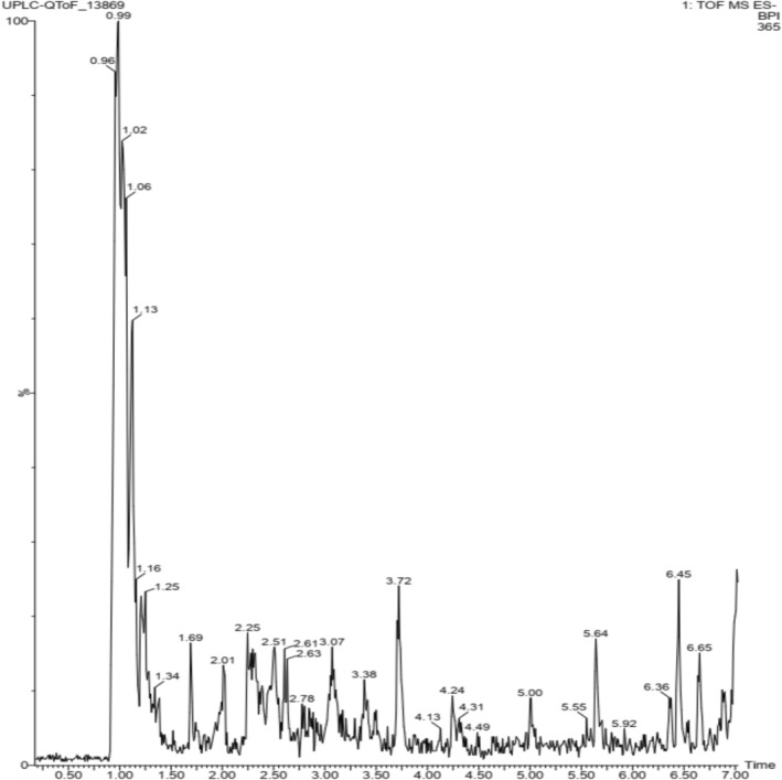
High definition mass spectrometry chromatogram (UPLC‐MS/MS) of the saponin‐enriched fraction ofSarcomphalus joazeirostem barks (FSJ), in negative ionic mode.
According to the pattern of MS fragments previously identified by Pu et al. (2017) and comparing these results with published literature. The nitrogen compound (C33H27N2O9) was identified as 5‐allyl‐1‐(2,3,4‐tris‐O‐benzoylpentofuranosyl)‐2,4(1 H,3 H)‐pyrimidinedione.
The compounds zizyphursolic acid (C31H19O4) and betulinic acid + OH (C30H46O4) (Figure1) are two natural triterpenoids found in plant species belonging to the genusSarcomphalus(Silva et al.2024).
Isospinosin (C24H39O19), spinosin (C24H39O19), and chalconaringenin‐di‐C‐hexoside (C27H31O15) were the compounds identified as flavonoids. The saponins identified were ziziphin (C51H79O18), jujuboside B (C52H84O21), jujuboside II (C52H83O21), jujubasaponin III (C50H79O19), jujubasaponin IV (C49H79O20), and zizyphus saponin I (C47H75O17). Additionally, the acid compounds identified were citric acid (C6H7O7), malic acid (C4H5O5), aconitic acid (C6H5O6), protocatechuic acid (C7H5O4), and guanidinosuccinic acid (C5H9N3O4) (Figure2).
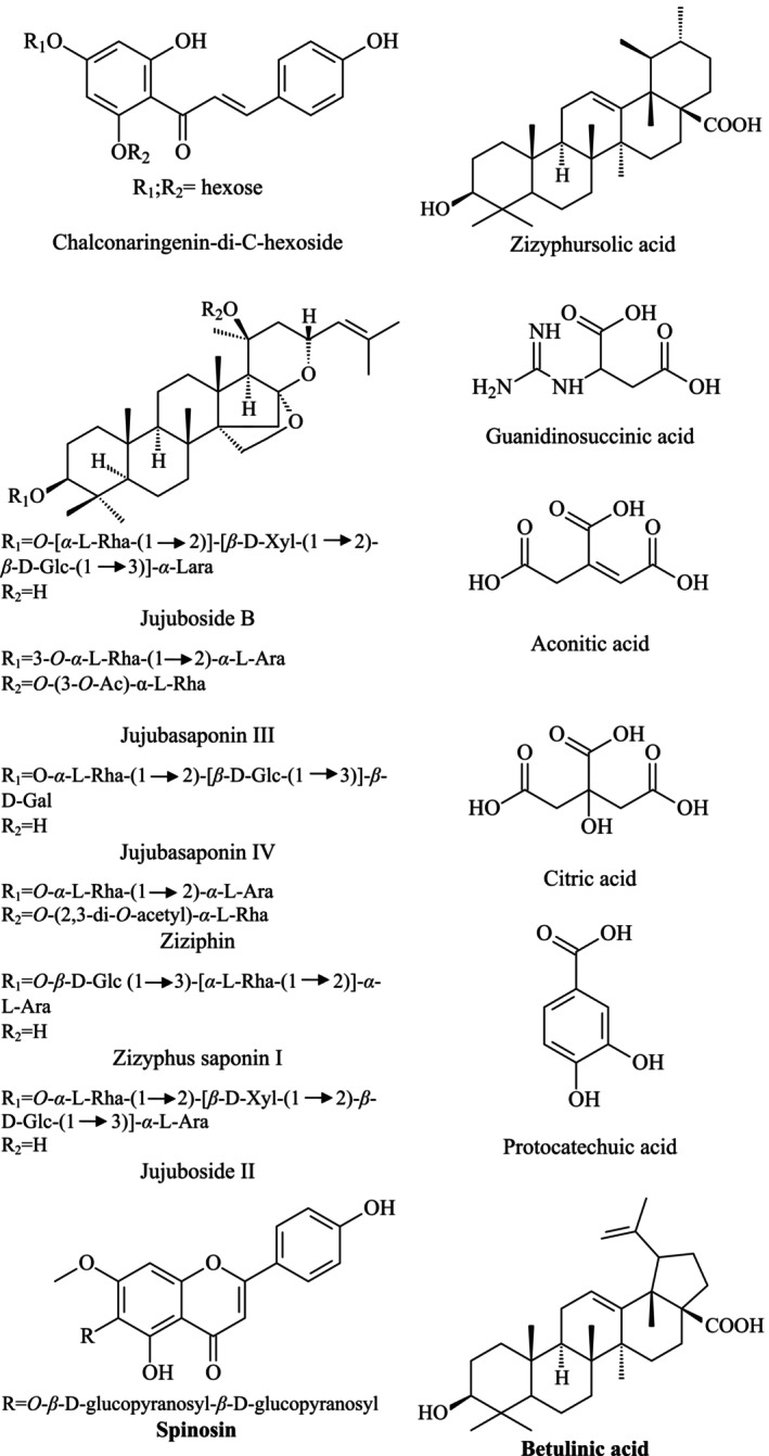
Chemical structure of the compounds identified in the fraction ofSarcomphalus joazeiro(FSJ).
After analysis to determine the MIC of the fraction (FSJ), it was not possible to observe inhibition of fungal growth at any of the concentrations tested, indicating that the fraction did not show antifungal activity when used alone. Therefore, due to the lack of significant inhibition of the fraction throughout the concentrations evaluated, the MFC was carried out only with fluconazole, which showed fungistatic action at a concentration of 512 μg/mL.
The 50% inhibition of the microorganism population (IC50) for the fraction (Table1) againstC. albicansshowed a high concentration (840.6 μg/mL) and obtained an IC50of 231.2 and 212.5 μg/mL againstC. tropicalisandC. krusei, respectively. The IC50of the fraction againstC. kruseiwas lower than that of FCZ. However, the antifungal (FCZ) showed an effect againstC. tropicalisat a concentration of 13.23 μg/mL. When the fraction was combined with fluconazole, antagonism was observed, with an increase in concentration againstC. krusei(318.3 μg/mL), as shown in Table1.
Table: 50% inhibitory concentrations (IC50) of microorganisms (μg/mL) by the fraction ofSarcomphalus joazeiro(FSJ).
Figure3shows the results of the cell viability of the fraction (FSJ) alone and in combination with fluconazole for the strains ofC. albicans,C. tropicalis, andC. krusei. Therefore, when the performance of the fraction alone is observed in relation to FCZ, it can be seen that FSJ showed an almost similar performance for theC. tropicalisandC. kruseistrains, in which a decrease in the percentage of microorganisms was not observed as a function of the increase in the concentration of the fraction.
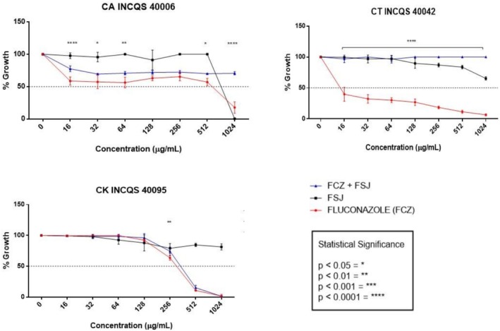
*Evaluation of the modifying effect of fluconazole action by the fraction. CA:Candida albicans; CT:Candida tropicalis; CK:Candida krusei; INCQS: National Institute for Quality Control in Health; FSJ: fraction fromSarcomphalus joazeiro; FCZ: fluconazole; ns: not significant; *p< 0.05; **p< 0.01; ***p< 0.001; ***p< 0.0001—statistical significance.
It is worth noting that the FSJ in question was used at its sub‐inhibitory concentration (where the matrix concentration of the fraction was divided by 8) so that the fraction could not only show its action against the yeasts tested but also contribute at this concentration as a possible potentiator of the action of fluconazole.
In these graphs, the performance of the fraction associated with fluconazole was similar for theC. albicansandC. tropicalisstrains. It was also observed that FSJ together with the antifungal FCZ showed a viability curve similar to that of the drug alone against theC. kruseistrain (Figure3).
The search for natural agents with bioactive properties has grown significantly, driven by the need to develop new alternatives of therapeutic importance, especially for the control of antimicrobial resistance (Lee et al.2017). In this context, saponins, plant compounds widely distributed in nature, have aroused interest due to their diverse biological activities, including antifungal properties (Nguyen et al.2020).
Saponins are able to form complexes with cell membrane constituents, thus altering cell permeability and leading to their destruction. According to certain studies, steroid saponins break the fungal cell membrane, making it permeable and allowing cellular components to escape. This interference can impede the vitality and growth of fungal species (Sikarwar et al.2023).
Flavonoids exhibit diverse biological activities, including antifungal effects against species of the Candida genus. They are compounds that often inhibit fungal growth through various mechanisms, including disruption of the plasma membrane, inhibition of cell wall formation, or the efflux‐mediated pumping system (Al Aboody and Mickymaray2020).
Secondary metabolites such as saponins, flavonoids and steroids are reported in extracts of the stem bark ofS. joazeiro(Mauro et al.2021). Phytochemical prospecting studies carried out on hydroalcoholic extracts of the stem bark, leaves and fruit ofS. joazeirohave also revealed the presence of saponins, steroids and triterpenoids (Melo et al.2012), as well as in the ethanolic extract of the leaves (Souza et al.2023) and stem bark (Araújo et al.2022). Chemical analysis using high performance liquid chromatography (HPLC‐DAD) identified phenolic acids in the hydroalcoholic extract ofS. joazeiroleaves (Brito et al.2015).
In a study carried out by Bezerra‐Neto (2019), in which the fungal susceptibility ofCandidaspp. isolates was evaluated against the enriched fraction obtained from the hydroethanolic extract ofS. joazeiro, a significant antifungal activity was found against isolates ofC. albicansandCandida glabrata, and the fraction was able to interfere with the expression of virulence factors of this group of yeasts.
Andrade et al. (2020), in a study evaluating the antifungal and modulating potential ofS. joazeirobark and leaf extracts, alone and in combination with fluconazole, against resistant species of theCandidagenus, found that both extracts inhibited fungal growth, as well as showing inhibitory concentrations similar to fluconazole.
In the study carried out by Ribeiro et al. (2013), the properties of saponins extracted from the bark ofS. joazeirowere evaluated, showing activity againstC. albicans(156 μg/mL), using the broth microdilution method. Silva et al. (2011) and Melo et al. (2012), respectively, showed that the ethanolic and hydroalcoholic extracts ofS. joazeirobark were active againstC. albicansusing the agar diffusion method.
TheS. joazeirospecies also stands out for its use in folk medicine to treat infections, dandruff, or seborrheic dermatitis. According to the indications for use of the species by the population, the most commonly used parts for therapeutic, medicinal, dental, and cosmetic purposes are bark, stem, and leaves, with different forms of preparation from syrup, infusion, maceration, decoction, baths, and others (Rego2019).
Conclusion
In conclusion, the results obtained in this study contribute to understanding the antifungal potential of the saponin enriched fraction of theS. joazeirospecies against fungal infections, especially those caused byCandidaspp. The data obtained from the chemical characterization of this fraction provides information on the identification of bioactive compounds present in theS. joazeirospecies, such as triterpenoids, flavonoids, acids, and saponins. Therefore, this work highlights important scientific information for future research into new alternatives from natural products to combat microbial resistance.
Disclosure
The authors declared that they did not use any AI tool in the text of this manuscript.
Significance Statement
This study contributes to understanding the antifungal potential of the saponin enriched fraction of theS. joazeirospecies against fungal infections, especially those caused byCandidaspp. The data obtained from the chemical characterization of this fraction provide information on the identification of bioactive compounds present in theS. joazeirospecies, such as triterpenoids, flavonoids, acids, and saponins.
References
- Al Aboody, M. S. , andS. Mickymaray. 2020. “Anti‐Fungal Efficacy and Mechanisms of Flavonoids. ”Antibiotics9, no. 2: 45. doi.org/10.3390/antibiotics9020045
- Andrade, J. C. , A. T. L. Santos, A. R. P. Silva, et al. 2020. “Phytochemical Characterization of theZiziphus joazeiroMart. Metabolites by UPLC‐QTOF and Antifungal Activity Evaluation. ”Cellular and Molecular Biology66, no. 4: 127–132.
- Andrade, J. C. , A. R. P. Silva, M. A. Freitas, et al. 2019. “Control of Bacterial and Fungal Biofilms by Natural Products ofZiziphus joazeiroMart. (Rhamnaceae). ”Comparative Immunology, Microbiology and Infectious Diseases65: 226–233. doi.org/10.1016/j.cimid.2019.06.006
- Andrade, J. C. , A. R. P. Silva, A. T. L. Santos, et al. 2019. “UPLC‐MS‐ESI‐QTOF Characterization and Evaluation of the Antibacterial and Modulatory Antibiotic Activity ofZiziphus joazeiroMart. Aqueous Extracts. ”South African Journal of Botany123: 105–112.
- Araújo, N. J. S. , A. R. P. Silva, M. S. Costa, et al. 2022. “Chemical Characterization UPLC‐ESI‐QToF‐MSE, Antibacterial and Antibiofilm Potential ofSarcomphalus joazeiro(MART. ) Hauenschild. ”Food Bioscience50: 102066.
- Bezerra‐Neto, F. A. 2019. “Caracterização do Efeito de Uma Fração Enriquecida em Saponinas Obtida deZiziphus joazeiroem Isolados deCandidaspp. Oriundos de Pacientes com Candidemia com Ênfase na Expressão dos Fatores de Virulência. ” Dissertação de Mestrado (Programa de Pós‐graduação em Ciências Farmacêuticas). Universidade Federal do Rio Grande do Norte, Brasil.
- Brito, S. M. O. , H. D. M. Coutinho, A. Talvani, et al. 2015. “Analysis of Bioactivities and Chemical Composition ofZiziphus joazeiroMart. Using HPLC‐DAD. ”Food Chemistry186: 185–191. doi.org/10.1016/j.foodchem.2014.10.031
- Brito, S. M. O. , A. O. B. P. B. Martins, M. R. C. Oliveira, et al. 2020. “Gastroprotective and Cicatrizing Activity of theZiziphus joazeiroMart. Leaf Hydroalcoholic Extract. ”Journal of Physiology and Pharmacology71: 429–436. doi.org/10.26402/jpp.2020.3.14
- Coutinho, H. D. M. , J. G. M. Costa, E. O. Lima, V. S. Falcão‐Silva, andJ. P. Siqueira‐Júnior. 2008. “Enhancement of the Antibiotic Activity Against a MultiresistantEscherichia colibyMentha arvensisL. and Chlorpromazine. ”Chemotherapy54, no. 4: 328–330. doi.org/10.1159/000151267
- Fonseca, V. J. A. , A. L. Braga, R. S. Almeida, et al. 2022. “Lectins ConA and ConM Extracted FromCanavalia ensiformis(L. ) DC andCanavalia rosea(Sw. ) DC Inhibit PlanktonicCandida albicansandCandida tropicalis. ”Archives of Microbiology204, no. 6: 346. doi.org/10.1007/s00203-022-02959-x
- Javadpour, M. M. , A. M. Juban, W. ‐C. J. Lo, et al. 1996. “De Novo Antimicrobial Peptides With Low Mammalian Cell Toxicity. ”Journal of Medicinal Chemistry39, no. 16: 3107–3113. doi.org/10.1021/jm9509410
- Lee, C. ‐R. , J. H. Lee, M. Park, et al. 2017. “Biology ofAcinetobacter baumannii: Pathogenesis, Antibiotic Resistance Mechanisms, and Prospective Treatment Options. ”Frontiers in Cellular and Infection Microbiology7: 55. doi.org/10.3389/fcimb.2017.00055
- Mauro, A. C. , B. D. Ribeiro, R. Garrett, et al. 2021. “Ziziphus joazeiroStem Bark Extract as a Green Corrosion Inhibitor for Mild Steel in Acid Medium. ”Processes9, no. 8: 1323.
- Melo, M. S. F. , C. Q. Rocha, M. H. Santos, et al. 2012. “Pesquisa de Bioativos com Atividade Antimicrobiana nos Extratos Hidroetanólicos do Fruto, Folha e Casca de Caule doZizyphus joazeiroMart. ”Revista da Universidade Vale do Rio Verde10, no. 2: 43–51.
- Morais‐Braga, M. F. B. , D. L. Sales, J. N. P. Carneiro, et al. 2016. “Psidium guajavaL. andPsidium brownianumMart ex DC. : Chemical Composition and Anti‐CandidaEffect in Association With Fluconazole. ”Microbial Pathogenesis95: 200–207. doi.org/10.1016/j.micpath.2016.04.013
- Nguyen, L. T. , A. C. Farcas, S. A. Socaci, et al. 2020. “An Overview of Saponins—A Bioactive Group. ”Bulletin of University of Agricultural Sciences and Veterinary Medicine Cluj‐Napoca: Food Science and Technology77, no. 1: 25–36.
- Oliveira, P. M. L. , A. M. Dantas, A. R. S. Morais, et al. 2020. “Juá Fruit (Ziziphus joazeiro) From Caatinga: A Source of Dietary Fiber and Bioaccessible Flavanols. ”Foodservice Research International129: 108745. doi.org/10.1016/j.foodres.2019.108745
- Pu, Y. , T. Ding, N. Zhang, et al. 2017. “Identification of Bitter Compounds From Dried Fruit ofZiziphus jujubacv. Junzao. ”International Journal of Food Properties20, no. 1: 26–35.
- Rego, E. L. 2019. “Estudo Etnobotânico da Utilização do JúaZiziphus joazeiroMart. (Rhamnaceae) na Comunidade do Sítio Serra Verde‐PE/Ethnobotanic Study on the Use of JúaZiziphus joazeiroMart. (Rhamnaceae) in the Serra Verde‐PE Community. ”Brazilian Journal of Health Review2, no. 6: 5801–5810.
- Ribeiro, B. D. , D. S. Alviano, D. W. Barreto, andM. A. Z. Coelho. 2013. “Functional Properties of Saponins From Sisal (Agave sisalana) and Juá (Ziziphus joazeiro): Critical Micellar Concentration, Antioxidant and Antimicrobial Activities. ”Colloids and Surfaces A: Physicochemical and Engineering Aspects436: 736–743.
- Rocha, T. S. , A. Lima, J. N. Silva, et al. 2022. “Vitamin C and Phenolic Antioxidants of Jua (Ziziphus joazeiroM. ) Pulp: A Rich Underexplored Brazilian Source of Ellagic Acid Recovered by Aqueous Ultrasound‐Assisted Extraction. ”Molecules (Basel, Switzerland)27, no. 3: 627. doi.org/10.3390/molecules27030627
- Sikarwar, M. S. , N. Borah, andM. R. Vyas. 2023. “Exploring the Antifungal Properties of Steroidal Saponins: An In‐Depth Analysis. ”Revista Electrónica de Veterinaria24, no. 2: 38–46.
- Silva, A. R. P. , M. S. Costa, N. J. S. Araújo, et al. 2024. “UPLC‐QTOF‐MS/MS Characterization of a Fraction Enriched in Saponins FromSarcomphalus joazeiroSpecies and Evaluation of the Antibacterial Activity Against Multidrug‐Resistant Bacteria. ”South African Journal of Botany165: 324–330.
- Silva, M. V. C. C. , L. M. Sousa, R. T. M. Ribeiro, et al. 2022. “Flora of Ceará, Brazil: Rhamnaceae. ”Rodriguésia73: e01692021.
- Silva, P. M. , L. P. B. Rocha, L. G. Arruda, et al. 2022. “Natural Products From the Brazilian Caatinga as a Sustainable Source of Phytocosmetics: A Review. ”Research, Society and Development11: e167111736940.
- Silva, T. C. L. , C. C. B. R. Almeida, J. Veras‐Filho, et al. 2011. “Atividades Antioxidante e Antimicrobiana deZiziphus joazeiroMart. (Rhamnaceae): Avaliação Comparativa Entre Cascas e Folhas. ”Revista de Ciências Farmacêuticas Básica e Aplicada32, no. 2: 193–199.
- Souza, A. B. , J. C. Andrade‐Pinheiro, J. B. Soares, et al. 2023. “Antibacterial Activity and Anxiolytic‐Like Effect ofZiziphus joazeiroMart. Leaves in Adult Zebrafish (Danio rerio). ”Fish and Shellfish Immunology Reports5: 100108. doi.org/10.1016/j.fsirep.2023.100108
- Vaou, N. , E. Stayropoulou, C. Voidarou, et al. 2021. “Towards Advances in Medicinal Plant Antimicrobial Activity: A Review Study on Challenges and Future Perspectives. ”Microorganisms9, no. 10: 2041. doi.org/10.3390/microorganisms9102041
Republished from the open web under CC-BY. Authors: da Silva ARP, do Socorro Costa M, Araújo NJS, Alencar GG, Moura TF, Fonseca VJA, Dos Santos ATL, Morais-Braga MFB, de Souza EO, Tavares JF, Ribeiro PRV, Andrade-Pinheiro JC, Coutinho HDM. Read the original.