Validation of a Pregnancy-Associated Glycoprotein-Based Lateral Flow Assay for Early Pregnancy Detection in Goats.
Background The significant cross-reactivity between bovine, ovine, and caprine pregnancy-associated glycoproteins (PAGs) enables the adaptation of bovine-specific diagnostics for use in other ruminants; consequently, the Alertys OnFarm Pregnancy Test (AOFPT)-a blood-based lateral flow assay-provides a rapid and practical solution for pregnancy detection directly at the animal's head under field conditions. Objectives This study evaluated to assess and validate the performance of AOFPT in goats at days 21 and 28 post-mating, by comparing the results with serum progesterone (P4) analysis and using transabdominal ultrasonography (TAUS) as the gold standard. Methods The study involved 85 Kilis goats, five months post-partum. Estrus was synchronized using an 11-day progestagen device, d-cloprostenol, and PMSG. Whole blood and serum samples were collected on Days 21 and 28 post-mating. AOFPT was performed on-farm immediately after collection. For validation, serum progesterone concentrations were measured via electrochemiluminescence immunoassay, and TAUS was performed on Days 35 and 42, with Day 42 findings serving as the gold standard. Results Results indicated that on Day 21, AOFPT sensitivity, specificity, positive predictive value, and negative predictive value were 79.4%, 93.8%, 98.2%, and 51.7%, respectively. By Day 28, these metrics reached 100%, 81.3%, 95.8%, and 100%. Statistical agreement between AOFPT and the reference method was K = 0.55 (82.14%) on Day 21 and K = 0.87 (96.43%) on Day 28 (p Conclusions AOFPT demonstrated accuracy and reliability closely matching P4 measurements and the reference method. This test provides a practical tool for early on-farm pregnancy diagnosis in goats, potentially enhancing reproductive management and productivity in dairy goat farms and large herds.
Introduction
Goat farming profitability depends on both milk and meat production, which is directly linked to reproductive efficiency. Selecting appropriate breeding and herd management strategies to improve reproductive performance—and thereby increase both the quantity and quality of animals—relies on early pregnancy diagnosis (Barbato et al.2022; Khanum et al.2008; Zamfirescu et al.2011). A reliable and appropriate diagnostic method enables timely re‐breeding, culling, or selective management of non‐pregnant animals (Karadaev2015) while facilitating appropriate nutritional protocols for pregnant does (El Amiri et al.2015; Sharma et al.2020b). Additionally, the widespread adoption of controlled breeding techniques, such as artificial insemination and out‐of‐season breeding, in large goat herds has increased the demand for an accurate, practical test for early pregnancy diagnosis (Salve et al.2016). Pregnancy diagnostic methods may be categorized as direct/indirect or functionally grouped into: (i) visual assessment, (ii) clinical examination, and (iii) laboratory testing (Barık and Yadav2022; Jainudeen and Hafez2000; Purohit2010). While some field‐applicable methods lack sufficient accuracy, others demonstrate high precision but require specialized equipment and technical expertise (Karadaev2015). The most commonly used methods include hormonal assays, analysis of pregnancy‐associated proteins, and ultrasonography (Karen et al.2001).
Pregnancy‐associated glycoproteins (PAGs) are a type of inactive aspartic protease that is produced exclusively by the placenta in ruminants (Balhara et al.2013; El Amiri et al.2015; Kaya et al.2016). After secretion by trophoectodermal cells of the placenta, PAGs migrate from foetal tissues and fuse with maternal uterine epithelial cells to form hybrid foeto‐maternal trinucleate cells (Cruz et al.2024). PAGs become detectable in the peripheral blood of a pregnant animal around the time when the extraembryonic membranes attach to the endometrium, coinciding with the migration of binucleate trophoblast cells to form the embryo–maternal syncytium and their fusion with endometrial cells (Wooding1984; Younis and Aboud2023). Therefore, PAGs serve as reliable biomarkers for both pregnancy status and fetoplacental function (Barbato et al.2022; Garbayo et al.2000; Sharma et al.2020b). Blood‐based assays for maternal serum PAGs have become more prevalent in recent years. It is stated that cross‐reactivity may occur due to N‐glycosylation similarities in PAG proteins isolated from cattle, sheep and goats (Garbayo et al.1998; Sousa et al.2006). So, commercial PAG tests use antibodies raised against bovine PAGs; however, due to structural similarities across ruminant PAG isoforms, cross‐reactivity occurs between bovine, ovine, and caprine PAGs. This cross‐reactivity enables the use of commercially available bovine tests for pregnancy detection in small ruminants and other ruminant species (Roberts2022).
The commercial IDEXX Rapid Visual Pregnancy Test (RVPT), which is the test based on enzyme‐linked immunoassay (ELISA) that can be run without ELISA instrumentation and read visually, on the principle of PAG detection, has been evaluated extensively in cattle (Akköse et al.2025; Mayo et al.2016; Rashmi et al.2020; Rice et al.2013) and sheep (Akköse2020; Chaves et al.2017; Chaves et al.2020; Roberts et al.2019; Steckeler et al.2018). In addition, studies using the Alertys OnFarm Pregnancy Test (AOFPT), which is a blood based lateral‐flow test that gives results in 5–20 min—employed in this study—in cattle were conducted by Akköse (2023), Szelenyi et al. (2024), Kline et al. (2024) and Safak et al. (2025). However, except for the investigations by Kim et al. (2020) and Akkaya Doğan and Köse (2022) used RVPT, no studies have evaluated commercially available blood based lateral‐flow test, in goats. Compared to AOFPT, RVPT requires laboratory use (Akkaya Doğan and Köse2022), while AOFPT, which operates on a blood‐based lateral flow principle and provides rapid results, can be easily used at the animal's head under field conditions (Akköse2023). Because RVPT analysis results are visually assessed by colour change, it is a test open to subjective evaluation (Akkaya Doğan and Köse2022).
Therefore, the aim of the present study was to assess and validate the performance of AOFPT—originally designed to detect PAG molecules in bovine serum—in goats at Days 21 and 28 post‐mating, by comparing the results with serum progesterone (P4) analysis and using transabdominal ultrasonography (TAUS) as the gold standard.
Materials and Methods
Animals and Synchronization Protocol
The animals for this study comprised 85 clinically healthy, fertile Kilis does on post‐partum 150 days and aged 3–5 years, with body weights ranging from 45 to 65 kg (55.17 ± 0.47; mean ± SEM), and 10 bucks with body weights ranging from 55 to 76 kg (67.41 ± 2.12; mean ± SEM), maintained by a producer of the Yayladağı district, Hatay. Animals were turned out to graze on dry forage and crop stubble from 04:00 to 09:00 h and from 16:00 to 20:00 h. After 20:00 h, they were returned to their pens and fed a ration consisting of a mixture of barley grain and hay. Drinking water was provided from both the municipal supply and a clean well source. Estrus synchronization in does was performed during the breeding season (August–September) using an 11‐day intravaginal sponge (Esponjavet, HIPRA, Spain). On the day of sponge removal, an intramuscular injection of 125 µg d‐cloprostenol (Gestavet Prost, HIPRA, Spain) and 500 IU PMSG (Oviser 500, HIPRA, Spain) was administered (Akkaya Doğan and Köse2022). Twenty‐four hours after sponge removal, bucks were introduced to the flock for 12 h, remaining with the does for 2‐h intervals; thereafter, the bucks were removed. Does exhibiting estrus were mated by hand. Following completion of matings, does were housed in a separate pen.
Blood Sample Collection
Following mating, on Days 21 (between 17:00 and 19:00) and 28 (between 17:00 and 19:00), the blood samples were collected from the jugular vein of each doe using a double‐ended needle (21 G × 1.5 inch; 0.8 × 38 mm) and a plastic vacutainer holder. Samples were drawn into vacuum tubes with gel (no anticoagulant) and into EDTA‐coated tubes (with anticoagulant). Each animal's ear tag number was recorded on the tube. Anticoagulated blood samples were used for AOFPT. Blood samples with no anticoagulant were centrifuged at 3000 rpm for 10 min at room temperature in the laboratory, and blood serum was separated. The separated serum was transferred into labelled Eppendorf tubes and stored at −20°C until P4 assay.
Whole Blood Pregnancy Test Procedure
The Alertys OnFarm Pregnancy Test was conducted on‐farm using whole blood samples without prior knowledge of each animal's pregnancy status, following the manufacturer's instructions (IDEXX, Westbrook, Maine, ABD). The EDTA tube was gently inverted once or twice, and the blood sample and wash solution were sequentially applied to the test well. After a 20‐min incubation, the presence of both the control (C) and test (T) lines indicated pregnancy, whereas appearance of only the C line indicated nonpregnancy.
P4 Measurement
Serum P4 concentrations were measured in the laboratory using an electrochemiluminescence immunoassay (ECL) on a fully automated analyser (Cobas E601, Roche, Switzerland). Does with serum P4 levels of ≥1.5 ng/mL on Day 21 were considered pregnant (Boscos et al.2003).
Pregnancy Diagnosis by Taus
Pregnancy examinations were performed twice: first at Day 35 and again at Day 42 post‐mating. All TAUS examinations were conducted by the same clinician using a real‐time ultrasound machine equipped with a 5 MHz convex probe (Falco, Pie Medical, The Netherlands). With the goat standing, the hair over the right paralumbar fossa ventral to the udder was clipped to serve as the scanning window (Dinç et al.1994). Positive diagnosis of pregnancy by TAUS on Day 35 is assured by imaging the embryo/foetus and foetal heartbeat surrounded by fluid (Dawson2002; Gonzalez et al.2004). A positive pregnancy diagnosis was recorded when a fluid‐filled uterus, placentomes, moving foetus, and foetal heartbeat were visualized. These findings were recorded, and the day 42 TAUS result was designated as the gold standard (Wojtasiak et al.2020).
Statistical Analysis
Descriptive statistics (count, percentage, mean, standard error of means, etc.) were calculated for the recorded variables. Agreement between the Day 21 and Day 28 pregnancy test results and the reference TAUS findings on Day 42 was assessed using Cohen's kappa coefficient. And none (0.0–0.20), minimal (0.21–0.39), weak (0.40–0.59), moderate (0.60–0.79), strong (0.8–0.90), or almost perfect (>0.90) agreement was accepted for kappa coefficients categories ([McHugh et al](#ref-McHugh et al).2012). Changes in P4 concentrations over time were analysed by a paired‐samplest‐test. P4 values were compared between pregnant and nonpregnant groups (based on day 42 TAUS) using the Mann–Whitney U test due to the normality was not hold. Statistical significance was set atp< 0.05 for all analyses. Data normality was evaluated using skewness and kurtosis values (±1.5). AOFPT characteristics (sensitivity, specificity, positive predictive value and negative predictive value) were calculated according to Kastelic2006. The accuracy of blood serum progesteron level in predicting the presence of the pregnancy were estimated by receiver operating characteristic (ROC) analysis by calculating area under the curve (AUC) and 95% confidence intervals. Low (0.5< AUC ≤0.7), moderate (0.7< AUC ≤0.9), or high (0.9< AUC ≤1) accuracy was accepted for AUC categories (Gardner and Greiner2006). All analyses were performed using SPSS Statistics for Windows, version 26.0 (International Business Machines Corporation‐IBM, Armonk, NY, USA).
Results
One of the goats in the study expired. Table1presents the results of the pregnancy‐associated glycoprotein test (AOFPT) on Days 21 and 28, the serum progesterone concentration (P4) on Days 21 and 28, and the transabdominal ultrasonography (TAUS) on Days 35 and 42. According to AOFPT findings, 14 false negatives and 1 false positive were determined on Day 21, and 3 false positives on day 28. According to P4 findings, 5 false positives were detected on Day 21 and 6 false positives on Day 28. In the TAUS examination on Day 35, although gestational sacs were observed in two goats, the absence of embryonic movement or heartbeat, which are necessary for a positive pregnancy diagnosis, resulted in a suspected diagnosis for these two goats on that day. On Day 42 TAUS examination, these two goats were diagnosed as nonpregnant due to lack of the pregnancy criteria aforementioned before.
Table: Goats' pregnancy‐related findings following the application of the examination techniques.
The pregnancy rate was found to be 81.0% based on the results of the 42nd day of the TAUS examination, which was approved as the study's gold standard. The AOFPT with the study's gold standard examination findings results on Day 21 was examined, and the kappa value was calculated as 0.55 at a 95% confidence interval. For the Day 21 AOFPT, the sensitivity, specificity, positive predictive value, negative predictive value, and accuracy rate were determined as 79.41%, 93.75%, 98.18%, 51.72% and 82.14%, respectively (Table2). The AOFPT results on Day 28 was the kappa value was calculated as 0.87 at a 95% confidence interval. For the Day 28 AOFPT, the sensitivity, specificity, positive predictive value, negative predictive value and accuracy rate were determined as 100%, 81.25%, 95.77%, 100.00% and 96.43%, respectively (Table3).
Table: Consistency of pregnancy‐associated glycoprotein test on Day 21.
Table: Consistency of pregnancy‐associated glycoprotein test on Day 28.
A comparison of serum progesterone concentrations on Day 21 and Day 28 according to the reference method examination findings is presented in Table4. The results of the 42nd TAUS indicated that pregnant goats exhibited significantly higher P4 values (median ± IQR) than non‐pregnant goats on the 21st (5.95 ± 2.60 vs. 0.14 ± 2.73) and 28th day (6.92 ± 2.93 vs. 0.84 ± 6.06) (p< 0.001).
Table: Comparison of progesterone concentrations (ng/mL) according to reference method findings.
According to the gold standard examination findings, serum progesterone concentration analysed on day 21, the AUC value was found to be 0.958, indicating high accuracy (Figure1and Table5). The cutoff value was determined to be >4.19 ng/mL. Sensitivity was 88.2%, and specificity was 87.5% (Table5). On Day 28, the AUC value was found to be 0.841 (Figure1and Table5), which was lower than that on Day 21. The cut‐off value was determined to be >6.13 ng/mL. Sensitivity was 75%, and specificity was 75% (Table5).
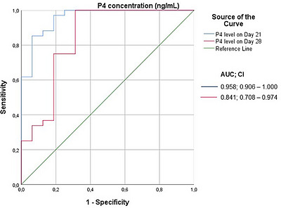
ROC curve of progesterone concentrations according to reference method findings.
Table: Ideal cut‐off values of progesterone concentrations.
AOFPT‐identified pregnant goats exhibited significantly higher P4 levels (p< 0.001) than non‐pregnant goats on Day 21 (5.98 ± 3.21 vs. 4.05 ± 5.46) and Day 28 (6.84 ± 3.06 vs. 0.75 ± 5.96) (Table6).
Table: Comparison of the P4 concentration (ng/mL) based on the 21st and 28th day test results.
Discussion
The present study aimed to evaluate and validate the performance of the AOFPT, a PAG‐based lateral flow test, for early pregnancy diagnosis in goats on Days 21 and 28 post‐mating. In the present study, even though statistical significance was detected (p< 0.001), the agreement between Day 21 AOFPT results and the reference test yielded a Kappa value of 0.55 (95% CI: 0.37–0.74), indicating weak agreement. The sensitivity of AOFPT on Day 21 was 79.41%, specificity was 93.75%, PPV was 98.18%, and NPV was 51.72% (Table2). On the other hands, for Day 28, the Kappa value between AOFPT and the reference method was 0.87 (95% CI: 0.74–1.00), indicating strong agreement (p< 0.001). The sensitivity of AOFPT was 100%, specificity was 81.25%, PPV was 95.77%, and NPV was 100% (Table3). These results demonstrate that the test correctly identified pregnancy in 95.77% of does that were pregnant and correctly excluded pregnancy in 100% of non‐pregnant does. Akköse (2023) noted that timely identification of nonpregnant cows facilitates earlier re‐insemination and emphasized that the primary goal of pregnancy diagnosis should be accurate detection of nonpregnant animals rather than simply identifying pregnant ones. In this context, Day 28 AOFPT results in the present study were highly successful in correctly identifying both pregnant and nonpregnant does.
In studies conducted by Akköse (2023) to determine the diagnostic accuracy of two commercial PAG tests (IDEXX On‐Farm Pregnancy Test [OFPT] and IDEXX Rapid Visual Pregnancy Test [RVPT]) for early pregnancy detection in dairy cows at Days 28–31 post‐insemination, OFPT demonstrated sensitivity, specificity, PPV, and NPV of 100%, 93.1%, 89.1% and 100%, respectively, whereas the RVPT demonstrated sensitivity, specificity, PPV, and NPV of 97.4%, 92.1%, 87.4% and 98.4%, respectively. Both tests exhibited excellent discrimination between pregnant and nonpregnant cows (both AUCs > 0.90). Szelenyi et al. (2024) reported sensitivity, specificity, PPV, and NPV values of 98.9%, 88.7%, 86.8% and 99.1%, and overall accuracy of 93.1% for OFPT on Days 28–35 post‐insemination. In heifers, the test showed 100% sensitivity and 81.6% specificity; in multiparous cows, sensitivity and specificity were 98.5% and 89.5%, respectively. Kline et al. (2024) validated OFPT by comparing the accuracy of three commercial PAG assays with transrectal ultrasonography; they reported a significant positive correlation between OFPT and transrectal ultrasonography (r= 0.77;p< 0.01) and a concordance rate of 92.4% between the two methods. In another study conducted in Awassi ewes also indicated that the test could be considered a practical, rapid, and reliable method for early pregnancy detection (sensitivity 95.55 %, accuracy 93.00 %, and kappa = 0.859) starting from the 28th day after mating (Uyanık et al.2026). In the present study, AOFPT performance on both days in goats was comparable to results reported in cattle and ewes, confirming its suitability for early pregnancy diagnosis.
In sheep and goats, PAG concentrations become detectable as early as Days 17–18 post‐breeding and reach 3–5 ng/mL around Days 21–22 (Sousa et al.1999). Salve et al. (2016) reported that in goats, PAG concentrations increased significantly (p< 0.01) from Day 16 of pregnancy through Days 20, 24 and 28. Zamfirescu et al. (2011) stated that PAG concentrations begin to increase from Day 16 post natural mating. Detection of the antigen and markedly elevated concentrations between Days 16 and 20 in goats coincides with the time when the trophoblast firmly adheres to the uterine wall and placentation begins. Therefore, the presence of PAGs in the dam's serum not only serves as a useful pregnancy diagnostic tool between Days 21 and 24 post‐conception but also provides information on embryonic and/or foetal viability (Gonzalez et al.2004). The presence of 14 false‐negative AOFPT results on Day 21 (Table1) in the present study may be explained by the gradually increasing PAG concentrations reported by Salve et al. (2016) between Days 20 and 28. Additionally, as noted in the same study, some goats may have had PAG concentrations below the detectable threshold, which could also account for 14 false‐negative results in the test outcomes.
P4 plays a critical role in implantation, embryonic development, and maintenance of pregnancy in mammals. In goats, the ovaries are the primary source of P4 throughout both the estrous cycle and pregnancy (Charallah et al.2010; Sharma et al.2020a). Serum P4 concentrations increase gradually as pregnancy progresses, then decline to baseline levels at the onset of parturition, abortion, or embryonic death (Barik and Yadav2022). Elevated P4 levels (above 1 ng/mL) indicate the presence of a functional corpus luteum and are generally associated with pregnancy or the diestrus phase of the cycle; however, they may also be observed under abnormal conditions, such as extended estrous cycles, early embryonic death, luteal cysts, or pseudopregnancy (Cruz et al.2024). Medan et al. (2004) reported a mean P4 concentration of 7.4 ± 0.5 ng/mL in pregnant goats on Day 21 with no statistical difference in P4 concentrations between pregnant and nonpregnant goats on Day 7 and found that on Day 21, P4 concentrations correlated with ultrasonographic findings, yielding accuracy rates of 80% for pregnant and 100% for nonpregnant goats. Gonzalez et al. (2004) reported that all pregnant goats had mean P4 levels of 8.42 ± 0.23 ng/ml on Day 22, whereas 22 nonpregnant goats (34.4%) exhibited P4 concentrations similar to pregnant goats (8.01 ± 0.75 ng/mL), resulting in a 34.4% false‐positive rate. Among those 22 nonpregnant goats, five had basal P4 levels on Day 26, whereas the remaining 17 maintained functional P4 levels (7.72 ± 0.83 ng/mL) on Day 26. The authors concluded that this may have been associated with early embryonic death, hydrometra, or luteal cysts that prolong the lifespan of the corpus luteum. Salve et al. (2016) reported that in goats, P4 concentrations increased significantly by day 8 post‐insemination and remained relatively stable until Day 12 of pregnancy, after which they increased significantly from Day 16 (3.76 ± 0.15 ng/mL) to Day 20 (6.25 ± 0.33 ng/mL), Day 24 (8.38 ± 0.26 ng/mL) and Day 28 (11.39 ± 0.40 ng/mL) (p< 0.01). Cruz et al. (2024) reported P4 concentration of 8.15 ± 1.05 ng/mL in pregnant goats on Day 28. In the present study, mean P4 concentrations in pregnant goats, based on the reference method, were 6.30 ± 0.27 ng/mL on Day 21 and 7.50 ± 0.28 ng/mL on Day 28. These results are consistent with those of Cruz et al. (2024), Gonzalez et al. (2004), Medan et al. (2004) and Salve et al. (2016). Serum P4 values in pregnant goats were significantly higher than those in nonpregnant goats on Days 21 and 28 (p< 0.001) (Table4). The P4 cut‐off concentrations for pregnancy prediction were determined to be >4.19 ng/mL on Day 21 and >6.13 ng/mL on Day 28 (Table5). The Day 21 P4 cut‐off provided higher specificity and sensitivity for predicting pregnancy (Figure1).
When non‐pregnant goats (based on the gold standard method) were evaluated individually, of the 16 non‐pregnant does, seven had P4 concentrations of <1.5 ng/mL on Day 21 and remained <1.5 ng/mL on Day 28; four had P4 levels of >1.5 ng/mL on Day 28; three had P4 concentrations of >1.5 ng/mL on Day 21 but returned to basal levels by Day 28; and two maintained P4 concentrations of >1.5 ng/mL on Day 28. In the first seven does, estrous cyclicity may not have been active; in four does, they may have been in the diestrus phase of a new estrous cycle; and in the remaining five, measurement error or other conditions, such as early embryonic death, hydrometra, or luteal cysts—which can extend the lifespan of the corpus luteum—may have been present, as noted by Gonzalez et al. (2004). Among these five does, one produced false‐positive AOFPT results on both Days 21 and 28, whereas the others produced false‐positive results only on Day 28. These data suggest that embryonic death may have occurred in these animals. Previous studies have reported that the half‐life of PAGs is approximately 7.5–8 days (Haugejorden et al.2006; Mialon et al.1993). Following embryonic death, a delayed decrease or persistence of high PAG concentrations may result in false‐positive AOFPT outcomes.
Ultrasonography is a valuable diagnostic imaging technique used in reproductive management of both small and large ruminants (Jones and Reed2017; Wojtasiak et al.2023). In small ruminants, anechoic fluid within the uterine lumen can be detected transabdominally on Days 25–28 (Devi et al.2019). However, the presence of fluid in the uterine lumen should not constitute the sole basis for confirming pregnancy; definitive diagnosis requires visualization of the amniotic vesicle and detection of the foetal heartbeat (Roberts2022). In goats, foetal heartbeats can be detected between Days 27 and 35 using a transabdominal approach (Suguna et al.2008). Placentomes also become visible via transabdominal ultrasonography around Day 40 of gestation (Erdoğan2012). Tekin and Köse (2022) reported that TAUS performed on Days 35 and 50 showed high concordance in ewes (κ = 0.864,p< 0.001), and TAUS performed on Day 35 accurately determined pregnancy status 93.93% of the time. Jones et al. (2016) reported that the transabdominal method had a sensitivity of 40% on Day 21 and 100% on Day 39 of pregnancy. Sensitivity increases after Day 40, when the uterus assumes an intraabdominal position. Therefore, the ideal time for pregnancy diagnosis via TAUS is considered to be between Days 40 and 75 (Fthenakis et al.2012; Lone et al.2016; Mali et al.2022). In the study TAUS was performed twice on Days 35 and 42, and Day 42 was considered the gold standard. In the present study, two does were provisionally diagnosed as suspicious at the first examination; during the second examination, these suspicious diagnoses were resolved, and the does were determined to be nonpregnant (Table1). Individual retrospective records showed that both of these does had P4 concentrations of >1.5 ng/mL on Day 28 and yielded false‐positive AOFPT results on Day 28. It was assessed that embryonic loss may have occurred in these animals.
Early pregnancy detection and determination of offspring number are critical for improving reproductive efficiency in goats. Accurate pregnancy diagnosis provides essential information for effective herd management practices (Suguna et al.2008). TAUS is effective for early pregnancy detection in goats; however, it requires training and expertise. On a herd‐by‐herd basis, this method may be less accessible for producers with limited resources or for large‐scale dairy operations, as ultrasonographic examination of many animals demands significant time, labour, skilled personnel and expensive equipment. In such cases, serological methods may prove more practical and efficient than ultrasonography (Lv et al.2021). Nonetheless, relying solely on circulating P4 concentrations to monitor pregnancy in goats can lead to misdiagnoses, because P4 levels may be influenced by pathophysiological conditions, such as luteal cysts, pseudopregnancy, or early embryonic loss (Charallah et al.2010).
In this context, serological tests that detect circulating PAGs, which are markers of fetoplacental unit presence, serve as useful diagnostic tools for early pregnancy detection under typical farm management conditions (Cruz et al.2024). The advantage of these tests is that they provide rapid results, do not require trained personnel or expensive equipment, and can be applied even in field conditions (Rial et al.2024). A limitation of the present study is that although AOFPT is simple, rapid, and does not require specialized labour (it provides on‐site results immediately after blood collection), it is a qualitative assay rather than a quantitative one. Consequently, it cannot measure PAG concentrations precisely or provide information on foetal viability or litter size.
In conclusion, the commercial AOFPT developed to detect PAG molecules in bovine serum, was observed to be a rapid, practical, and effective method for early pregnancy diagnosis for to improve reproductive performance and herd management strategies in goats, especially following the post‐mating 28th day. However, if the results are false negative or false positive, it is recommended that the test be repeated or the animal be re‐examined by using the gold‐standard TAUS test. Moreover, according to the authors' knowledge, this study seems to be the initial comparative evaluation of serum P4 measurement and the OnFarm Pregnancy Test versus TAUS for early pregnancy diagnosis in goats.
Author Contributions
Ayşe Merve Köse: conceptualization, methodology, investigation, writing (original draft, review and editing).Mehmet Can Kara: methodology, investigation, formal analysis, writing (original draft).
Funding
The authors have nothing to report.
Ethical Statement
This study was conducted with the approval of the Hatay Mustafa Kemal University Rectorate Animal Experiments Local Ethics Committee's decision numbered 2022/05‐02.
Conflicts of Interest
The authors declare no conflicts of interests.
Acknowledgement
This study was summarized from the first author's M.Sc. dissertation. For their support to the study, we thank Veterinary Medicine M. Murat GÖKÇE and Veterinary Medicine Abdurrezzak TAŞATAN, staff of the Diagen A.Ş. Besides, we would also like to thank IDEXX Laboratories for their support in providing the Alertys OnFarm Pregnancy Test.
Data Availability Statement
The data may be provided on reasonable request.
Associated Data
Data Availability Statement
The data may be provided on reasonable request.
References
- Akkaya Doğan, A. , and Köse A. M.. 2022. “Evaluation of Bovine Visual ELISA Test for Detection of Pregnancy‐Associated Glycoproteins in Early Pregnancy Diagnosis in Goats.” Small Rumin Res 212: 106722. 10.1016/j.smallrumres.2022.106722. doi.org/10.1016/j.smallrumres.2022.106722
- Akköse, M. 2020. “Evaluation of a Boivine Rapid Visual PAG ELISA Test and Transabdominal Ultrasonography for Early Pregnancy Diagnosis in Awassi Sheep.” KSU J Agric Nat 23: 1366–1372. 10.18016/ksutarimdoga.vi.668707. doi.org/10.18016/ksutarimdoga.vi.668707
- Akköse, M. 2023. “Comparative Evaluation of Two Commercial Pregnancy‐Associated Glycoproteins Tests for Early Detection of Pregnancy in Dairy Cattle.” Theriogenology 200: 11–17. 10.1016/j.theriogenology.2023.01.022. doi.org/10.1016/j.theriogenology.2023.01.022
- Akköse, M. , Kurban M., Gökçe M. M., and Vural M. R.. 2025. “Assessment of the Diagnostic Accuracy of a Pregnancy‐Associated Glycoproteins Test for Early Pregnancy Diagnosis in Dairy Heifers.” Reproduction in Domestic Animals = Zuchthygiene 60: e70050. 10.1111/rda.70050. doi.org/10.1111/rda.70050
- Balhara, A. K. , Gupta M., Singh S., Mohanty A. K., and Singh I.. 2013. “Early Pregnancy Diagnosis in Bovines: Current Status and Future Directions.” Scientific World Journal 2013: 958540. 10.1155/2013/958540. doi.org/10.1155/2013/958540
- Barbato, O. , Menchetti L., Brecchia G., and Barile V. L.. 2022. “Using Pregnancy‐associated Glycoproteins (PAGs) to Improve Reproductive Management: From Dairy Cows to Other Dairy Livestock.” Animals 12: 2033. 10.3390/ani12162033. doi.org/10.3390/ani12162033
- Barık, S. , and Yadav S.. 2022. “Trends in Development of Pregnancy Diagnosis Techniques in Ruminants.” e‐planet 20: 105–114.
- Boscos, C. M. , Samartzi F. C., Lymberopoulos A. G., Stefanakis A., and Belibasaki S.. 2003. “Assessment of Progesterone Concentration Using Enzymeimmunoassay for Early Pregnancy Diagnosis in Sheep and Goats.” Reproduction in Domestic Animals = Zuchthygiene 38: 170–174. 10.1046/j.1439-0531.2003.00407.x. doi.org/10.1046/j.1439-0531.2003.00407.x
- Charallah, S. , Amirat Z., Sulon J., Khammar F., and Beckers J. F.. 2010. “Pregnancy‐associated Glycoprotein and Progesterone Concentrations During Pregnancy Failure in Bedouin Goat From the Southwest of Algeria.” Reproduction in Domestic Animals = Zuchthygiene 45: e231–e238. 10.1111/j.1439-0531.2009.01511.x. doi.org/10.1111/j.1439-0531.2009.01511.x
- Chaves, C. M. S. , Costa R. L. D., Duarte K. M. R., Beltrame R. T., and Quirino C. R.. 2020. “Evaluation of a Cattle Rapid Test for Early Pregnancy Diagnosis in Sheep.” Tropical Animal Health and Production 52: 1345–1349. 10.1007/s11250-019-02130-7. doi.org/10.1007/s11250-019-02130-7
- Chaves, C. M. S. , Costa R. L. D., Duarte K. M. R., Machado D. C., Paz C. C. P., and Beltrame R. T.. 2017. “Visual ELISA for Detection of Pregnancy‐associated Glycoproteins (PAGs) in Ewe Serum.” Theriogenology 97: 78–82. 10.1016/j.theriogenology.2017.04.026. doi.org/10.1016/j.theriogenology.2017.04.026
- Cruz, C. , Margatho G., Simões M., and Simões J.. 2024. “The Utilization of a Pregnancy‐associated Glycoprotein Profile and PAG/P4 Ratio Biomarker for the Diagnosis of Pseudopregnancy in Dairy Goats.” Veterinary Sciences 11: 574. 10.3390/vetsci11110574. doi.org/10.3390/vetsci11110574
- Dawson, L. 2002. “Pregnancy Diagnosis in Goats.” In: Proceedings of the 17th Annual Goat Field Day, Langston University, 27April 2002, 41–44.
- Devi, N. B. , Bhuyan D., and Das A.. 2019. “Application of Ultrasonic Doppler Technique for Pregnancy Diagnosis in Local Assam Goat.” Journal of Pharma Innovation 8: 1088–1091.
- Dinç, D. A. , Taşal İ., Erdem H., Semacan A., and Aral S.. 1994. “Koyunlarda Transabdominal Ultrasonografi Ile Yavru Sayımı.” Eurasian Journal of Veterinary Sciences 10: 81–83.
- El Amiri, B. , Sousa N. M., Alvarez‐Oxiley A., Hadarbach D., and Beckers J. F.. 2015. “Pregnancy‐Associated Glycoprotein (PAG) Concentration in Plasma and Milk Samples for Early Pregnancy Diagnosis in Lacaune Dairy Sheep.” Research in Veterinary Science 99: 30–36. 10.1016/j.rvsc.2014.12.016. doi.org/10.1016/j.rvsc.2014.12.016
- Erdoğan, G. 2012. “Ultrasonic Assessment During Pregnancy in Goats‐a Review.” Reproduction in Domestic Animals = Zuchthygiene 47: 157–163. 10.1111/j.1439-0531.2011.01873.x. doi.org/10.1111/j.1439-0531.2011.01873.x
- Fthenakis, G. C. , Arsenos G., Brozos C., et al. 2012. “Health Management of Ewes During Pregnancy.” Animal Reproduction Science 130: 198–212. 10.1016/j.anireprosci.2012.01.016. doi.org/10.1016/j.anireprosci.2012.01.016
- Garbayo, J. M. , Green J. A., Manikkam M., et al. 2000. “Caprine Pregnancy‐associated Glycoproteins (PAG): Their Cloning, Expression, and Evolutionary Relationship to Other PAG.” Molecular Reproduction and Development 57: 311–322. 10.1002/1098-2795(200012)57:4<311::AID-MRD2>3.0.CO;2-F. doi.org/10.1002/1098-2795(200012)57:4<311::AID-MRD2>3.0.CO;2-F
- Garbayo, J. M. , Remy B., Alabart J. L., et al. 1998. “Isolation and Partial Characterization of a Pregnancy‐associated Glycoprotein family From the Goat Placenta.” Biology of Reproduction 58: 109–115. 10.1095/biolreprod58.1.109. doi.org/10.1095/biolreprod58.1.109
- Gardner, I. A. , and Greiner M.. 2006. “Receiver‐operating Characteristic Curves and Likelihood Ratios: Improvements Over Traditional Methods for the Evaluation and Application of Veterinary Clinical Pathology Tests.” Veterinary Clinical Pathology 35, no. 1: 8–17. 10.1111/j.1939-165x.2006.tb00082.x. doi.org/10.1111/j.1939-165x.2006.tb00082.x
- Gonzalez, F. , Cabrera F., Batista M., et al. 2004. “A Comparison of Diagnosis of Pregnancy in the Goat via Transrectal Ultrasound Scanning, Progesterone, and Pregnancy‐associated Glycoprotein Assays.” Theriogenology 62: 1108–1115. 10.1016/j.theriogenology.2003.12.009. doi.org/10.1016/j.theriogenology.2003.12.009
- Haugejorden, G. , Waage S., Dahl E., Karlberg K., Beckers J. F., and Ropstad E.. 2006. “Pregnancy Associated Glycoproteins (PAG) in Postpartum Cows, Ewes, Goats and Their Offspring.” Theriogenology 66: 1976–1984. 10.1016/j.theriogenology.2006.05.016. doi.org/10.1016/j.theriogenology.2006.05.016
- Jainudeen, M. R. , and Hafez E. S. E.. 2000. “Pregnancy diagnosis.” In Reproduction in Farm Animals, edited by Hafez ESE and Hafez B (eds), 395–404. Lippincott Williams & Wilkins.
- Jones, A. K. , Gately R. E., McFadden K. K., Zin S. A., Govoni K. E., and Reed S. A.. 2016. “Transabdominal Ultrasound for Detection of Pregnancy, Fetal and Placental Landmarks, and Fetal Age Before Day 45 of Gestation in the Sheep.” Theriogenology 85: 939–945. 10.1016/j.theriogenology.2015.11.002. doi.org/10.1016/j.theriogenology.2015.11.002
- Jones, A. K. , and Reed S. A.. 2017. “Benefits of Ultrasound Scanning During Gestation in the Small Ruminant.” Small Rumin Res 149: 163–171. 10.1016/j.smallrumres.2017.02.008. doi.org/10.1016/j.smallrumres.2017.02.008
- Karadaev, M. 2015. “Pregnancy Diagnosis Techniques in Goats—A Review.” Bulgarian Journal of Veterinary Medicine 18: 183–193. 10.15547/bjvm.837. doi.org/10.15547/bjvm.837
- Karen, A. , Kovacs P., Beckers J. F., and Szenci O.. 2001. “Pregnancy Diagnosis in Sheep: Review of the Most Practical Methods.” Acta Veterinaria Brno 70: 115–126.
- Kastelic, J. P. 2006. “Critical Evaluation of Scientific Articles and Other Sources of Information: An Introduction to Evidence‐based Veterinary Medicine.” Theriogenology 66, no. 3: 534–542. 10.1016/j.theriogenology.2006.04.017. doi.org/10.1016/j.theriogenology.2006.04.017
- Kaya, M. S. , Köse M., Bozkaya F., Mutlu H., Uçar E. H., and MO A.ı. 2016. “Early Pregnancy Diagnosis Using a Commercial ELISA Test Based on Pregnancy‐associated Glycoproteins in Holstein‐Friesian Heifers and Lactating Cows.” Turkish Journal of Veterinary & Animal Sciences 40: 694–699. 10.3906/vet-1602-41. doi.org/10.3906/vet-1602-41
- Khanum, S. A. , Hussain M., and Kausar R.. 2008. “Progesterone and Estradiol Profiles During Estrous Cycle and Gestation in Dwarf Goats (Capra hircus).” PakistanVeterinary Journal28: 1–4.
- Kim, K. W. , Lee E. D., Lee J., Kim D. K., Lee S. S., and Lee S. H.. 2020. “Artificial Insemination and Delivery Rate of Crossbred Goat Using Frozen‐thawed Semen.” Journal of the Korea Academia‐Industrial Cooperation Society 21: 181–186. 10.5762/KAIS.2020.21.10.181. doi.org/10.5762/KAIS.2020.21.10.181
- Kline, A. C. , Menegatti Zoca S., Epperson K. M., et al. 2024. “Evaluation of Pregnancy Associated Glycoproteins Assays for on Farm Determination of Pregnancy Status in Beef Cattle.” PLoS One 19: e0306325. 10.1371/journal.pone.0306325. doi.org/10.1371/journal.pone.0306325
- Lone, S. A. , Gupta S. K., Kumar N., et al. 2016. “Recent Technologies for Pregnancy Diagnosis in Sheep and Goat: An Overview.” International Journal of Environmental Science and Technology 5: 1208–1216.
- Lv, M. J. , Li H., and Zi X. D.. 2021. “Assessment of Estrous Synchronization Protocols and Pregnancy Specific Protein b Concentration for the Prediction of Kidding Rate in Lezhi Black Goats.” Small Ruminant Research 195: 106299. 10.1016/j.smallrumres.2020.106299. doi.org/10.1016/j.smallrumres.2020.106299
- Mali, A. B. , Mehar R. M., Kapane S. H., and Hadimani M. R.. 2022. “Scanning the Future: Veterinary Reproductive Ultrasonography.” Anim Reproduction Update 2: 82–89. 10.48165/aru.2022.2105. doi.org/10.48165/aru.2022.2105
- Mayo, L. M. , Moore S. G., Poock S. E., Silvia W. J., and MC L.. 2016. “Technical Note: Validation of a Chemical Pregnancy Test in Dairy Cows That Uses Whole Blood, Shortened Incubation Times, and Visual Readout.” Journal of Dairy Science 99, no. 9: 7634–7641. 10.3168/jds.2016-11224. doi.org/10.3168/jds.2016-11224
- McHugh, M. L. 2012. “Interrater Reliability: the kappa Statistic.” Biochemia Medica 22, no. 3: 276–282.
- Medan, M. , Watanabe G., Absy G., Sasaki K., Sharawy S., and Taya K.. 2004. “Early Pregnancy Diagnosis by Means of Ultrasonography as a Method of Improving Reproductive Efficiency in Goats.” The Journal of Reproduction and Development 50: 391–397. 10.1262/jrd.50.391. doi.org/10.1262/jrd.50.391
- Mialon, M. M. , Camous S., Renand G., Martal J., and Menissier F.. 1993. “Peripheral Concentrations of a 60‐kDa Pregnancy Serum Protein During Gestation and After Calving and in Relationship to Embryonic Mortality in Cattle.” Reproduction, Nutrition, Development 33: 269–282. doi.org/10.1051/rnd:19930309
- Purohit, G. 2010. “Methods of Pregnancy Diagnosis in Domestic Animals: The Current Status.” Webmed Central 1: 2–26.
- Rashmi, S. , Sahadev A., Swamy M. N., et al. 2020. “Comparative Evaluation of Early Pregnancy Diagnostic Methods in Dairy Cows.” International Journal of Livestock Research 10: 71–77. 10.5455/ijlr.20200109120845. doi.org/10.5455/ijlr.20200109120845
- Rial, C. , Hussain I., Hoff R., et al. 2024. “Development and Evaluation of a Lateral Flow‐based Portable Optical System for Determination of the Pregnancy Status of Dairy Cows.” Journal of Dairy Science 107: 8642–8653. 10.3168/jds.2024-24899. doi.org/10.3168/jds.2024-24899
- Rice, A. M. , Fabbricante M. J., Koller S. I., Lemieux L., and Welles P.. 2013. “Performance of an IDEXX Bovine Pregnancy Test Adapted for Visual‐read Results and Designed for in Clinic Use.” In: Proceedings of the Forty‐Sixth Annual Conference, American Association of Bovine Practitioners, 19‐21 September 2013, 199.
- Roberts, J. 2022. “Pregnancy Diagnosis in Small Ruminants.” In: Proceedings of the Fifty‐Fifth Annual Conference, American Association of Bovine Practitioners, 22‐24 September 2022, 156–158.
- Roberts, J. , May K., Ajani O., and Kaneene J.. 2019. “A Comparison of Pregnancy Diagnosis Methods in Commercial Sheep Using Lambing as a Gold Standard.” Clinical Theriogenology 11: 107–113.
- Safak, T. , KÇ T.ü, Yİ A., Özen T. Y., and Yilmaz Koc O.. 2025. “Evaluation of the Accuracy and Performance of Two Commercial Pregnancy‐associated Glycoprotein Tests for Early Pregnancy Detection in Cows.” Veterinary Medicine and Science 11: e70226. 10.1002/vms3.70226. doi.org/10.1002/vms3.70226
- Salve, R. R. , Ingole S. D., Nagvekar A. S., Bharucha S. V., and Dagli N. R.. 2016. “Pregnancy Associated Protein and Progesterone Concentrations During Early Pregnancy in Sirohi Goats.” Small Ruminant Research 141: 45–47. 10.1016/j.smallrumres.2016.07.003. doi.org/10.1016/j.smallrumres.2016.07.003
- Sharma, N. , Singh S. P., and Bharadwaj A.. 2020a. “Temporal Changes in Circulating Progesterone and Pregnancy Associated Glycoprotein Concentrations in Jakhrana Goats With Failed Pregnancy.” Indian Journal of Animal Sciences 90: 861–864.
- Sharma, N. , Singh S. P., and Bharadwaj A.. 2020b. “Changes in Milk and Plasma Progesterone and Pregnancyassociated Glycoprotein and Their Relationships With the Foetal Number During Early Pregnancy in Jakhrana Goats.” Indian Journal of Animal Sciences 90: 1589–1593.
- Sousa, N. M. , Ayad A., Beckers J. F., and Gajewski Z.. 2006. “Pregnancy‐associated Glycoproteins (PAG) as Pregnancy Markers in the Ruminants.” Journal of Physiology and Pharmacology 57: 153–171.
- Sousa, N. M. , Garbayo J. M., and Beckers J. F.. 1999. “Pregnancy Associated Glycoproteins and Progesteron Profiles During Pregnancy and Postpartum in Native Goat From the North‐East of Brazil.” Small Ruminant Research 32: 137–147. 10.1016/S0921-4488(98)00171-0. doi.org/10.1016/S0921-4488(98)00171-0
- Steckeler, P. , Weber F., Zerbe H., Rieger A., and Voigt K.. 2018. “Evaluation of a Bovine Visual Pregnancy Test for the Detection of Pregnancy‐associated Glycoproteins in Sheep.” Reproduction in Domestic Animals = Zuchthygiene 54: 280–288. 10.1111/rda.13356. doi.org/10.1111/rda.13356
- Suguna, K. , Mehrotra S., Agarwal S. K., et al. 2008. “Early Pregnancy Diagnosis and Embryonic and Fetal Development Using Real Time b Mode Ultrasound in Goats.” Small Ruminant Research 80: 80–86. 10.1016/j.smallrumres.2008.10.002. doi.org/10.1016/j.smallrumres.2008.10.002
- Szelényi, Z. , Lipthay I., Sánta A., Lénárt L., Répási A., and Szenci O.. 2024. “Pregnancy Evaluation With a Point‐of‐care Pregnancy Test in Dairy Cattle.” Theriogenology 214: 201–205. 10.1016/j.theriogenology.2023.10.014. doi.org/10.1016/j.theriogenology.2023.10.014
- Tekin, T. C. , and Köse A. M.. 2022. “Investigation of the Effectiveness of Ultrasonography in Determining Pregnancy and the Number of Fetuses on the 35th Day of Pregnancy in Awassi Sheep.” Journal of Advances in VetBio Science and Techniques 7: 143–152. 10.31797/vetbio.1091526. doi.org/10.31797/vetbio.1091526
- Uyanık, G. , Yüksel M., Abay M., et al. 2026. “The Field Evaluation of the Diagnostic Performance of a Commercial Pregnancy‐associated Glycoprotein‐based Lateral Flow Assay for Early Pregnancy Detection in Ewes.” Veterinary Journal 316: 106584. 10.1016/j.tvjl.2026.106584. doi.org/10.1016/j.tvjl.2026.106584
- Wojtasiak, N. , Stankiewicz T., Błaszczyk B., and Udała J.. 2023. “Ultrasound Parameters of Embryo Fetal Morphometry and Doppler Indices in the Umbilical Artery During the First Trimester of Pregnancy in Goats.” Pakistan Veterinary Journal 43: 97–102. 10.29261/pakvetj/2022.067. doi.org/10.29261/pakvetj/2022.067
- Wojtasiak, N. , Stankiewicz T., and Udała J.. 2020. “Ultrasound Examination of Pregnancy in the Domestic Goat (Capra hircus)‐a Review.” Animal Science Genetics 16: 65–78. 10.5604/01.3001.0014.2019. doi.org/10.5604/01.3001.0014.2019
- Wooding, F. B. 1984. “Role of Binucleate Cells in Fetomaternal Cell Fusion at Implantation in the Sheep.” American Journal of Anatomy 170: 233–250. 10.1002/aja.1001700208. doi.org/10.1002/aja.1001700208
- Younis, L. , and Aboud Q.. 2023. “Evaluate the Effectiveness of Pregnancy‐associated Glycoprotein and Progesterone in Predicting the Gestational Status in Goats.” University of Thi‐Qar Journal of Agricultural Research 12: 201–210. 10.54174/utjagr.v12i1.252. doi.org/10.54174/utjagr.v12i1.252
- Zamfirescu, S. , Anghel A., Nadolu D., and Dobrin N.. 2011. “Plasmatic Profiles of Pregnancy‐Associated Glycoprotein and Progesterone Levels During Early Pregnancy in Carpathian Goat.” Annals of RSCB 16: 50–53.
Republished from the open web under CC-BY. Authors: Kara MC, Köse AM. Read the original.